

Markers for prenatal diagnosis and monitoring
a prenatal diagnosis and monitoring technology, applied in the field of prenatal diagnosis and monitoring, can solve the problems of limited application of this technology, increased complexity of fetal dna-based analysis, and invasive conventional methods,
- Summary
- Abstract
- Description
- Claims
- Application Information
AI Technical Summary
Benefits of technology
Problems solved by technology
Method used
Image

Examples
example 1
Genetic Loci on Chromosome 21 or 18 with Expression in Placental Tissues
Methods
Subjects
[0079]Placental tissue and blood samples were collected with informed consent from pregnant women during the first trimester, who attended the Department of Obstetrics and Gynecology at the Prince of Wales Hospital, Hong Kong. The study was approved by the Clinical Research Ethics Committee.
Sample Preparation for Microarray Analysis
[0080]Five first-trimester placental tissue samples were obtained from pregnant women by chorionic villus sampling (CVS) before therapeutic terminations. Fetal karyotypes in all cases were subsequently confirmed to be normal. The placental tissue samples were stored in RNAlater™ (Ambion®, Austin, Tex.) immediately upon collection and kept at −80° C. until RNA extraction. Six milliliters of maternal peripheral blood were collected concurrently at the time of tissue collection and stored in PAXgene™ Blood RNA Tubes (PreAnalytiX, Hombrechtikon, Switzerland). Total RNA from...
example 2
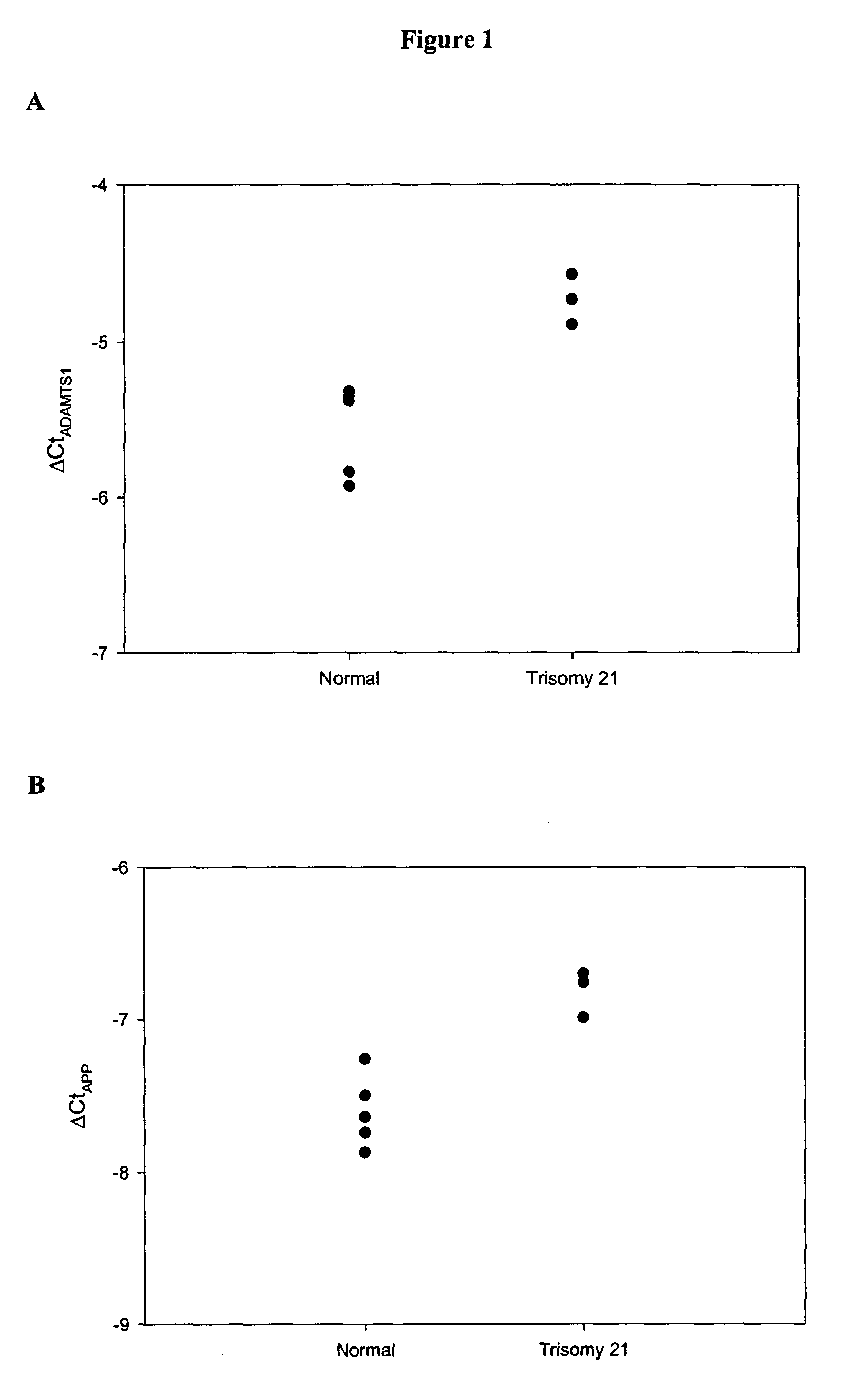
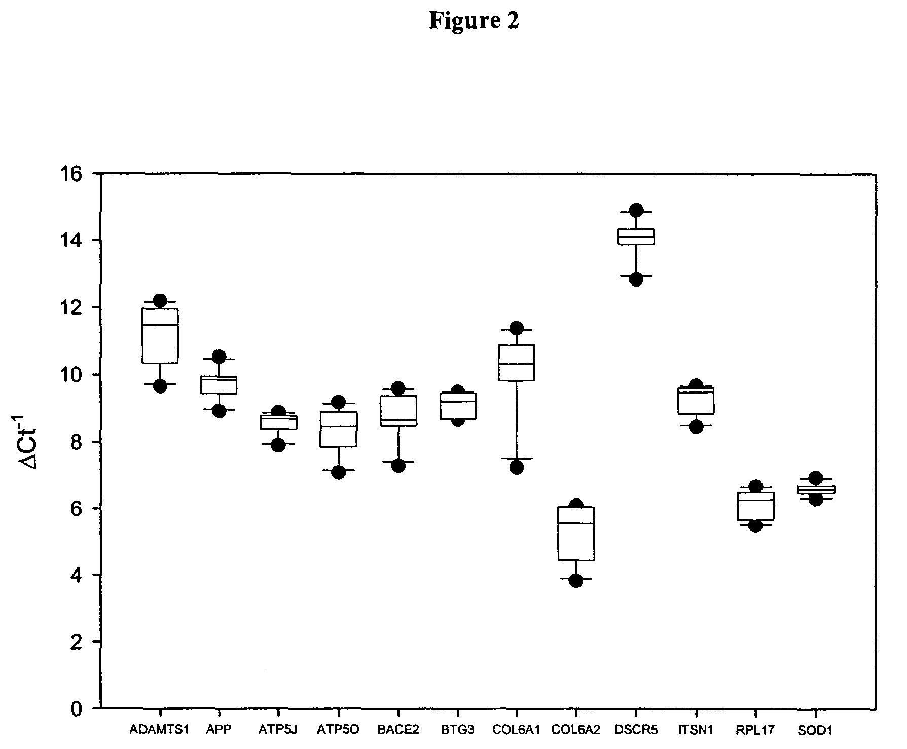
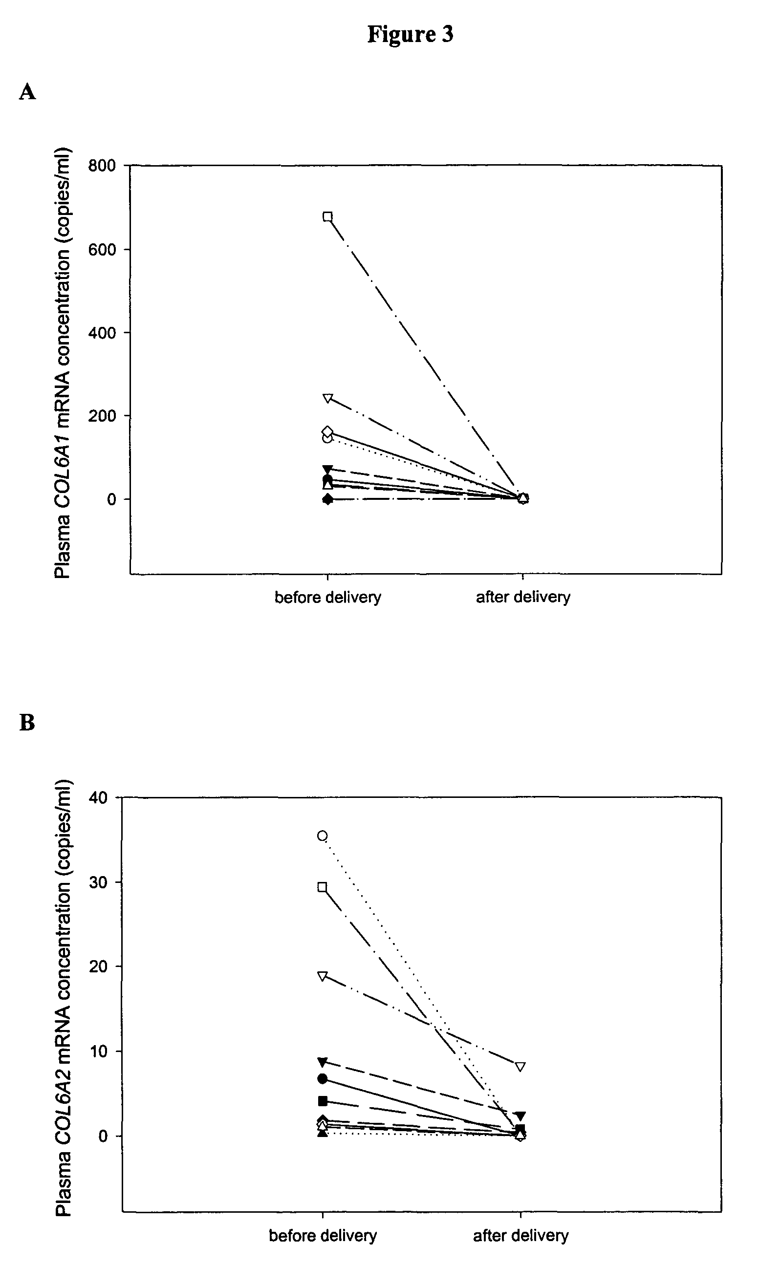
Genes with Increased Expression in Placentas of Trisomy 21 Pregnancies Compared with that of Normal Pregnancies
Methods
Subjects
[0096]All placental tissue and blood samples in this study were collected with informed consent from women in the first trimester of pregnancy, who attended the Department of Obstetrics and Gynecology at the Prince of Wales Hospital, Hong Kong. The study was approved by the Clinical Research Ethics Committee.
[0097]In the first part of the study, placental tissue gene expression profiles of both the normal and trisomy 21 pregnancies were identified by oligonucleotide microarray. First-trimester placental tissue samples were obtained from pregnant women by chorionic villus sampling (CVS). Five women with normal pregnancies (gestational age range: 10-12 weeks) and three pregnant women conceived with trisomy 21 fetuses (gestational age range: 12-13 weeks) were recruited with the respective fetal karyotype subsequently confirmed. In the second part of the study, t...
example 3
Genes with Aberrant Expression in Placentas of Pregnancies Affected by Preeclampsia Compared with that of Normal Pregnancies
Methods
Subjects
[0111]All placental tissue and blood samples in this study were collected with informed consent from women in the third trimester of pregnancy, who attended the Department of Obstetrics and Gynecology at the Prince of Wales Hospital, Hong Kong. The study was approved by the Clinical Research Ethics Committee.
[0112]In the first part of the study, placental tissue gene expression profiles of both normal and preeclamptic (PET) pregnancies were identified by oligonucleotide microarray. Placental tissues from 5 PET pregnant women (gestational age range: 37-40 weeks) and 5 healthy pregnant women (gestational age range: 38-40 weeks) were obtained immediately after cesarean section. Peripheral blood was collected immediately before delivery. In the second part of the study, the gene expression profiles generated from the oligonucleotide microarray experi...
PUM
| Property | Measurement | Unit |
|---|---|---|
| temperature | aaaaa | aaaaa |
| volume | aaaaa | aaaaa |
| diastolic blood pressure | aaaaa | aaaaa |
Abstract
Description
Claims
Application Information