
Apparatus and method for near-field imaging of tissue

Active Publication Date: 2010-05-25
VAN DER WEIDE DANIEL WARREN
View PDF29 Cites 15 Cited by
  • Summary
  • Abstract
  • Description
  • Claims
  • Application Information

AI Technical Summary

Benefits of technology

[0005]Short electrical impulses (in the range of 1 to 100 ps) have correspondingly broad frequency spectra, ranging from the microwave through the millimeter-wave regime. Alternatively, frequency-swept or single-frequency sources covering this range or a subset thereof can provide the required stimulus to perform spectroscopic imaging. Electromagnetic energy in this range has shown efficacy in spectroscopic imaging for detecting both surface and sub-surface anomalies, since, for example, tumors and scars of the skin show strong and specific absorption and dispersion in the 0.001-300 GHz range. Gaining spatial resolution at dimensions much smaller than those normally dictated by the wavelengths being used is done with an array of near-field antennas (e.g. coaxial probe tips). Such arrays channel the electromagnetic energy to and from the object (e.g. tissue surface or sub-surface) enabling a new and informative image of the tissue or cells to be formed. With visible (e.g. fiber-optic) imaging co-located with the array, two types of images can be collected simultaneously and overlaid on a computer screen for a more accurate diagnosis. This can be an aid in diagnosing the type and extent of potentially malignant tumors of the skin. Furthermore, the visible imaging ability can enable an accurate mapping of the skin surface so that an irregular scan of the skin will nevertheless build up a fully registered image.
[0006]The present invention consists of an array of near-field antennas that can image tissue or cells using the reflection of both visible light and micro- and millimeter-wave signals, which can result from electrical pulses, swept continuous-wave wave (CW) signals, or a combination thereof. Such micro- and millimeter-wave dielectric contrast between cancerous and healthy tissue has been resolved on freshly excised skin tumors using these techniques. While visible light helps physicians diagnose the presence of many tumors, the sub-surface condition of the skin or other tissue, hence the extent of the potential tumor, is difficult to assess, and must usually be diagnosed by surgery, which may require successive biopsies. By using an array of near-field probes, which can be co-located with a means for imaging in the visible or sub-visible illumination, the present invention offers a unique tool for diagnosis of tumor boundaries, reducing the pain and disfigurement of surgery, or speeding the achievement of an accurate diagnosis.
[0007]Skin cancer (basal cell carcinoma or BCC) is not only the most common of all cancers, but also is a rapidly growing disease, especially in the United States, where since 1973 the incidence rate for melanoma has more than doubled. While melanoma accounts for only 4% of skin cancers, it causes 79% of deaths from the disease, and 54,200 new cases in the U.S. will be diagnosed in 2003 (American Cancer Society, 2003). Diagnosis of BCC is done visually and with Mohs surgery, which involves repeated removal of tissue and examination under a microscope. Diagnosing the presence of a tumor and knowing its extent without performing surgery would save money, pain and even lives. The need for accurate pathology of specimens in general is extremely high, and even small increments in accuracy will result in lives saved.
[0010]The present technology is based on electronic circuits, and has already proven itself in a variety of other spectroscopic applications. Broadband (as opposed to single-wavelength) imaging has the chief advantage of flexibility: differences in skin composition, even on a single patient, can lead to different diagnoses if only a single-wavelength or narrowband source is used. A source with a broad range of frequencies maximizes the opportunity to detect the signature of a surface and even a sub-surface skin anomaly. Furthermore, time-gating of either a real or a synthetic pulse can enable sub-surface spectroscopic imaging while ignoring the return from the surface. With computer-based control, the present technology allows building of a “trainable” detection system with the capability to ultimately learn the signatures of different skin conditions. Furthermore, the advantages of the present approach to screening over other purely-visible imaging techniques include smaller size, lower cost, and potential for integration directly with other imaging modalities such as ultrasound and visible, infrared and ultraviolet imaging techniques such as cameras.
[0011]The present invention may be carried out to make broadband dielectric reflection measurements of skin. Spectral measurements can be performed to link the dielectric response to composition of the target. Imaging array techniques can be utilized to improve the detection and location of the target, since fiber-optic light pipes can be used as endoscopes. The integrated-circuit nature of the technology enables arrays to be built as extensions of single-pixel proofs-of-principle.

Problems solved by technology

While visible light helps physicians diagnose the presence of many tumors, the sub-surface condition of the skin or other tissue, hence the extent of the potential tumor, is difficult to assess, and must usually be diagnosed by surgery, which may require successive biopsies.

Method used


Image

  • Apparatus and method for near-field imaging of tissue
  • Apparatus and method for near-field imaging of tissue
  • Apparatus and method for near-field imaging of tissue

Examples


Embodiment Construction

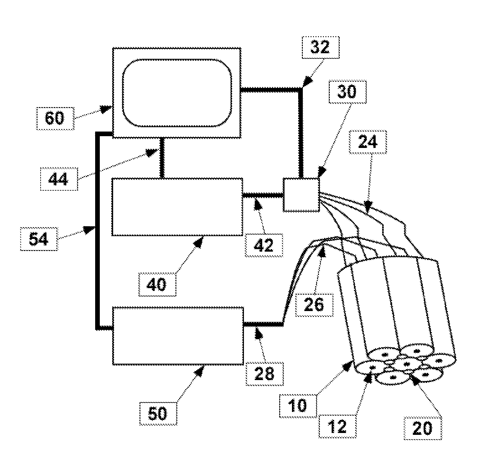
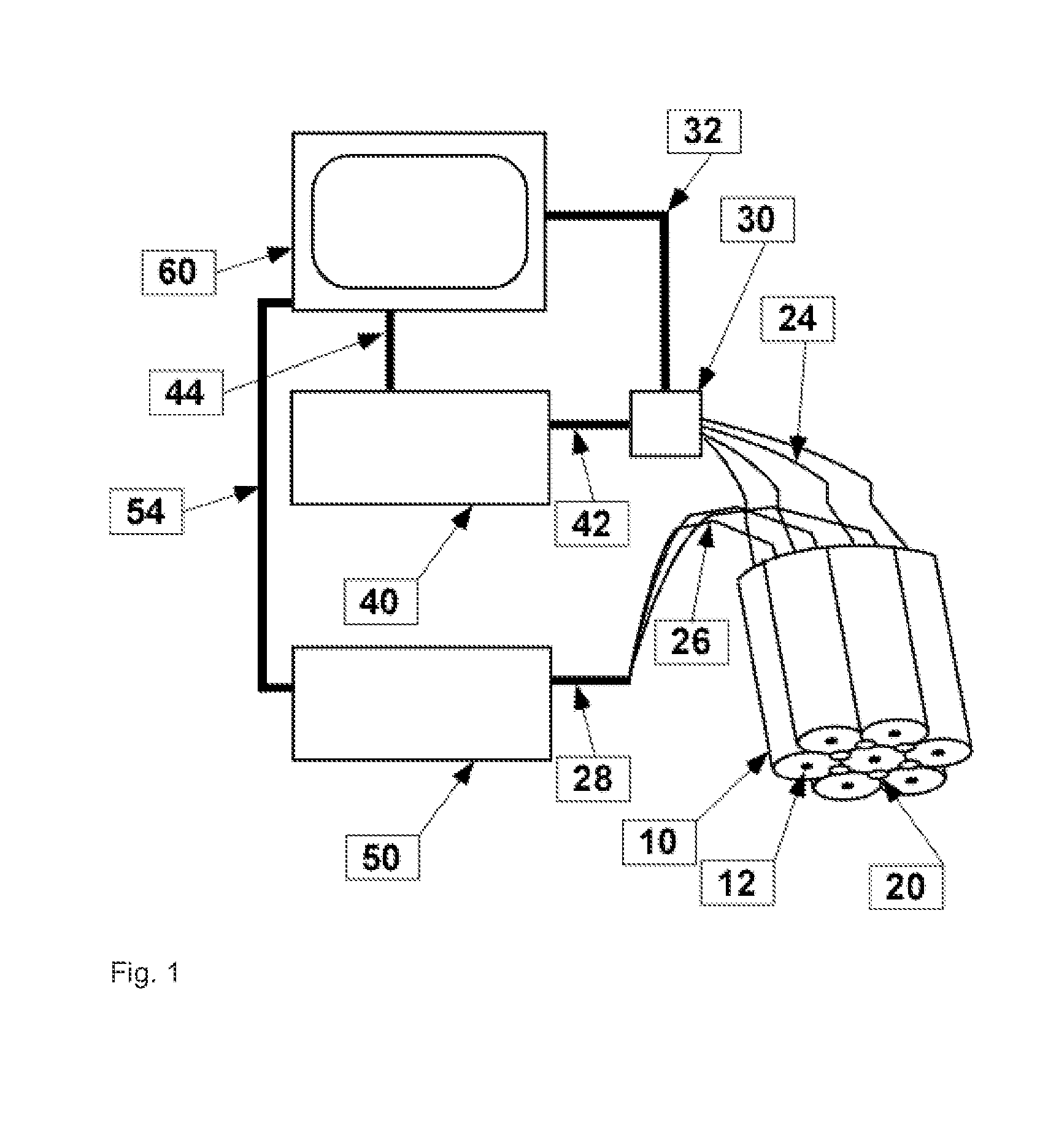
[0019]As illustrated in FIG. 1, an array consisting of one or a plurality of near-field antennas or probes such as open-circuited coaxial transmission lines is connected to computer-controlled instrumentation to effect a scanning spectroscopic imaging system. Each coaxial probe consists of an outer conductive shield 10 and inner conductor 12. Fiber-optic light pipes 20 may be interposed and co-located with the coaxial probes to provide simultaneous optical image collection and mapping of the object. The coaxial cables are connected to broadband microwave and millimeter-wave stimulus / response instrumentation 40 via cables 24, and can be multiplexed by a switch array or power combiner / splitter 30. Alternatively, the present invention provides for the use of compact and inexpensive sources and detectors of this broadband energy, eliminating the need for the multiplexer 30. The fiber-optic light pipes 20 are connected to optical instrumentation such as a light source and camera 50 via f...


PUM


Abstract

Using pulsed or continuous-wave sources, broadband electromagnetic energy generally in the 10 MHz to 300 GHz range is applied through one or a plurality of near-field antennas such as coaxial probe tips. The electromagnetic energy reflected from the surface or transmitted through the near surface of the object (e.g. surface of skin or other tissue or cells) is detected, and the ratio of the test measurement to that from normal tissue is recorded and presented to determine the degree of dielectric contrast, hence inhomogeneity. This degree of contrast is used both on its own and in conjunction with simultaneously acquired optical images to map the boundary of an organ inhomogeneity such as a tumor. The bundle of antennas may be scanned over a surface of the object on a pixel-by-pixel basis to determine the spectra of the sample on a pixel-by-pixel basis, allowing a two dimensional display of the absorption spectra to be provided.

Description

CROSS-REFERENCE TO RELATED APPLICATIONS[0001]This application claims the benefit of U.S. provisional application 60 / 475,181 filed Jun. 2, 2003 entitled Apparatus and Method for Near-Field Imaging of Tissue, hereby incorporated by reference.FIELD OF THE INVENTION[0002]This invention pertains generally to the field of inspection and identification systems for diseases of epithelial tissue or for pathology of tissue preparations.BACKGROUND OF THE INVENTION[0003]While visible light and microscopy helps physicians diagnose the presence of many diseases such as carcinomas, pre-carcinomas and other tumors and related skin diseases, as well as pathologies of tissue and cell preparations (e.g. Pap smears), identification of the disease state is still subject to ambiguity. Having both visible (or fluorescent) and alternative imaging means overlaid on the same presentation would enable the physician to more accurately diagnose equivocal presentations. Alternatively, physicians or technicians w...

Claims


Application Information

Patent Timeline
IPC IPC(8): A61B5/05A61B5/103
CPCA61B5/0062A61B5/05A61B5/6843A61B5/441A61B5/0507
Inventor VAN DER WEIDE, DANIEL WARREN
Owner VAN DER WEIDE DANIEL WARREN
Learn More