Method for imaging the mechanical properties of tissue

a mechanical property and tissue technology, applied in the field of diagnostic imaging, can solve the problems of only measuring static properties of tissue, elastography methods, and method is sensitive to noise and measurement bias, and achieve the effect of reliable results

Active Publication Date: 2010-06-08
THE UNIV OF BRITISH COLUMBIA
View PDF29 Cites 25 Cited by
  • Summary
  • Abstract
  • Description
  • Claims
  • Application Information

AI Technical Summary

Benefits of technology

[0035]A novel approach for calculating tissue properties considers the measurements as inputs and outputs of a linear dynamic system that models the tissue dynamics. Such a method and system can be constructed from a variety of possible components, including conventional low intensity ultrasound with external vibrators. The result is a unique medical imaging technique that can rapidly and robustly measure new aspects of the dynamics properties of tissue.
[0037]The calculation of the mechanical properties is based on a novel approach of creating a specific model of the tissue, and calculating the parameters of the model. By using an excitation containing multiple frequencies and robust system identification techniques, this new imaging approach is extremely effective at calculating the underlying properties of the tissue.
[0041]The first approach uses a lumped parametric model that is derived from finite-element techniques. The finite element model is composed of mass elements connected by springs and dampers. Preferably it is based on realistic models from the field of biomechanics. The method of identifying the parameters of the lumped model is based on fitting the model to the measurements of the motions at each of the elements. The set of identified parameters at each of the measured locations are then used to form an image for display. The effectiveness of this approach is based on the use of multiple measurements from an excitation containing a range of frequencies to produce reliable results in the presence of noise.
[0042]The second approach is based on transfer functions, and has the advantage that no a priori modelling assumptions are required other than linearity. Using the same excitation of tissue motion in the previous approach, a transfer function is calculated between tissue motions at adjacent spatial locations by considering one location to be the input and the adjacent location to be the output of a linear dynamic system. Both the magnitude and phase of the transfer function is obtained. Like the first approach, it is the combination of measurements at multiple time instances and an excitation that has significant frequency content that leads to the transfer function method providing reliable tissue properties results in the presence of measurement noise. After obtaining the transfer function, one or more properties of the transfer function can be computed for display.

Problems solved by technology

The major disadvantage of all static elastography methods is that they measure only static properties of tissue.
In addition, because static elastography uses information at a single frequency (zero Hz or dc), the method is sensitive to noise and measurement bias.
In all four of these dynamic imaging patents, measurements are made of the velocity and resonance behavior of the tissue, but they are not based on modelling the underlying properties of the tissue, such as the elasticity, viscosity and density that produces the resonance behavior.
Moreover, the measurements must be made with an excitation of only one frequency at a time, making the acquisition of data slow.
The requirement that localized shear waves be used constitutes a significant drawback.
This reduces the speed of forming a complete image.
And again, the use of high intensity focused ultrasound poses a possible hazard to the patient.
The drawbacks of this approach include the need for specialized equipment for both producing the oscillating point force and measuring the emissions.
This reduces the speed of forming a complete image.
Both localized methods—tissue response and acoustic emissions—produce images of one or more aspects of the tissue response to the dynamic excitations, but do not identify a specific model of the tissue dynamics.
Thus, this method is restricted to using excitations with periodic amplitudes to be able to reach equilibrium, and where the ratios of the frequencies is an integer.
The requirement that the excitation consist of carefully controlled frequencies and phases in synchronization with magnetic resonance imaging means that a very complicated system is needed compared to other techniques.
Another limitation is the need to reach an equilibrium state before measurements can begin.
While this method does measure dynamic properties of tissue (relative viscosity), it suffers from a number of drawbacks.
The speed of the repeated measurements is limited by the need for the tissue to relax from the step force.
Second, the identification technique Viola et al. use to compute relative stiffness and viscosity relies upon the step response of tissue.
It is well known to experts in parameter identification (see for example L. Ljung, “System Identification, Theory For The User”, Prentice Hall, 1999) that such an approach can fail in the presence of noise.
Furthermore, there may be local minima.
So in summary, the static elastography methods (both local and global excitation methods) are incapable of measuring the dynamic properties of tissue.
The dynamic elastography methods with local excitation have shown an ability to measure some dynamic properties, but the local nature of excitation makes the imaging procedure slow.
The current dynamic methods with global excitation are also slow because of the need to either synchronize with the imaging device after equilibrium, or sweep through a range of excitation frequencies.
Moreover, no dynamic elastography method (including the dynamic methods with either local or global excitations) has so far proposed a method of excitation combining multiple frequency components together, so that robust system identification techniques can be employed to identify the tissue properties.

Method used


Image

  • Method for imaging the mechanical properties of tissue
  • Method for imaging the mechanical properties of tissue
  • Method for imaging the mechanical properties of tissue

Examples


Embodiment Construction

[0062]The following description describes an apparatus and method of imaging the properties of human or animal tissue.

Overview of the Approach

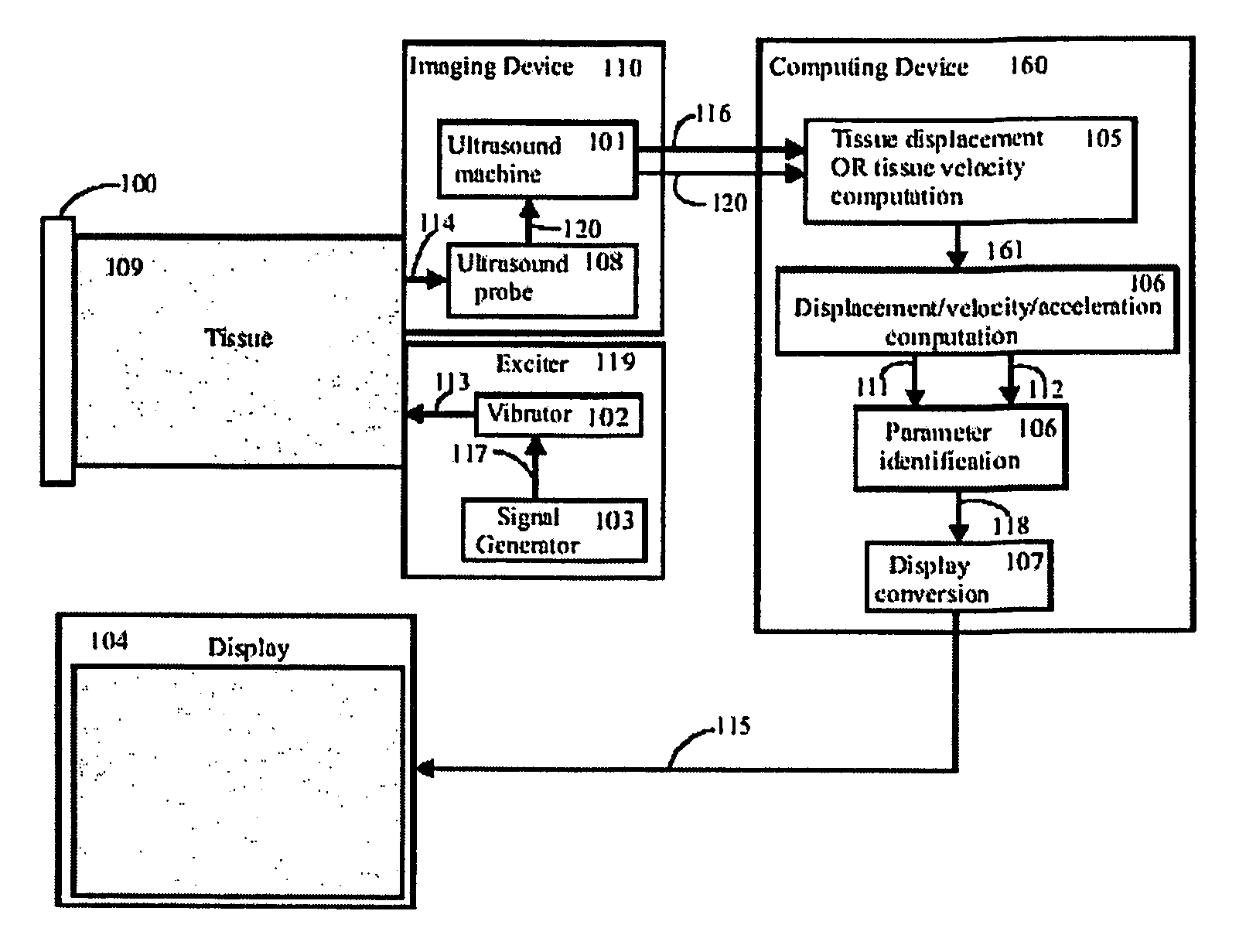
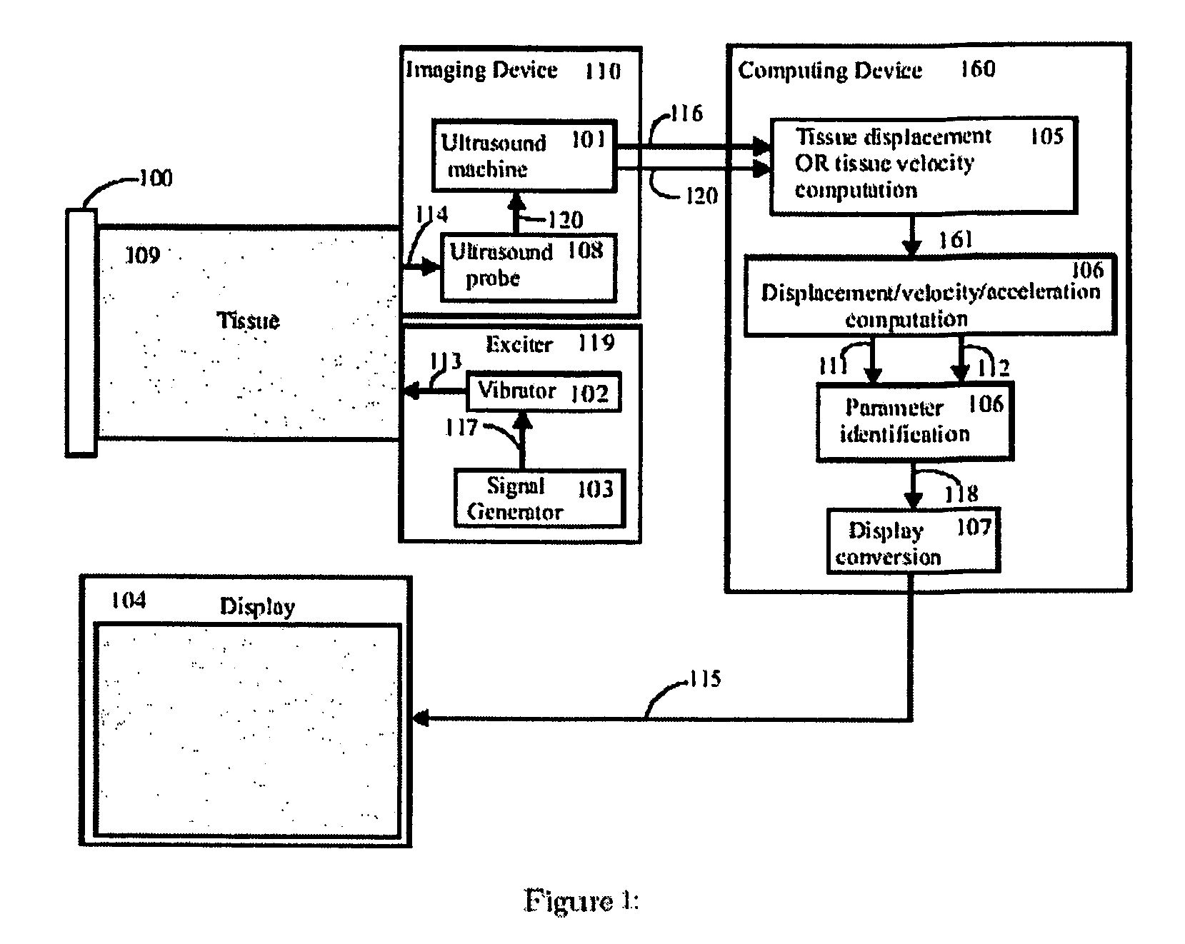
[0063]An embodiment of the present invention is shown in FIG. 1. Broadly, the system has an exciter 119 to create mechanical waves in the tissue, an imaging system 110 to capture tissue displacements and / or velocities from the wave motion, a computing device 160 that calculates tissue viscoelastic parameters from the captured tissue displacements and / or velocities, and a display 104 for displaying information based on the resulting set of parameters.

[0064]The viscoelastic parameters that are computed by 160 are derived by considering specific models of the tissue being excited. The use of an appropriate model of the tissue, together with the identification of the parameters of this model, makes this new imaging approach extremely effective.

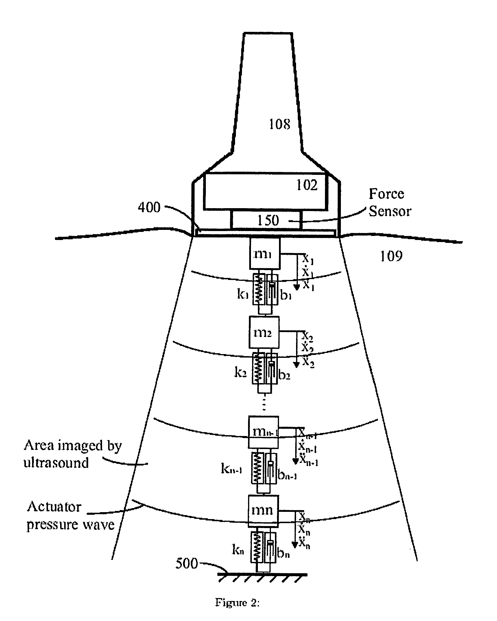
[0065]As shown in FIG. 2 and FIG. 3, the computing block 160 makes use of a tissue model that includes a ...


PUM


Abstract

An imaging system comprises a device to excite mechanical waves in elastic tissue, a device for measuring the resulting tissue motion at a plurality of locations interior to the tissue at a number of time instances, a computing device to calculate the mechanical properties of tissue from the measurements, and a display to show the properties according to their location. A parameter identification method for calculating the mechanical properties is based on fitting a lumped dynamic parametric model of the tissue dynamics to their measurements. Alternatively, the mechanical properties are calculated from transfer functions computed from measurements at adjacent locations in the tissue. The excitation can be produced by mechanical vibrators, medical needles or structures supporting the patient. The measurements may be performed by a conventional ultrasound imaging system and the resulting properties displayed as semi-transparent overlays on the ultrasound images.

Description

REFERENCE TO RELATED APPLICATIONS[0001]This application claims the benefit of U.S. Provisional Application No. 60 / 510,528, filed 14 Oct. 2003.BACKGROUND OF THE PRESENT INVENTION[0002]1. Field of the Invention[0003]This invention relates to diagnostic imaging in general, and to measurement of the mechanical properties of tissue in particular.[0004]2. Description of the Related Art[0005]The basis of medical imaging is the measurement of a property of tissue that varies with tissue composition. Medical images are formed by displaying intensities as a function of these properties measured at multiple locations in the body. From such images, a depiction of anatomy or pathology is gained. Each different imaging modality in common use, such as X-ray, computed tomography, ultrasound and magnetic resonance imaging, measures a different property of tissue.[0006]Mechanical properties of tissue are important indicators of disease potential. Indeed, palpation techniques are commonly used by medi...

Claims


Application Information

Patent Timeline
Patent Type & Authority Patents(United States)
IPC IPC(8): A61B8/00A61B5/103A61B8/08G01S7/52G01S15/00G01S15/89
CPCA61B5/0048A61B8/08G01S15/8977G01S7/52042A61B8/485A61B8/0825A61B8/488G01S15/895A61B5/0051
Inventor SALCUDEAN, SEPTIMIU E.ROHLING, ROBERT N.TURGAY, EMRE
Owner THE UNIV OF BRITISH COLUMBIA
Why Eureka
Try Eureka