Method for using an MRI compatible biopsy device with detachable probe

a biopsy device and compatible technology, applied in the field of imaging assisted tissue sampling, can solve the problems of affecting the accuracy of the biopsy, so as to achieve accurate and efficient results

Active Publication Date: 2010-08-03
DEVICOR MEDICAL PROD
View PDF45 Cites 35 Cited by
  • Summary
  • Abstract
  • Description
  • Claims
  • Application Information

AI Technical Summary

Benefits of technology

[0020]The invention overcomes the above-noted and other deficiencies of the prior art by providing a method whereby a core biopsy is accurately and efficiently performed even with only intermittent Magnetic Resonance Imaging (MRI) scans. The stereotopically derived coordinates of the suspi...

Problems solved by technology

The type of biopsy used depends mainly on various factors present in the patient, and no single procedure is ideal for all cases.
However, existing medical biopsy sampling devices use small, multi-lumen probes extensively fabricated mostly if not entirely from metal.
Typically these metal probes are electrically conductive and often magnetically weak, which interferes with their use under MRI guidance.
The electrically conductive and magnetically weak nature of metal probes often work to create field distortions, called artifacts, on the image.
The image of the lesion will show the metal probe, and this is problematic because the image of the probe can obscure the image of the lesion.
The small sample size of conventional biopsy needles also presents a significant limitation due to the increase in the duration of the procedure.
This limits the number of samples that can be retrieved using conventional spring-loaded core biopsy needles under direct imaging guidance.
A further problem not infrequently encountered during core needle biopsy is the development of a hematoma at the biopsy site during the procedure.
An accumulating hematoma can be problematic during MRI-guided biopsy because residual contrast agent circulating in the hematoma can mimic enhancement in a suspicious lesion.
In addition, the accumulation of air at the biopsy site can cause susceptibility artifacts that can potentially interfere with the fat-suppre...

Method used


Image

  • Method for using an MRI compatible biopsy device with detachable probe
  • Method for using an MRI compatible biopsy device with detachable probe
  • Method for using an MRI compatible biopsy device with detachable probe

Examples


Embodiment Construction

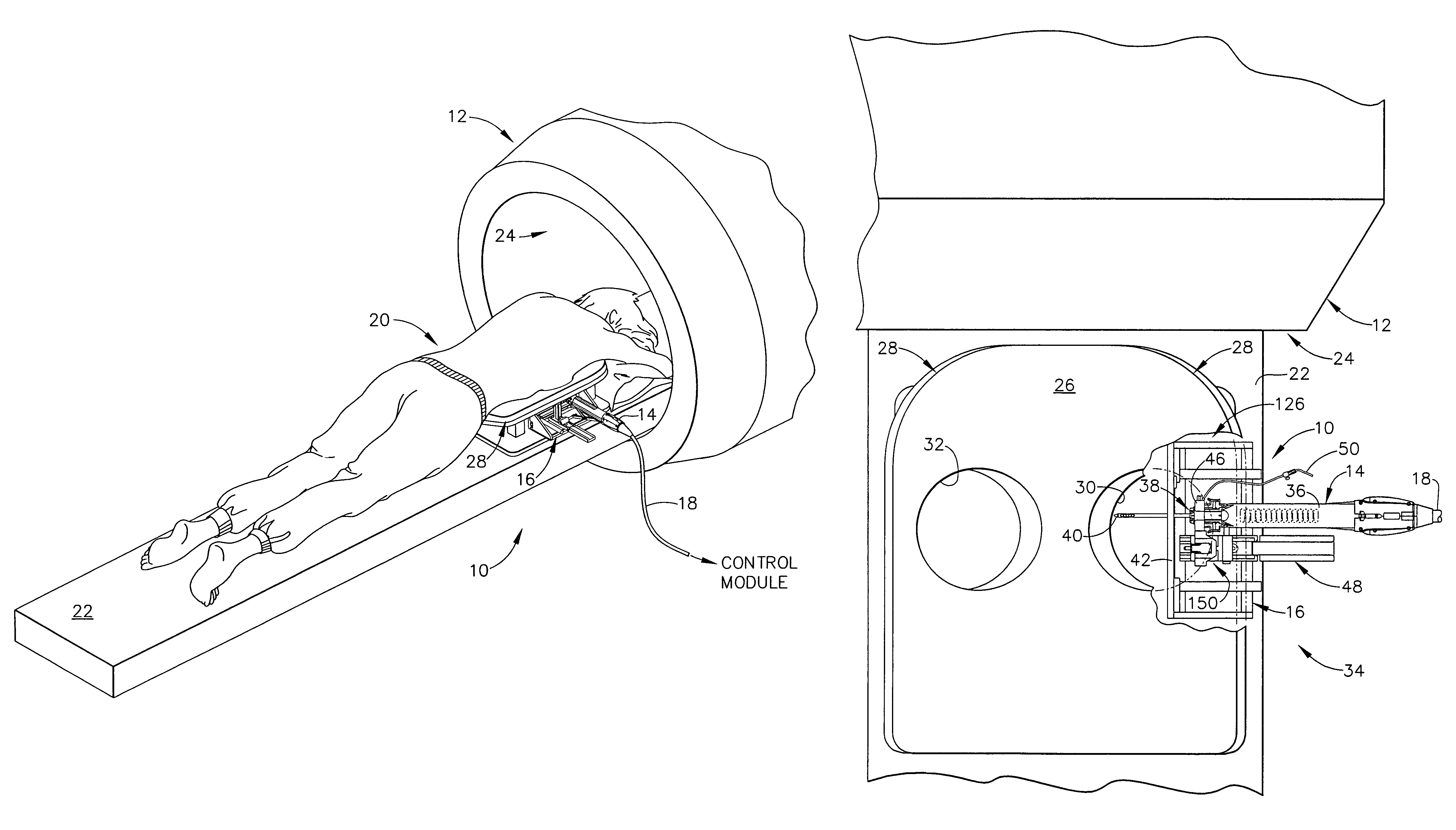
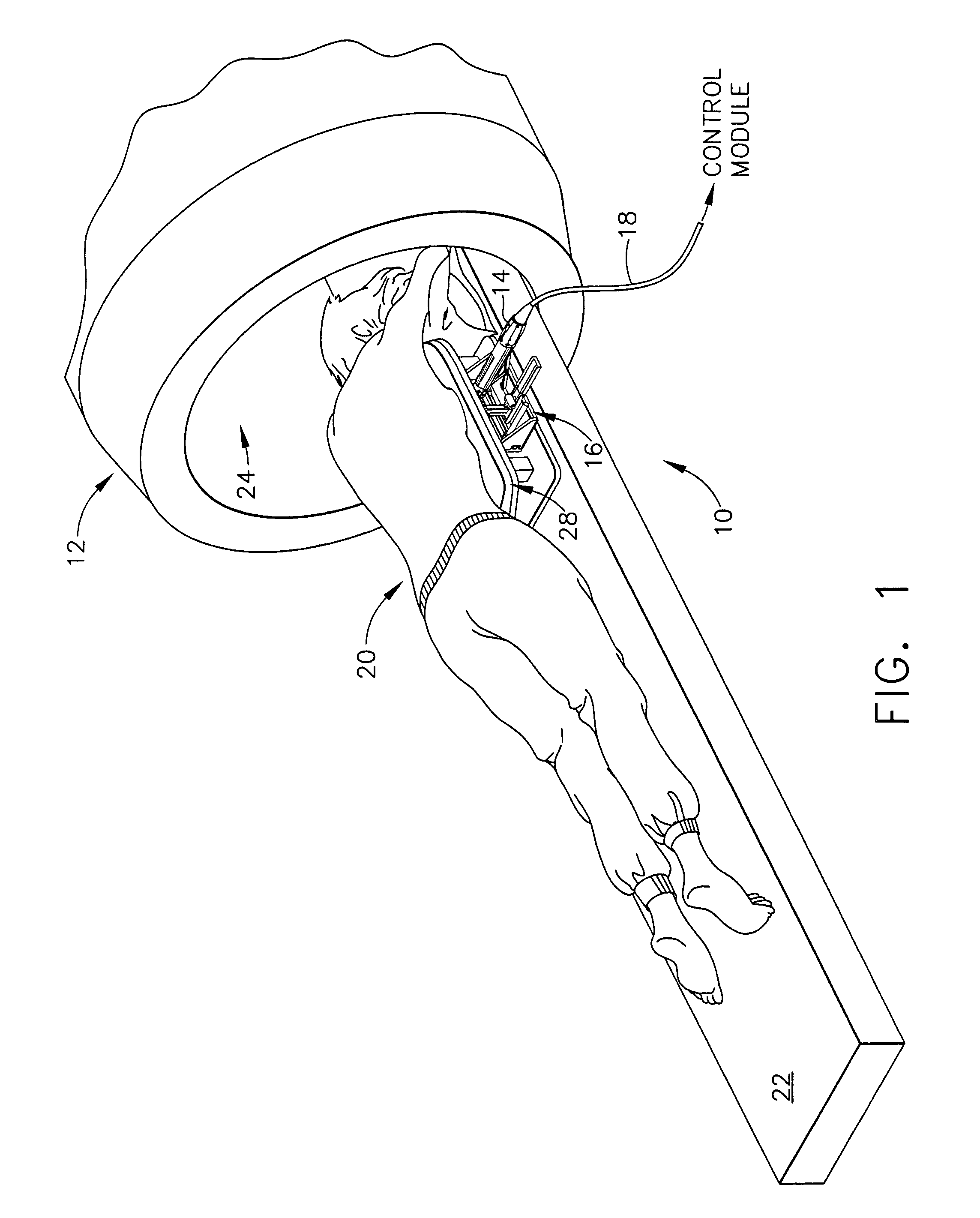
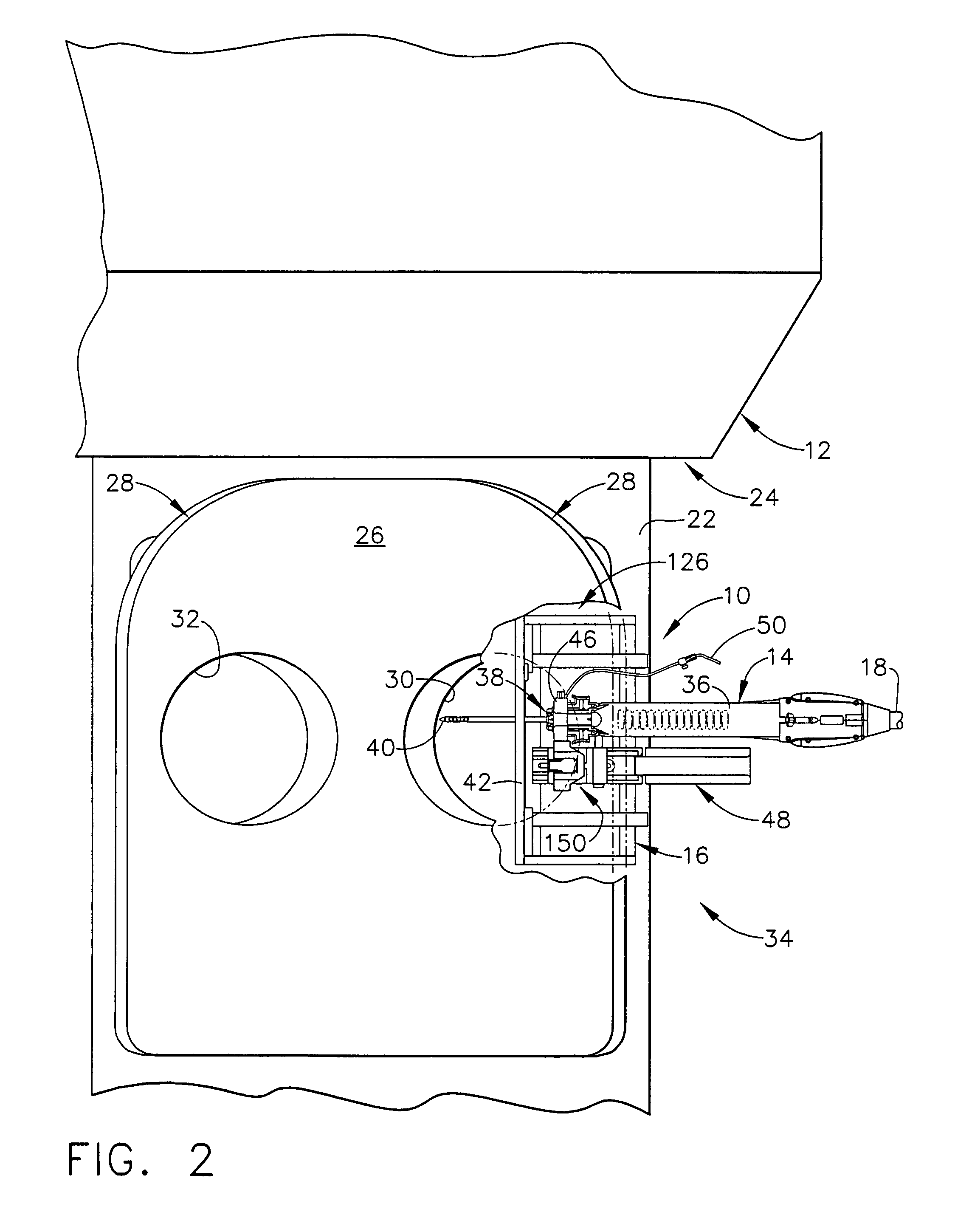
[0047]FIG. 1 depicts a core biopsy instrument system 10 that is vacuum assisted, detachable, and compatible with use in a Magnetic Resonance Imaging (MRI) machine, such as the depicted closed MRI machine 12. In the illustrative embodiment, the core biopsy instrument system 10 includes an MRI-compatible biopsy tool 14 that is selectably attached to a localization mechanism or fixture 16 to accurately and rapidly perform core biopsies of breast tissue with a minimum of insertions of a biopsy probe. A control module (not shown) senses encoder position signal and switch signals from the biopsy tool 14 and provides mechanical and vacuum power to the biopsy tool 14 via power cord 18.

[0048]With reference to FIGS. 1-2, a patient 20 is lying prone upon a patient support table 22, depicted in FIG. 1 as removed from a magnet bore 24 of the MRI machine 12. The patient's chest rests upon a top surface 26 of a chest support 28, the top surface 26 having openings 30, 32 for allowing the patient's ...


PUM


Abstract

A method for performing Magnetic Resonance Imaging (MRI) guided core biopsies is tendered more accurate and efficient by precisely positioning a disengaged probe assembly with respect to a localization fixture attached to a breast coil platform. The precise position is defined by MRI stereotopic location of suspicious tissue with respect to a fiducial marker on the localization fixture. With the probe inserted, dual lumens in the probe assembly are used for drainage or insertion of fluids as well as inserting diagnostic and therapeutic tools. Core biopsies are performed by engaging a biopsy instrument handle containing a cutter, with the localization fixture providing support and position to the handle. Repeated MRI scans are facilitated by the ability to disengage the handle without risk of displacing the probe assembly from the biopsy site.

Description

CROSS REFERENCE TO RELATED APPLICATIONS[0001]The present application hereby claims the benefit of the provisional patent application of the same title, Ser. No. 60 / 374,807 filed on Apr. 23, 2002. The present application is related to co-pending and commonly-owned applications filed on even date herewith entitled “LOCALIZATION MECHANISM FOR AN MRI COMPATIBLE BIOPSY DEVICE” to Hibner et al. and “AN MRI COMPATIBLE BIOPSY DEVICE WITH DETACHABLE PROBE” to Hibner et al., the disclosure of both is hereby incorporated by reference in their entirety.FIELD OF THE INVENTION[0002]The present invention relates, in general to a method of imaging assisted tissue sampling and, more particularly, to an improved method for positioning a biopsy probe with respect to a magnetic resonance imaging (MRI) breast coil for acquiring subcutaneous biopsies and for removing lesions.BACKGROUND OF THE INVENTION[0003]The diagnosis and treatment of patients with cancerous tumors, pre-malignant conditions, and other...

Claims


Application Information

Patent Timeline
IPC IPC(8): A61B5/05A61B5/055A61B10/00G01R33/28A61B10/02A61B17/34A61B19/00
CPCA61B10/0275A61B17/3403A61B2017/00911A61B2019/5236A61B2019/5454A61B2090/374A61B2090/3954
Inventor HIBNER, JOHN ANTHONYFREEMAN, LYNETTA JEANSEBERN, ELIZABETH LYNNPYZOHA, JESSICA MARYMCCOY, TERRY DARNELL
Owner DEVICOR MEDICAL PROD
Why Eureka
Try Eureka