
Therapeutic and diagnostic uses of antibody specificity profiles

a technology of antibody specificity and therapeutic use, which is applied in the field of therapeutic and diagnostic use of antibody specificity profiles, can solve the problems of astonishing complexity of the immune system, difficult diagnosis and therapy, and diversity

Inactive Publication Date: 2010-08-31
THE BOARD OF TRUSTEES OF THE LELAND STANFORD JUNIOR UNIV
View PDF31 Cites 3 Cited by
  • Summary
  • Abstract
  • Description
  • Claims
  • Application Information

AI Technical Summary

Benefits of technology

[0011]The invention also provides a method for the identification of patients likely to develop a more severe form of disease, enabling selection of more aggressive therapy based on a patient's antibody specificity profile.
[0013]In yet another aspect of the invention, there are methods for determining the antibody specificity profile for monitoring therapeutic response in patient receiving treatment for immune-related disorders. Therapeutic responses are assessed based on alterations in the antibody specificity profile including changes in antibody targets (i.e. the antigen or epitope profile), changes in antibody titers, changes in antibody isotypes, and changes in large-scale patterns of antibody recognition. In another embodiment, antibody specificity profiles can be utilized to predict adverse outcomes in individual patients, thereby enabling selection of alternative therapies.

Problems solved by technology

The astonishing complexity of the immune system is its greatest strength.
But this diversity has its drawbacks.
Under these circumstances, the complexity of the system makes diagnosis and therapy extremely difficult.
For most autoimmune diseases, atopic states and undesired immune responses, no effective diagnostic blood tests or therapeutic agents exist.
Since this treatment is not antigen specific, the result is a global decrease in immune function, resulting in susceptibility to infection and disease.
Achieving diagnosis is particularly difficult where there is a strong T cell component to the autoimmune disease, due to the T cell receptor recognition of antigen only when bound to an appropriate major histocompatibility protein.
Although precise identification of the cognate antigen for T cells remains challenging (see Altman et al.

Method used


Image

  • Therapeutic and diagnostic uses of antibody specificity profiles
  • Therapeutic and diagnostic uses of antibody specificity profiles
  • Therapeutic and diagnostic uses of antibody specificity profiles

Examples


example 1

Antigen Array Characterization of the Specificity of the Autoantibody Response in EAE in Rats and Mice

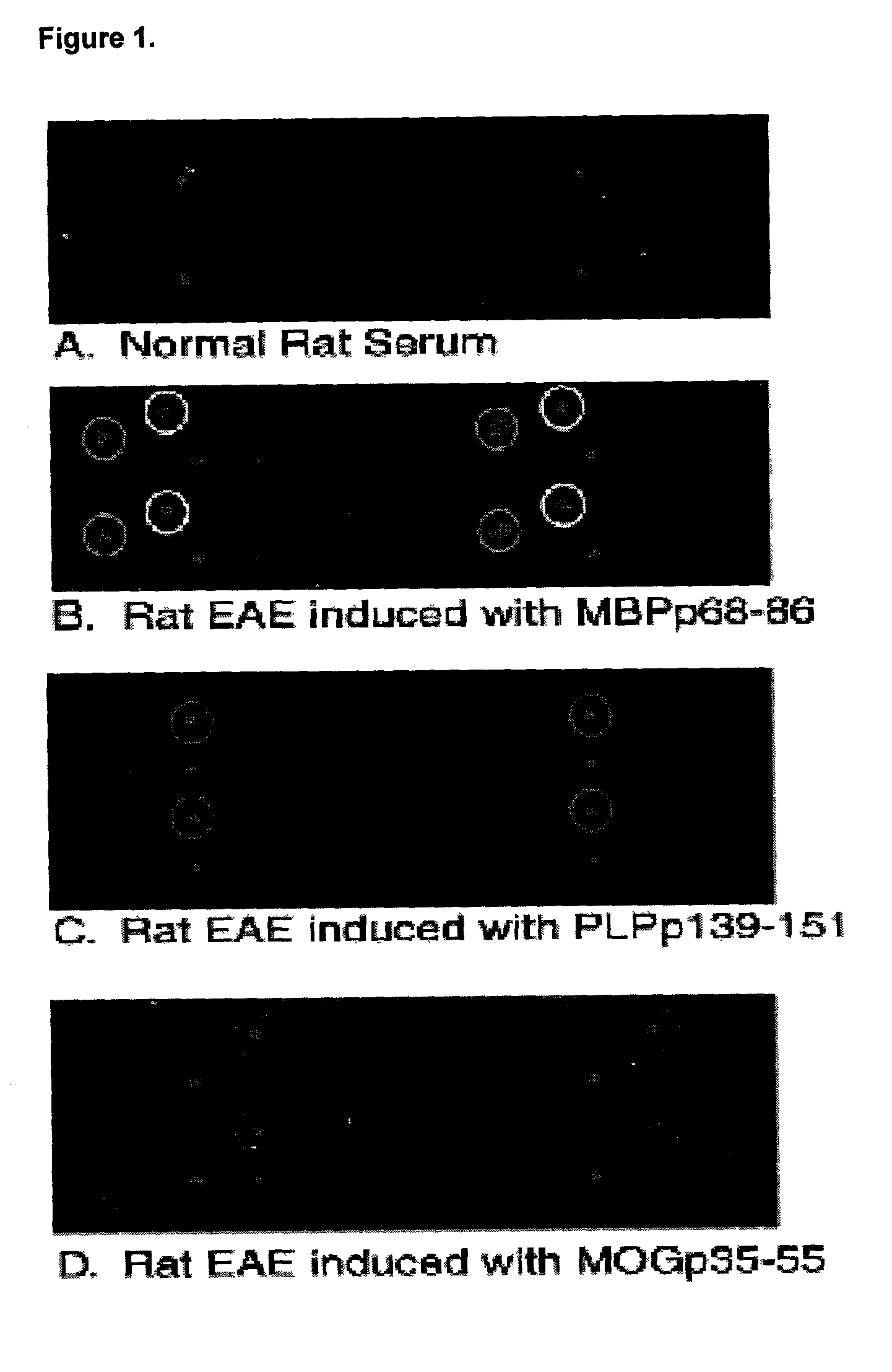
[0117]To determine the autoantibody specificity profile in the serum of animals with EAE, an assay was performed testing the binding of serum antibodies to a protein microarray.

[0118]Antigen arrays were generated by spotting the immunodominant myelin peptide epitopes and purified native myelin basic protein, as listed in Table 1. FIGS. 1A-D are scanned imaged of arrays probed with serum from a normal control rat (A) and 3 Lewis rats induced to develop EAE with different myelin protein peptides in complete Freund's adjuvant (B-D), followed by Cy-3 labeled anti-rat Ig secondary antibody.

[0119]Quantitative computer analysis of the scanned images is presented in Table 1. The reported numbers represent the ratio of the fluorescence intensity of antigens recognized by serum derived from animals with EAE relative to control serum adjusting for background levels such that ratios greater or ...

example 2

Determination of the Patient Antibody Specificity Profile in Autoimmune Rheumatic Diseases

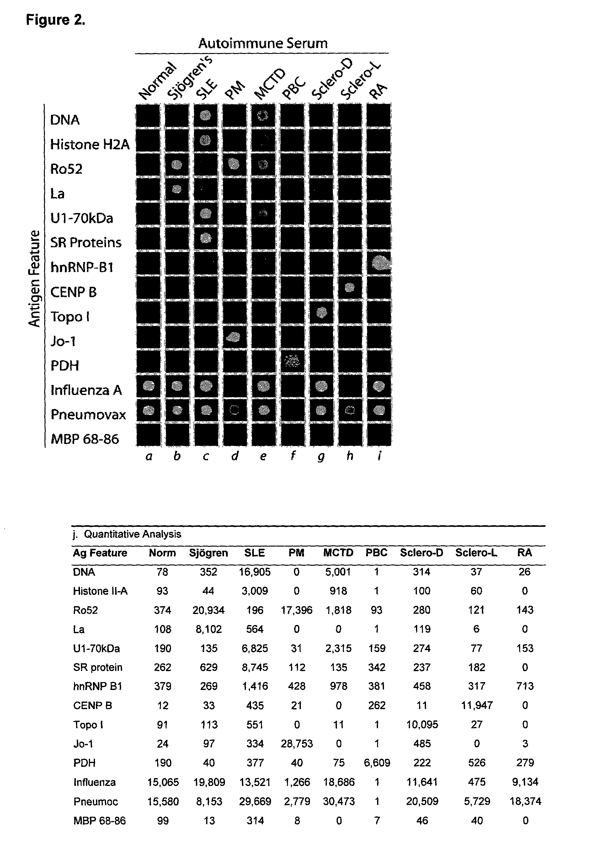
[0121]Autoantigen arrays identify autoantibody profiles characteristic of eight distinct human autoimmune rheumatic diseases. Representative examples of autoantigen array analysis of over 50 highly-characterized autoimmune serum samples are presented (FIG. 2).

[0122]Ordered autoantigen arrays were generated by spotting 192 distinct putative autoantigens in quadruplicate sets using a robotic microarrayer to create a 1152-feature rheumatic disease autoantigen array. Spotted antigens include: 36 recombinant or purified proteins including Ro52, La, histidyl-tRNA synthetase (Jo-1), SR proteins, histone H2A (H2A), Sm-B / B′, the 70 kDa and C component of the U1 small nuclear ribonucleoprotein complex (U1-70 kDa, U1snRNP-C), Sm-B / B′, hnRNP-B1, Sm / RNP complex, topoisomerase I (topo I), centromere protein B (CENP B), and pyruvate dehydrogenase (PDH); six nucleic acid-based putative antigens including sever...

example 3

Determination of the Autoantibody Specificity Profile from the Cerebral Spinal Fluid from Human Multiple Sclerosis Patients

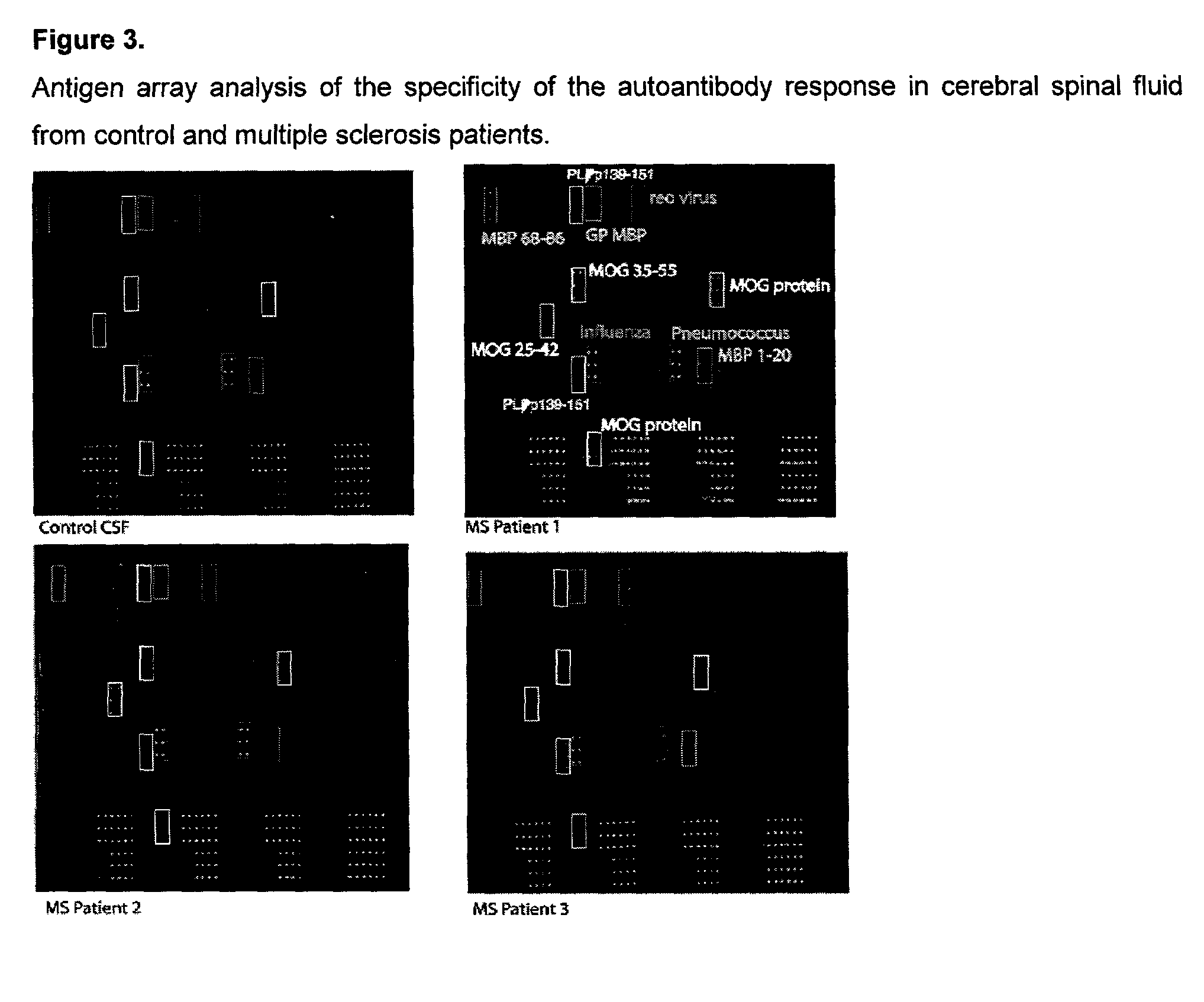
[0125]Antigen arrays containing myelin proteins and peptides were used to characterize the specificity of the autoantibody response in cerebral spinal fluid samples derived from human patients with multiple sclerosis (FIG. 3). The arrays in FIG. 3 demonstrate detection of cerebral spinal fluid autoantibodies specific for whole myelin basic protein (MBP), MBP peptide 1-20 (corresponding to amino acids 1-20 of MBP), MBP peptide 68-86, whole myelin oligodendrocyte protein (MOG), MOG peptide 35-55, and proteolipid protein (PLP) peptide 139-151. As in Example 2, such information can be used for diagnosis and to develop, monitor and select specific therapy.

[0126]2400-spot ‘myelin proteome’ arrays were produced by spotting these putative myelin antigens using a robotic microarrayer, probed with cerebral spinal fluid followed by Cy-3-labeled anti-human Ig secondary anti...


PUM

PropertyMeasurementUnit
Massaaaaaaaaaa
Timeaaaaaaaaaa
Timeaaaaaaaaaa

Abstract

This invention provides a method for determining the antibody specificity profile in an individual. This specificity profile reveals the individual's immune response to multiple antigens and / or epitopes of autoantigens, allergens, graft antigens, etc. The antibody specificity profile is determined through the binding of patient samples comprising antibodies to the arrays. The array can comprises antigens and epitopes. The invention also provides the means and methods for determining antigen or epitope specificity profiles that can be used in the development of either generic and individualized diagnosis and treatment for immune related diseases, including autoimmune disease, allergy and graft rejection.

Description

BACKGROUND OF THE INVENTION[0001]The astonishing complexity of the immune system is its greatest strength. The 1012-1014 possible antibody specificities, the delicate interplay between the various regulatory and effector cells, the restriction of T cell responses according to MHC antigens; all these contribute to the ability of the host to effectively react against infectious agents and other antigens perceived as foreign. But this diversity has its drawbacks. Mistakes happen: the target of a response may turn out to be a normal self protein; inflammatory responses are misregulated; and normal responses are undesirably directed against grafts and transplanted cells. Under these circumstances, the complexity of the system makes diagnosis and therapy extremely difficult.[0002]For most autoimmune diseases, atopic states and undesired immune responses, no effective diagnostic blood tests or therapeutic agents exist. For example, current therapeutic strategies are often based on system-w...

Claims


Application Information

Patent Timeline
IPC IPC(8): G01N33/00A61K39/00G01N33/53A61K39/35A61P1/18A61P3/10A61P11/06A61P19/02A61P25/28A61P29/00A61P37/00A61P37/06A61P37/08A61P43/00C12N15/09G01N33/564G01N33/68G01N37/00
CPCA61K39/0008G01N33/564G01N33/6854G01N2800/102G01N2800/24G01N2800/52A61P1/18A61P11/06A61P19/02A61P25/28A61P29/00A61P37/00A61P37/06A61P37/08A61P43/00A61P5/50A61P3/10
Inventor ROBINSON, WILLIAM H.HIRSCHBERG, DAVID L.STEINMAN, LAWRENCERUIZ, PEDRO JOSEUTZ, PAUL J.GARREN, HIDEKI
Owner THE BOARD OF TRUSTEES OF THE LELAND STANFORD JUNIOR UNIV
Learn More