Neuronal progenitor cells from hippocampal tissue and a method for isolating and purifying them

a technology of neural progenitor cells and hippocampal tissue, which is applied in the field of separating cells and hippocampal neural progenitor cells, can solve the problems of the failure of the mature brain to generate new neurons, the relative scarcity of adult brain tissue, and the inability of the damaged brain to function as structurally significant self-repair

Inactive Publication Date: 2010-08-31
CORNELL RES FOUNDATION INC
View PDF23 Cites 2 Cited by
  • Summary
  • Abstract
  • Description
  • Claims
  • Application Information

AI Technical Summary

Benefits of technology

[0017]Extension of this approach to include fluorescent transgenes under the control of stage- and phenotype-specific promoters (both of which are intended to be covered by reference to “cell-specific” promoters herein) allows even more specific separations to be performed, for example, of hippocampal neural progenitors over a range of developmental stages. This strategy permits sufficient enrichment for in vivo implantation of the defined and separated progenitor pools, as well as for in vitro analyses of phenotypic specification and growth control.
[0018]By providing a means of identifying hippocampal neural progenitor cells while alive, even when present in small numbers in mixed populations, the use of fluorescent transgenes driven by cell type-selective promoters such as the Tα1 tubulin promoter and the nestin enhancer will allow the specification of phenotype to be studied and perturbed on the single cell level, an approach that had previously only been feasible on larger populations. Indeed, when used in conjunction with post-transfection fluorescence-activated cell sorting (FACS), this strategy may permit the enrichment and purification of any cell type for which stage- or phenotype-specific promoters are available.
[0019]The method of the present invention provides a new strategy for the isolation and purification of hippocampal neural progenitor cells, from the adult brain. These cells may be used in both basic analyses of precursor and stem cell growth control, as well as in more applied studies of their transplantability and engraftment characteristics. Generally, by providing a means to identify and enrich neural precursor cells from adult brain, this strategy may allow a significant acceleration in the study of precursor and stem cell biology, as well as providing native unpassaged adult precursor cells in sufficient number for implantation studies. As such, this approach may spur the development of induced adult neurogenesis as a viable therapeutic modality for the structural repair of the damaged central nervous system.

Problems solved by technology

The damaged brain is largely incapable of functionally significant structural self-repair.
This is due in part to the apparent failure of the mature brain to generate new neurons (Korr, 1980; Sturrock, 1982).
However, the absence of neuronal production in the adult vertebrate forebrain appears to reflect not a lack of appropriate neuronal precursors, but rather their tonic inhibition and / or lack of post-mitotic trophic and migratory support.
A major impediment to both the analysis of the biology of adult neural precursors, and to their use in engraftment and transplantation studies, has been their relative scarcity in adult brain tissue, and their consequent low yield when harvested by enzymatic dissociation and purification techniques.
As a result, attempts at either manipulating single adult-derived precursors or enriching them for therapeutic replacement have been difficult.
However, hippocampal progenitor cells have never been isolated.
Yet despite the widespread incidence of hippocampal neurogenesis in adult animals, native hippocampal progenitor cells have never been separated and purified as such, in rodents or humans.
As a result, no assessment of the abundance, factor-responsiveness, or regenerative capacity of these cells has been possible.

Method used


Image

  • Neuronal progenitor cells from hippocampal tissue and a method for isolating and purifying them
  • Neuronal progenitor cells from hippocampal tissue and a method for isolating and purifying them
  • Neuronal progenitor cells from hippocampal tissue and a method for isolating and purifying them

Examples


example 1

Materials and Methods

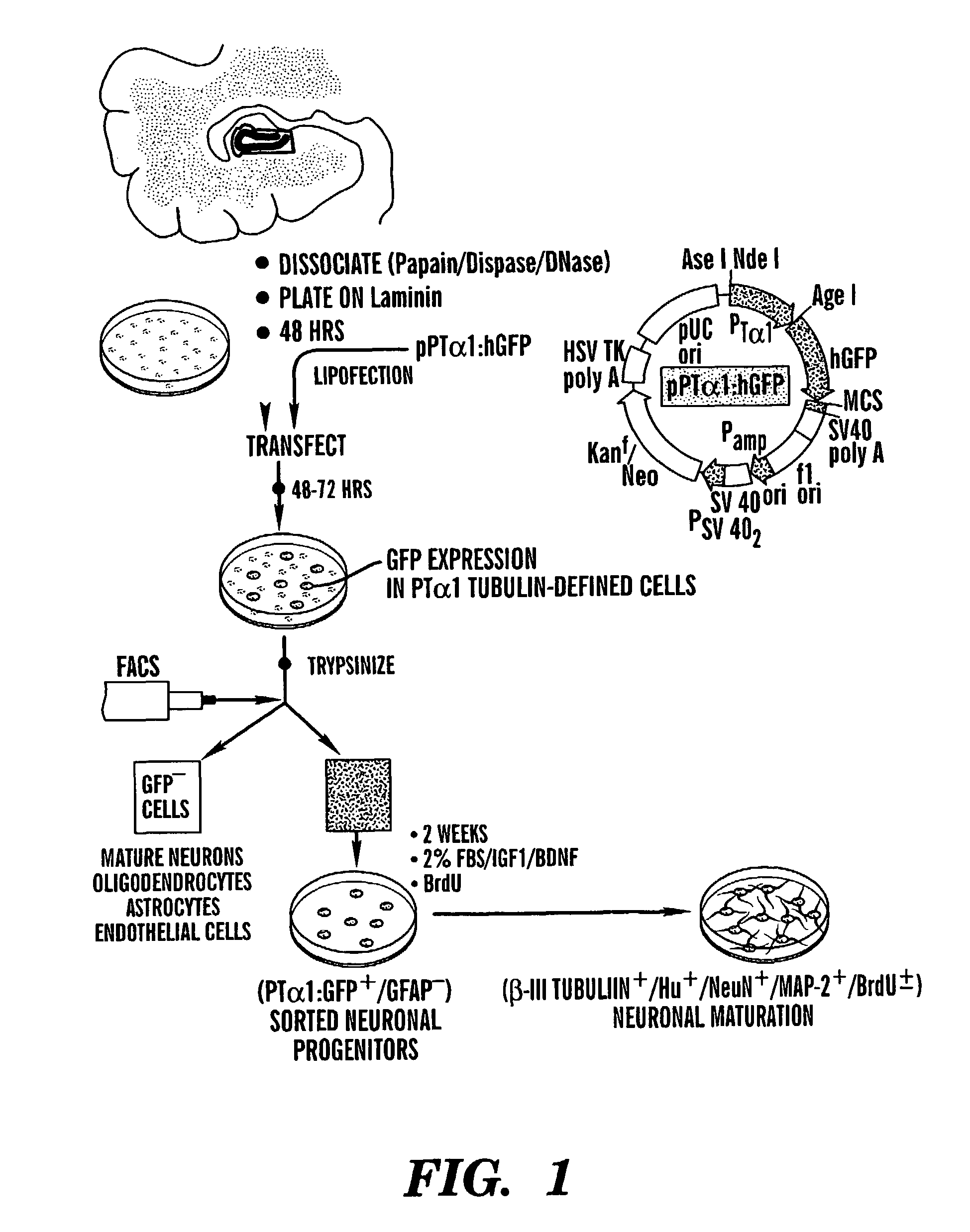
Adult Human Hippocampal Dissociation and Culture

[0056]Adult human brain tissue was obtained in the course of temporal lobectomy, as described (Pincus et al., 1998b; Kirschenbaum et al., 1994). The hippocampus was dissected free of the ventricular surface, and the dentate gyrus then dissected from the rest of the tissue. This sample was cut into pieces of roughly 2 mm on edge, rinsed twice with PIPES (120 mM NaCl, 5 mM KCl, 25 mM glucose, 20 mM PIPES), and dissociated in papain / DNase, as described (Roy et al., 1999; Doetsch et al., 1999). The dispersed cells were collected and rinsed with DMEM / F12 / N2 containing 20% platelet-depleted FBS (PD-FBS, Cocalico, Reamstown, Pa.) to stop the enzymatic dissociation, and resuspended at 1×10⁷ cells / ml in DMEM / F12 / N2 with 2% PD-FBS and 10 ng / ml FGF-2 (Sigma, St. Louis, Mich.). The cells were plated at 0.1 ml / dish into 35 mm Falcon Primaria plates coated with laminin (2 μg / cm2), at 37° C. in 5% CO2. Four hours later, an additi...

example 2

[0064]Neurons can be Harvested in High-Yield from the Adult Human Dentate Gyrus

[0065]To characterize mitotic cell types harvestable from the adult human hippocampus, papain dissociates of surgically-resected hippocampus were obtained from eight patients. All were males, ranging from 5 to 63 years old. Four patients had temporal lobe resections for medication-refractory epilepsy (Pincus et al., 1998b); two were subjected to decompressive resection during or after extra-axial meningioma removal, one sample was taken during aneurysm repair, and one during decompression for traumatic edema. In each, the dentate gyrus was dissected free of the temporal ventricular zone, and the two removed separately. The dentate was dissociated using papain / Dnase (Palmer et al., 1997); the resultant cultures were raised in DMEM / F12 / N2, with 20 ng / ml FGF2 and 2% platelet-depleted fetal bovine sera (PD-FBS). A mitotic marker, bromodeoxyuridine (BrdU; 10 μg / ml) was added at 6 hours in vitro. Cultures were ...

example 3

Persistent Neurogenesis by Mitotic Progenitors in Adult Hippocampal Cultures

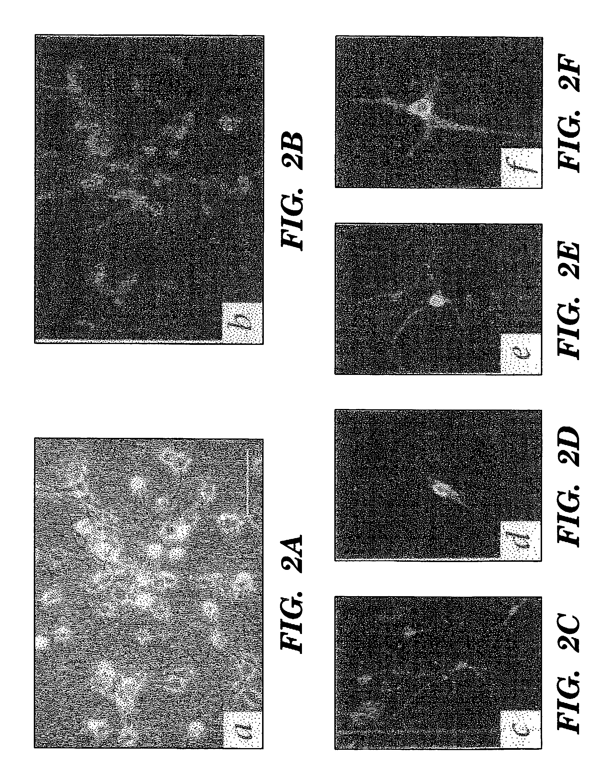
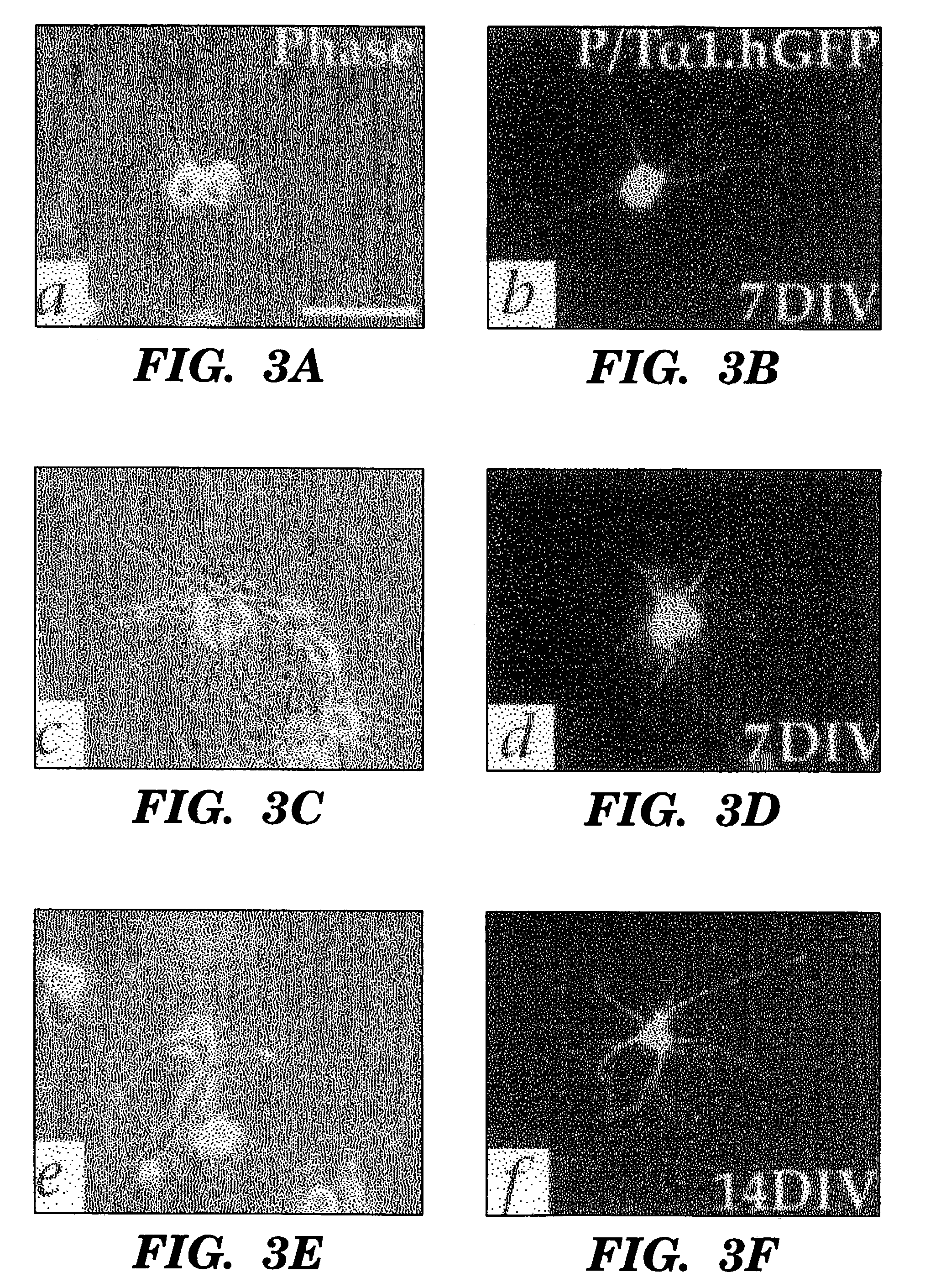
[0067]A substantial proportion of the antigenically-confirmed neurons incorporated BrdU from the culture media, indicating their genesis in vitro (FIGS. 2A-F). BrdU+ / βIII-tubulin+ neurons constituted 25.4±5.9% of all βIII-tubulin-defined neurons at 7 DIV. At this same time, BrdU+ / MAP-2+ cells were rare, consistent with the relatively late appearance of MAP-2 after neuronal mitogenesis. But by 14 DIV, 3.0±1.0% of all cells in these cultures expressed MAP-2, and of these, 22.5±7.3% were BrdU+.

[0068]The persistence of neurogenesis in these dissociates was also manifested by the sustained neuronal incorporation of BrdU in cultures to which the marker was first added after five days in culture. Among plates first exposed to BrdU on day 5 in vitro and fixed on day 7, 8.2±1.9% of the cells expressed TuJ1; this was roughly identical to the 8.9±2.8% incidence of TuJ1+ neurons in the plates exposed to BrdU from the ou...


PUM

PropertyMeasurementUnit
timeaaaaaaaaaa
diameteraaaaaaaaaa
diameteraaaaaaaaaa

Abstract

The present invention relates to an enriched or purified preparation of isolated hippocampal neural progenitor cells and progeny thereof. The present invention also relates to a method of separating neural progenitor cells from a mixed population of cell types from hippocampal tissue. This method includes selecting a promoter which functions selectively in the neural progenitor cells, introducing a nucleic acid molecule encoding a fluorescent protein under control of said promoter into all cell types of the mixed population of cell types from hippocampal tissue, allowing only the neural progenitor cells, but not other cell types, within the mixed population to express said fluorescent protein, identifying cells of the mixed population of cell types that are fluorescent, which are restricted to the neural progenitor cells, and separating the fluorescent cells from the mixed population of cell types, wherein the separated cells are restricted to the neural progenitor cells.

Description

[0001]This application claims the benefit of U.S. Provisional Patent Application Serial No. 60 / 176,677, filed Jan. 18, 2000, which is hereby incorporated by reference.[0002]The subject matter of this application was made with support from the United States Government under grants RO1 NS29813 and RO1 NS33106 of the National Institutes of Health.FIELD OF THE INVENTION[0003]The present invention relates generally to hippocampal neural progenitor cells and a method of separating cells of interest, in particular neural progenitor cells, from hippocampal tissue.BACKGROUND OF THE INVENTION[0004]Throughout this application various publications are referenced, many in parenthesis. Full citations for these publications are provided at the end of the Detailed Description. The disclosures of these publications in their entireties are hereby incorporated by reference in this application.[0005]The damaged brain is largely incapable of functionally significant structural self-repair. This is due i...

Claims


Application Information

Patent Timeline
Patent Type & Authority Patents(United States)
IPC IPC(8): C12N5/00C12N15/00C12N5/02C12N5/10C12N5/071C12N5/0797C12N15/85
CPCC12N5/0623C12N15/85C12N2510/00C12N2510/04C12N2830/002C12N2830/008C12N2830/30C12N2830/85
Inventor GOLDMAN, STEVEN A.
Owner CORNELL RES FOUNDATION INC
Why Eureka
Try Eureka