
Methods and apparatus for treating infarcted regions of tissue following acute myocardial infarction

By employing a delivery catheter with a flow control mechanism to manage blood flow through the coronary sinus, the method effectively increases the residence time of stem cells in the infarcted heart tissue, addressing the inefficiencies of conventional injection methods and promoting tissue regeneration.

Active Publication Date: 2010-10-26
COOK MEDICAL TECH LLC
View PDF10 Cites 28 Cited by
  • Summary
  • Abstract
  • Description
  • Claims
  • Application Information

AI Technical Summary

Benefits of technology

This approach significantly enhances the embedding of stem cells in the infarcted heart tissue by slowing blood flow through the coronary sinus, increasing the likelihood of successful tissue regeneration and improving heart function post-myocardial infarction.

Problems solved by technology

Myocardial infarction is caused by an abrupt occlusion of a coronary artery resulting in interruption of circulation to the affected muscle.
Unfortunately, conventional methods of separating stem cells from autologous material, such as bone marrow, typically require several hours of laboratory processing and culturing—far too long to provide effective near-term treatment for an acute myocardial infarction.
Accordingly, even where direct injection of stem cells into the cardiac vasculature is possible, such a process would be impractical after the blockage has been opened, as the dilated myocardial blood vessels would permit the injected stem cells to be swept through the vessels so quickly that only a small percentage of the stem cells could embed in the tissue.

Method used


Image

  • Methods and apparatus for treating infarcted regions of tissue following acute myocardial infarction
  • Methods and apparatus for treating infarcted regions of tissue following acute myocardial infarction
  • Methods and apparatus for treating infarcted regions of tissue following acute myocardial infarction

Examples


Embodiment Construction

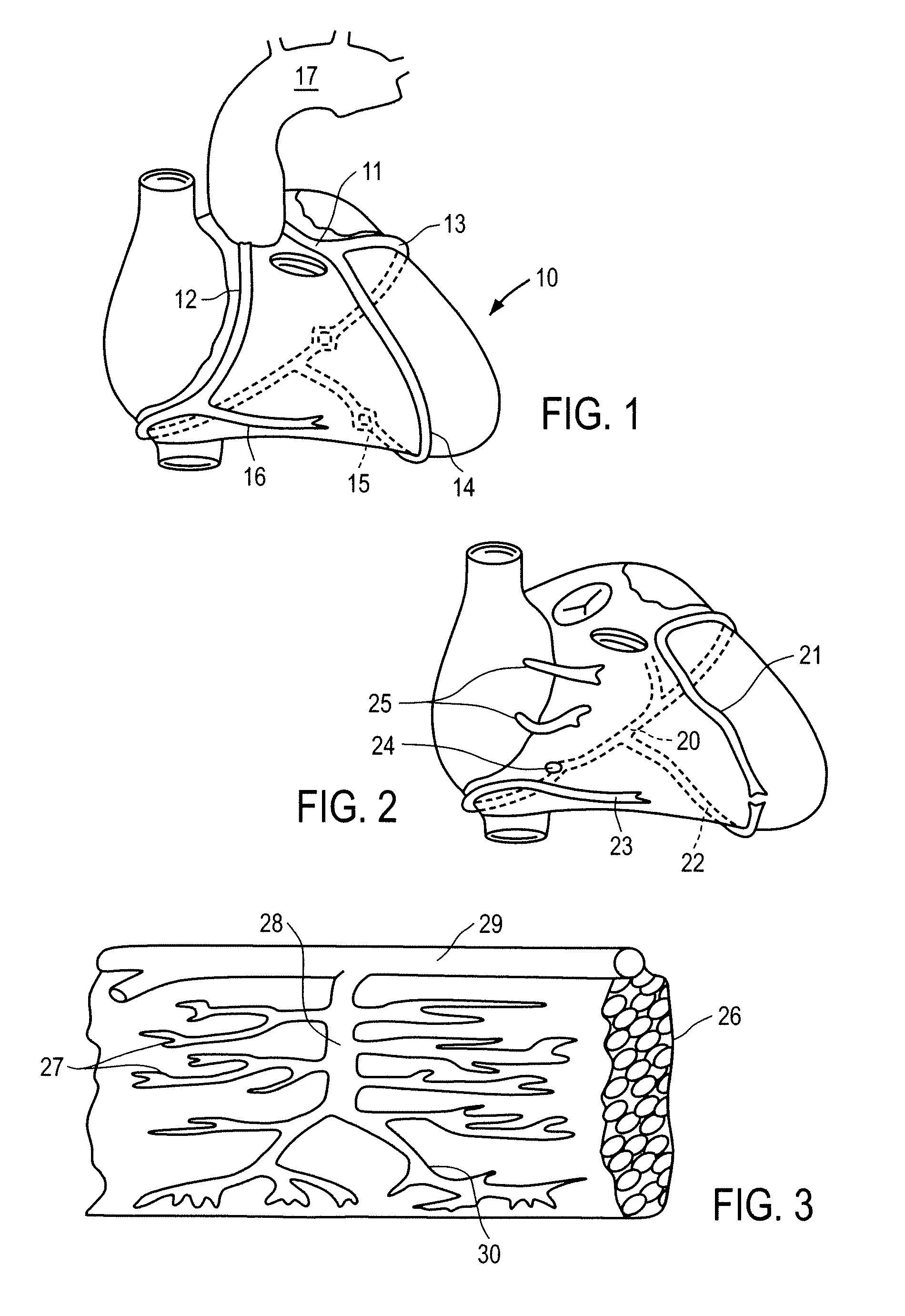
[0037]FIGS. 1 to 3 depict the coronary arterial and venous systems of a human heart. In FIG. 1, the myocardium of heart 10 is nourished by left coronary artery 11 and right coronary artery 12. Left coronary artery 11 comprises circumflex branch 13 and left anterior descending coronary artery 14; right coronary artery 12 comprises descending posterior branch 15 and marginal branch 16. Left and right coronary arteries 11 and 12 emanate from aorta 17.

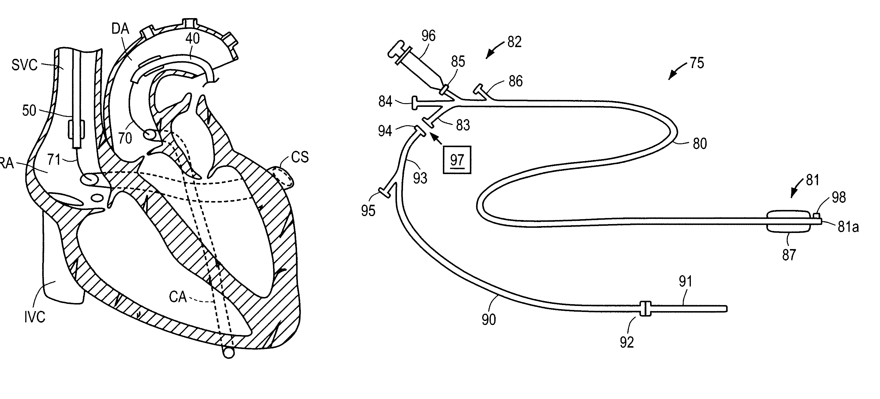
[0038]Referring to FIG. 2, the cardiac venous system of heart 10 comprises coronary sinus 20, which provides drainage for great cardiac vein 21, middle cardiac vein 22, and small cardiac vein 23. Deoxygenated blood flowing into coronary sinus 20 exits via coronary sinus ostium 24 into the right atrium. The venous system further includes anterior cardiac veins 25 that drain directly into the right atrium.

[0039]With respect to FIG. 3, myocardium 26 has a lattice of capillaries 27 that drain deoxygenated blood into intramyocardial veins 28. F...


PUM


Abstract

Apparatus and methods are provided for treating an infarcted region of myocardium following acute myocardial infarction by reducing the rate of blood flow through infarcted region, and then injecting a solution containing stem cells, endothelial progenitor cells or mediators of stem cell mobilization, migration and attachment into the effected region so that the stem cells become embedded therein and promote tissue regeneration. In a first embodiment, delivery of the stem cells, endothelial progenitor cells or mediators of stem cell mobilization, migration and attachment is accomplished using a catheter that relies upon antegrade flow through the coronary artery and a flow control catheter placed in the coronary sinus. In an alternative embodiment, the delivery catheter delivers the stem cells in a retrograde manner through the coronary sinus, and the delivery catheter further comprises an occlusion balloon for controlling outflow through the coronary sinus ostium.

Description

FIELD OF THE INVENTION[0001]The present invention relates to apparatus and methods for injecting and embedding autologous body material, including stem cells, endothelial progenitor cells or mediators of stem cell mobilization, migration and attachment into an infarcted region of the patient's heart.BACKGROUND OF THE INVENTION[0002]Myocardial infarction is caused by an abrupt occlusion of a coronary artery resulting in interruption of circulation to the affected muscle. It is now known that most myocardial infarctions occur when a blockage in the coronary artery ulcerates creating a milieu for clot formation. The cause of this change in plaque morphology (ulceration) is unclear but may be related to an inflammatory cascade or some other yet to be determined “trigger”. The term now used to describe this transition in coronary lesion morphology preceding myocardial infarction is “vulnerable plaque”. Rarely coronary artery occlusion may be caused by clot from a remote location (paradox...

Claims


Application Information

Patent Timeline
Patent Type & Authority Patents(United States)
IPC IPC(8): A61M31/00
CPCA61M25/0084A61M25/10A61M2025/0002
Inventor BATES, MARK C.
Owner COOK MEDICAL TECH LLC
Learn More