Monoclonal antibodies to HIV-1 and methods of using same
a technology of monoclonal antibodies and antibodies, applied in the field of monoclonal antibodies to hiv1, can solve the problems of no vaccine effective, no cure for acquired immune deficiency syndrome, and the pandemic of hiv is continuing worldwide, and achieves the effect of preventing the spread
- Summary
- Abstract
- Description
- Claims
- Application Information
AI Technical Summary
Benefits of technology
Problems solved by technology
Method used
Image
Examples
example 1
Isotype Determination and Epitope Mapping of 9F12 and 10F2 Vpr Antibodies
[0134]The 9F12 and 10F2 antibodies were characterized with respect to their isotype and with respect to the antigens on Vpr that they bind.
[0135]Isotype determination was made using an ELISA based format. Each well of a 96-well microtiter plate was coated with anti-mouse immunoglobulin antibodies. The 9F12 and 10F2 anti-Vpr antibodies were then added to separate, coated test wells and captured by the anti-mouse antibodies. Specific anti-mouse isotyping antibodies were then introduced and allowed to bind to the mouse-anti-mouse antibody complex. Finally, an peroxidase-tagged antibody that reacts specifically with the anti-isotyping antibodies was added, and colorimetric measurements indicated the immunoglobulin isotype of the sample.
[0136]Results of the isotyping experiments are shown in Table 1. Both the 9F12 and 10F2 antibodies are IgG1 kappa.
[0137]
TABLE 1IgG1IgG2aIgG2bIgG3IgAIgMIgκIgλType 9F121.795.022.01.055...
example 2
Immunoaffinity Capillary Electrophoresis Assay for the Detection of Vpr
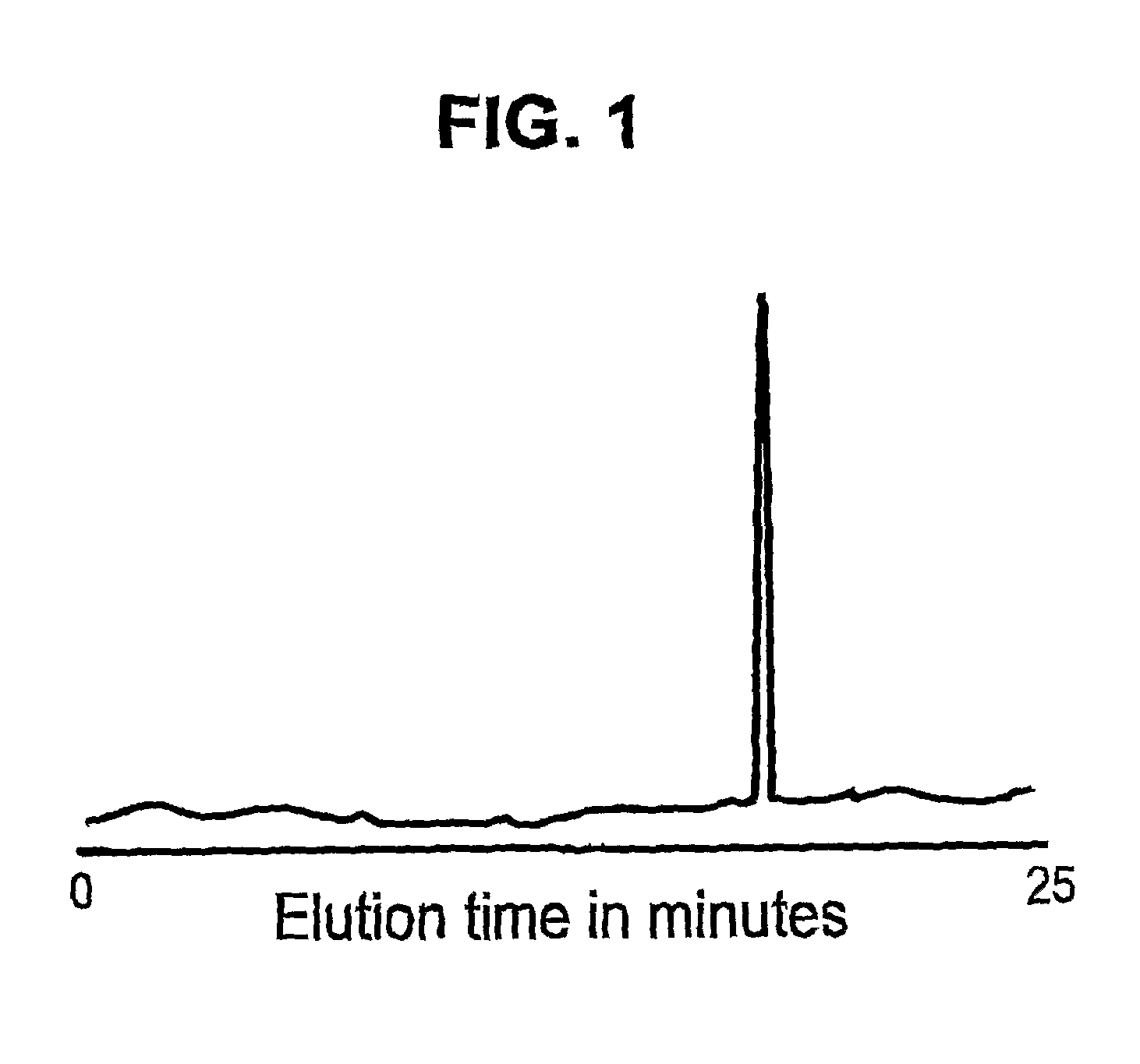
[0142]Immunoaffinity capillary electrophoresis (ICE) was used to detect and quantitate HIV-1 Vpr in a sample. 9F12 anti-Vpr antibody was immobilized on the internal wall of the first 5-cm of a 100-cm fused silica capillary. Samples containing Vpr were labeled with a red laser dye (AlexaFluor633 from Molecular Probes, Oregon) and 50-nanoliters of the labeled sample was introduced into the capillary by vacuum injection. The injected sample was allowed direct contact with the immobilized antibody coating for 10 minutes. Non-bound material was purged by the application of a neutral pH phosphate buffer wash, and the bound material was recovered by electro-elution in phosphate buffer at pH 1.5 for 25 minutes at 100-μA constant current with on-line LIF detection. FIG. 1 illustrates an electropherogram of Vpr showing the results of immunoaffinity capillary electrophoresis of a sample containing Vpr versus immobilized 9F1...
example 3
Measuring Vpr in the Plasma and Urine of Patients with HIV-1 Infection
[0143]Infectious HIV-1 in plasma and urine samples was rendered inactive by the addition of Panvirocide, which contains glutaraldehyde and three detergents, nonoxynol, Brij-23, NP-40. Patient samples were assayed for Vpr using the monoclonal antibody 9F12 in an immunocapillary electrophoresis assay as described in Example 1. The standard for calibration was synthetic Vpr 1-96 peptides, diluted in normal patient plasma.
[0144]Results of the experiments showed that among 4 healthy volunteers, all had <0.5 ng / ml Vpr in plasma. Among the 9 HIV-1 infected patients, median plasma Vpr concentration was 3 ng / ml, with a range from 1.3 to 171 ng / ml, and the median urine Vpr was 2 ng / ml, with a range of 0.1 to 34 ng / ml.
PUM
| Property | Measurement | Unit |
|---|---|---|
| temperature | aaaaa | aaaaa |
| temperature | aaaaa | aaaaa |
| pH | aaaaa | aaaaa |
Abstract
Description
Claims
Application Information
 - R&D
- Intellectual Property
- Life Sciences
- Materials
- Tech Scout
- Unparalleled Data Quality
- Higher Quality Content
- 60% Fewer Hallucinations
Browse by: Latest US Patents, China's latest patents, Technical Efficacy Thesaurus, Application Domain, Technology Topic, Popular Technical Reports.
© 2025 PatSnap. All rights reserved.Legal|Privacy policy|Modern Slavery Act Transparency Statement|Sitemap|About US| Contact US: help@patsnap.com

