Method of increasing immunological effect

a technology of immunological effect and immunological imbalance, applied in the field of methods, can solve the problems of serious problems such as inability to correct immune imbalance, and inability to actually do harm, so as to prevent apoptosis of lymphocyte/hematopoietic populations, reduce the effect of t cell apoptosis and reduce the number of effectors

Active Publication Date: 2011-08-09
BROOKLYN IMMUNOTHERAPEUTICS LLC
View PDF3 Cites 24 Cited by
  • Summary
  • Abstract
  • Description
  • Claims
  • Application Information

AI Technical Summary

Benefits of technology

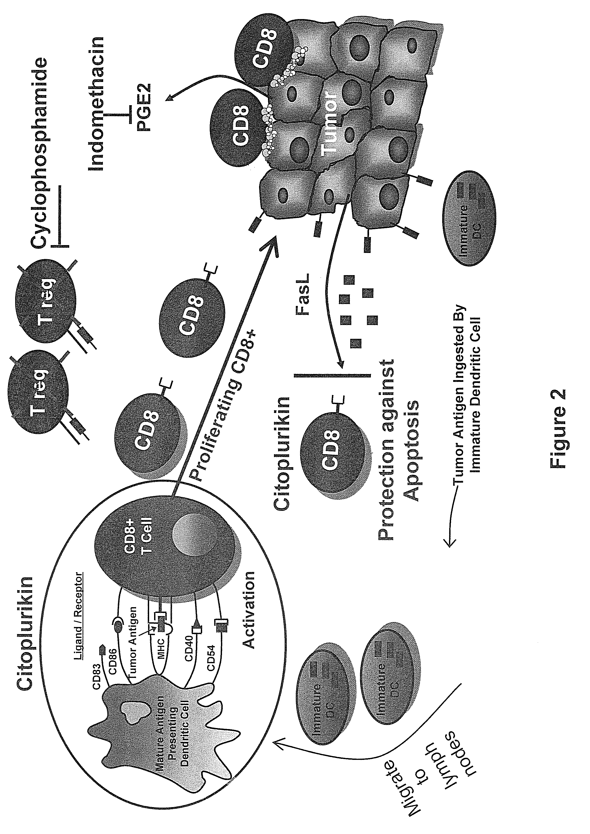
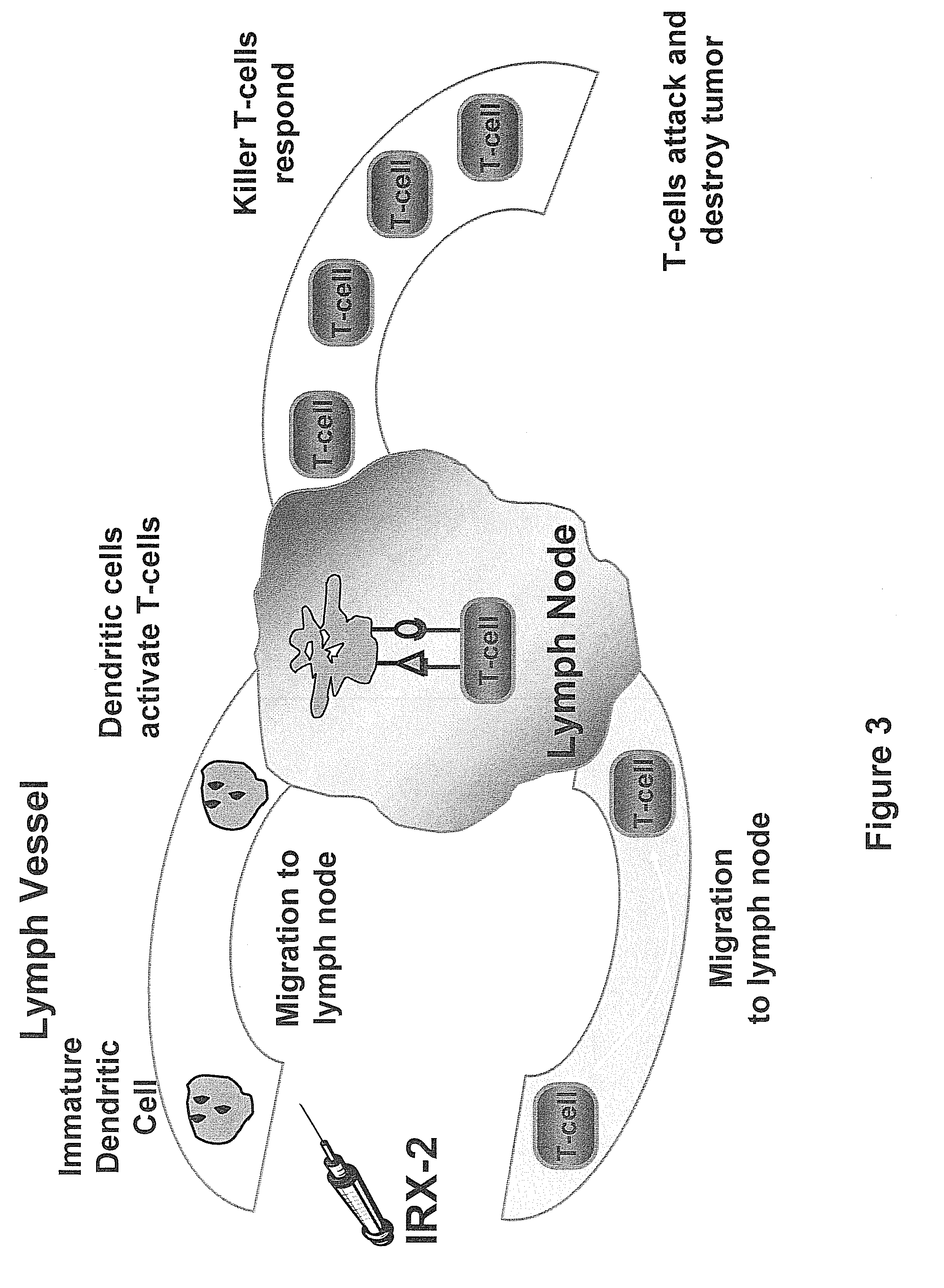
[0125]Overall, IRX-2 unsuppresses and potentiates all aspects of the cellular and humoral arms of the immune system to attack various immune targets. Any immune incompetent disease state (cancer, AIDS, and others as previously described above) can now be reversed by unsuppressing and potentiating the immune system through IRX-2. IRX-2 functions as a “symphony” rather than just a single “instrument” in that the specific combination of cytokines of IRX-2 effect multiple parts of the immune system, as opposed to prior art therapeutics which, while being combinations of components, either augment generation of effectors or prevent their apoptosis, i.e. only work on a single part of the immune system. Each part of the immune system is a gatekeeper of one effect experienced by IRX-2 administration. Each of these parts of the immune system is required in order to attack an immune target. FIGS. 2 and 3 depict the processes potentiated by IRX-2 therapy. Immature dendritic cells must become mature in order to activate naïve T cells. Production of naïve T cells also must be induced so that they can be presented with antigen by the mature dendritic cells. Both the naïve T cells and the dendritic cells must migrate to the regional lymph node in order for antigen to be presented to the naïve T cells by the dendritic cells. Once activated, the T cells must be protected from apoptosis so that they can differentiate into killer T cells and attack the immune target. B cells also mature into plasma cells to aid in attacking the immune target. Administration of IRX-2 augments all of these processes, providing a competent immune system that is ready to attack any immune target.
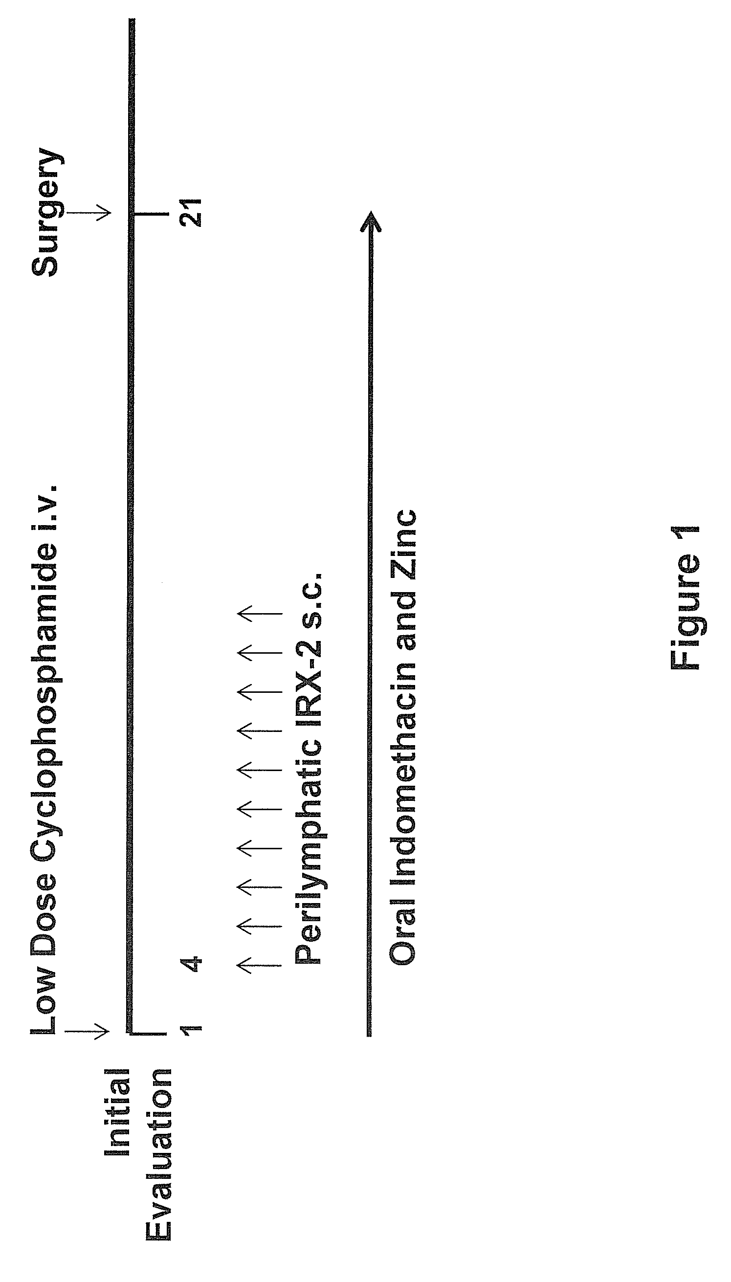
[0126]Administration of the primary cell derived biologic in vivo is the same as the vaccine+IRX-2 or IRX-2 alone immunotherapy disclosed in the previously mentioned patents related to IRX-2. IRX-2 is preferably injected perilymphatically over a 10 day regimen at 115 Units per injection, but can also be injected with other methods further described below. Alternatively, other regimes can be used wherein the IRX-2 is administered intermittently. For example, it can be administered three days a week or five out of seven days a week. As shown below, IRX-2 inhibits apoptosis over a range of concentrations: from 1:1 to 1:10 dilution of the IRX-2 liquid (i.e. dilution of the IRX-2 in the media in which it was produced).
[0127]Preferably, the IRX-2 is injected around lymphatics that drain into lymph nodes regional to a lesion, such as a tumor or other persistent lesion. Perilymphatic administration at draining nodes is critical. Peritumoral injection has been associated with little response, increase in the mitotic index of the tumor, even progression and is thus contraindicated. A ten (10) day injection scheme is optimal and a twenty (20) day injection protocol, while effective clinically, tends to reduce TH1 response and likely shifts towards a less desirable TH

Problems solved by technology

For example, dysfunction and apoptosis of these cells in the tumor-bearing host creates an immune imbalance that cannot be corrected by immunotherapies aimed only at activation of anti-tumor immune responses.
Apoptosis is an integral process during development; however, in certain instances it can actually do harm.
For example, apoptosis of lymphocyte/hematopoietic populations, including T cells, can be a serious problem during cancer therapy-related chemotherapy and/or radiation therapy.
Collectively, these mechanisms create a poisonous environment, which explains the failure of immunotherapy approaches in the past.
However, there have been many difficulties in finding an effective immunotherapy and understanding its mechanism of action.
Because there was little or no insight into the cause of these failures, no consistent mechanism of action emerged.
In contrast to defined antigen-based therapeutic cancer vaccines where antigen-specific reactivity can be measured, rejection antigens have not been discovered in H&NSCC, thus limiting the ability to measure antigen-specific reactivity after IRX-2 therapy.
Furthermore, while IRX-2 was shown to be effective in th

Method used


Image

  • Method of increasing immunological effect
  • Method of increasing immunological effect
  • Method of increasing immunological effect

Examples


example 1

Example 1

IRX-2 Protects Both Jurkat T Cells and Primary T Lymphocytes from Cell Death Mediated by a Variety of Apoptosis-Inducing Agents

[0150]To determine whether IRX-2 protects T cells from apoptosis mediated by tumor-derived microvesicles (MV), CD8+ FasL-sensitive Jurkat cells were pre-incubated with a 1:3 dilution of IRX-2 (approximately 4 ng / mL or 90 IU / mL IL-2) for 24 hours and subsequently treated them with 10 μg of tumor-derived MV (10 μg), CH-11 (400 ng / mL) or staurosporine (1 μg / mL) for 3 hours. As shown in Applicants' previous studies, the co-incubation of Jurkat cells with MV caused marked apoptosis, demonstrated by enhanced Annexin V binding (FIGS. 4A and 4C) and binding of FITC-VAD-FMK indicative of caspase activation (FIGS. 4B and 4D). Dead cells (7-AAD+) were excluded and the gate was set on 7-AAD negative CD8+ Jurkat cells. Upon pre-incubation of Jurkat T cells with IRX-2, the MV-induced apoptosis, as detected by both assays, was significantly reduced (FIGS. 4A-4D).

[...

example 2

IRX-2-Mediated Protection from Apoptosis is Time- and Concentration-Dependent

[0154]To better understand the protective effects of IRX-2 on T cells, CD8+ Jurkat cells were pre-incubated with IRX-2 (fixed dilution of 1:3=90 IU / ml IL-2) for incrementally longer time periods (0-24 hours) or with increasing concentrations of IRX-2 (as indicated) for a fixed 24 hour period and subsequently treated with MV (10 μg) for 3 hours (FIGS. 5A and 5B, respectively). Apoptosis was assessed using FITC-VAD-FMK staining of activated caspases by flow cytometry. IRX-2 was found to block MV-induced apoptosis, and this inhibition was time-dependent, as extending the time of the pre-incubation with IRX-2 intensified its protective effects. A maximal inhibition was observed after 24 hours of MV treatment (FIG. 5A). Pre-incubation of T cells with different IRX-2 concentrations showed a dose-dependent inhibition of apoptosis caused by MV (FIG. 5). At the highest possible concentration (i.e., undiluted IRX-2),...

example 3

Comparison of the Protective Effect of IRX-2 with the Effect of the Survival Cytokines IL-7 and IL-15

Caspase-Activation in Jurkat CD8+ Cells after Treatment with Tumor-MV

[0156]Jurkat CD8+ cells were plated at a density of 300,000 cells / 100 μL / well in a 96 well-plate and incubated for 24 hours with IRX-2 (1:3 final concentration), IL-7 (10 ng / mL), IL-15 (10 ng / mL), or both cytokines (10 ng / ML each), respectively. The cells were treated for 3 hours with PCI-13 / FasL-MV (15 μg). Jurkat CD8+ cells heated for 10 minutes at 56 degrees C. were used as a positive control. The cells were harvested, washed in 1 mL PBS, resuspended in 500 μL PBS, and stained with 5 μM VAD-FITC at 37 degrees C. for 20 minutes. Then the cells were washed in PBS and stained for 15 minutes for CD8-PE-Cy5. After washing, the cells were fixed in 1% PFA and analyzed by multiparametric flow cytometry.

[0157]The percent of activated caspase-VAD-FITC binding CD8+ Jurkat cells were determined for each treatment group as sh...


PUM

PropertyMeasurementUnit
Timeaaaaaaaaaa
Timeaaaaaaaaaa
Areaaaaaaaaaaa

Abstract

A method of increasing immunological effect in a patient by administering an effective amount of a primary cell derived biologic to the patient, inducing immune production, blocking immune destruction, and increasing immunological effect in the patient. Methods of treating an immune target, treating a tumor, immune prophylaxis, and preventing tumor escape.

Description

CROSS-REFERENCE TO RELATED APPLICATIONS[0001]This application claims the benefit of priority under 35 U.S.C. Section 119(e) of U.S. Provisional Patent Application No. 60 / 990,759, filed Nov. 28, 2007, and U.S. Provisional Patent Application No. 61,056,925, filed May 29, 2008, which is incorporated herein by reference.BACKGROUND OF THE INVENTION[0002](1) Field of the Invention[0003]The present invention relates to therapy of the immune system. In particular, the present invention relates to a primary cell derived biologic and methods of using the same to modify potentiation of the immune system.[0004](2) Description of Related Art[0005]In a functioning and competent immune system, immature dendritic cells ingest antigens and migrate to the lymph nodes, where they mature. The resulting mature dendritic cells are then able to activate naïve T cells, creating antigen-specific cytotoxic T cells that then proliferate, enter the circulation, and search out and kill the antigenic target. Thi...

Claims


Application Information

Patent Timeline
IPC IPC(8): A61K45/00A01N25/00
CPCA61K31/4439A61K33/00A61K45/06Y10S514/885A61K2300/00A61P17/02A61P3/02A61P31/00A61P31/18A61P35/00A61P37/04A61P43/00
Inventor HADDEN, JOHN W.WHITESIDE, THERESA L.CZYSTOWSKA, MARGARETAEGAN, JAMES E.SIGNORELLI, KATHY L.BRANDWEIN, HARVEY
Owner BROOKLYN IMMUNOTHERAPEUTICS LLC
Why Eureka
Try Eureka