Medical hyperspectral imaging for evaluation of tissue and tumor

a hyperspectral imaging and tumor technology, applied in the field of hyperspectral imaging analysis, can solve the problems of not translating into, contributing to significant local morbidity, cost of care and anxiety,

Active Publication Date: 2012-11-27
HYPERMED IMAGING
View PDF42 Cites 52 Cited by
  • Summary
  • Abstract
  • Description
  • Claims
  • Application Information

AI Technical Summary

Problems solved by technology

Early studies suggested that LR does not translate into death from disease.4 However, recent data showing lower LR rates and survival benefit by adding radiation therapy to mastectomy for patients with higher stage cancers indicate the potential importance of freedom from local recurrence.5 In addition, LR contributes to significant local morbidity, usually requiring a mastectomy.
Finally LR contributes to the cost of care and anxiety for the patient.
Despite these issues, 20-60% of patients undergoing BCT are found to have positive margins requiring additional surgical procedures, either re-excisional lumpectomies, or mastectomy.6 These additional procedures result in increased cost, increased anxiety for the patient, and, importantly, a delay in initiation of important systemic chemotherapy or radiation therapy.
At operation, the surgeon attempts to completely resect the cancer with negative microscopic margins, but faces several difficulties.
However, the microscopic extent of disease is difficult to gauge.
Frozen section analysis of breast biopsy margins is difficult and unreliable, because the fat content of breast tissue results in difficulty in sectioning frozen specimens.
This is difficult, and impractical in most institutions, but also does not provide real time information while the patient is in the operating room.
The former method does not allow re-resection of a specific positive margin and results in resection of a larger volume of breast tissue since the cavity side with a positive margin is not known.
With both approaches, the ink may creep into crevices, resulting in falsely positive margins.
With the multi-colored approach, inks may run together, resulting in confusion as to the location of a specific positive margin.
Many surgeons perform wide excisions, potentially resulting in significant breast deformity that is added to by the breast shrinkage associated with radiation therapy.
This delay results in patient anxiety and longer time to completion of the patient's surgical treatment.
The accuracy of sentinel node evaluation by frozen section is problematic,6 with a significant false negative risk, when compared to the final report.
Patients already treated for lymphoma are at the greatest risk.
Often, the resection of several additional margins is needed to completely remove the cancer and, during this time, the surgeon and patient are idle in the operating room.
Currently, there is no brain-like algorithm that can process vast amounts of spectral data, to facilitate the assessment of physiological conditions such as tissue viability or to make diagnoses or decisions in real life situations such as during surgery or critical care in the emergency room.
The volume of information contained in spectroscopic images makes standard data processing techniques time consuming and cumbersome.
Another disadvantage is that '964 also requires prior library of spectra for different tissue types, classifies each pixel based on correlation to the library samples.

Method used


Image

  • Medical hyperspectral imaging for evaluation of tissue and tumor
  • Medical hyperspectral imaging for evaluation of tissue and tumor
  • Medical hyperspectral imaging for evaluation of tissue and tumor

Examples


Embodiment Construction

Hyperspectral Imaging System

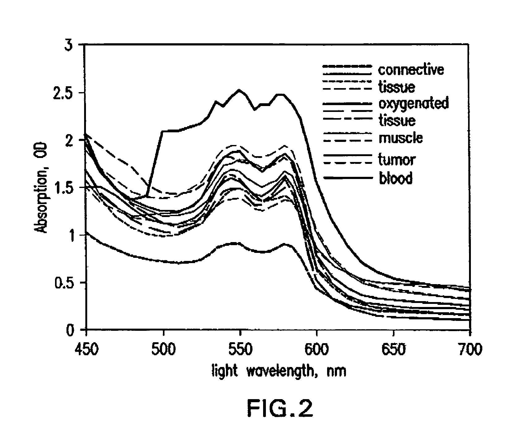
[0052]It has surprisingly been discovered that tissues can be assessed specifically for the detection of cancer or tumor beds during surgical excisions. In the present invention, new methods and tissue specific algorithms are presented that allow the assessment of tissue viability specifically to the detection of cancer, or tumor beds during surgical excision. Cancer detection can include the detection of solid cancers and precancers, and blood borne cancers such as lymphoma. Cancer detection can also include differentiating tumor from normal tissue and assessing the malignancy or aggressiveness or “grade” of tumor present. One problem is that often (e.g. during surgery) it is necessary to assess the tissue composition and oxygenation over a large area and in real or near-real time.

[0053]MHSI solves that problem by processing the hyperspectral cubes in near real-time and presenting a high-resolution, pseudo-color image where color varies with tissue type ...


PUM


Abstract

Apparatus and methods for hyperspectral imaging analysis that assists in real and near-real time assessment of biological tissue condition, viability, and type, and monitoring the above over time. Embodiments of the invention are particularly useful in surgery, clinical procedures, tissue assessment, diagnostic procedures, health monitoring, and medical evaluations, especially in the detection and treatment of cancer.

Description

REFERENCE TO RELATED APPLICATIONS[0001]The present application claims priority to U.S. Provisional Patent Application Ser. No. 60 / 631,135 entitled Hyperspectral Imaging in Medical Applications, filed Nov. 29, 2004, Ser. No. 60 / 667,678 entitled Hyperspectral Imaging in Breast Cancer, filed on Apr. 4, 2005, and Ser. No. 60 / 732,146 entitled Hyperspectral Analysis for the Detection of Lymphoma, filed on Nov. 2, 2005, which are hereby incorporated by reference.FIELD OF THE INVENTION[0002]The invention is directed to a hyperspectral imaging analysis that assists in real and near-real time assessment of biological tissue condition, viability, and type, and monitoring the above over time. Embodiments of the invention are particularly useful in surgery, clinical procedures, tissue assessment, diagnostic procedures, health monitoring, and medical evaluations.BACKGROUND OF THE INVENTION[0003]In 2005, 212,000 new cases of breast cancer are expected, and approximately 40,000 women will die of th...

Claims


Application Information

Patent Timeline
Patent Type & Authority Patents(United States)
IPC IPC(8): A61B6/00
CPCA61B5/0059A61B5/416A61B5/418G01J3/02G01J3/0224G01J3/0264G01J3/10G01J3/2823G01N21/21G01N21/31A61B5/0091A61B5/02007A61B5/4842A61B5/7425A61B5/415G01N21/359G01N21/35G01N2021/3137G01N2021/3155G01N2021/317A61B5/742
Inventor PANASYUK, SVETLANA V.FREEMAN, JENNY E.PANASYUK, ALEXANDER A.
Owner HYPERMED IMAGING
Why Eureka
Try Eureka