Insulin-like growth factor 1 receptor binding peptides
a growth factor and receptor technology, applied in the direction of peptide/protein ingredients, dna/rna fragmentation, fusion polypeptide, etc., can solve the problems of degradation of therapeutics, many therapeutics fail in vivo testing,
- Summary
- Abstract
- Description
- Claims
- Application Information
AI Technical Summary
Benefits of technology
Problems solved by technology
Method used
Image

Examples
example 1
Identification of IGF1R Binding Peptides
Phage Panning
[0062]The pIX phage libraries displaying random peptides were generated according to methods described in US Pat. Appl. No. US2010 / 0021477, and used as a source of human IGF1R binding peptides. This library was panned in solution against a biotinylated form of purified soluble IGF1R (sIGF1R) having a carboxy-terminal hexahistidine tag (R&D Systems, Minneapolis, Minn.) for three rounds. Biotinylation of sIGF1R was done using the EZ-Link No-Weigh Sulfo-NHS-LC-Biotin Microtubes (Pierce, Rockford, Ill.). Because of the large size of sIGF1R (˜330 kDa), Tetralink Avidin beads were used for round 1 selection due to their ˜10 fold greater binding capacity than Dynal magnetic beads.
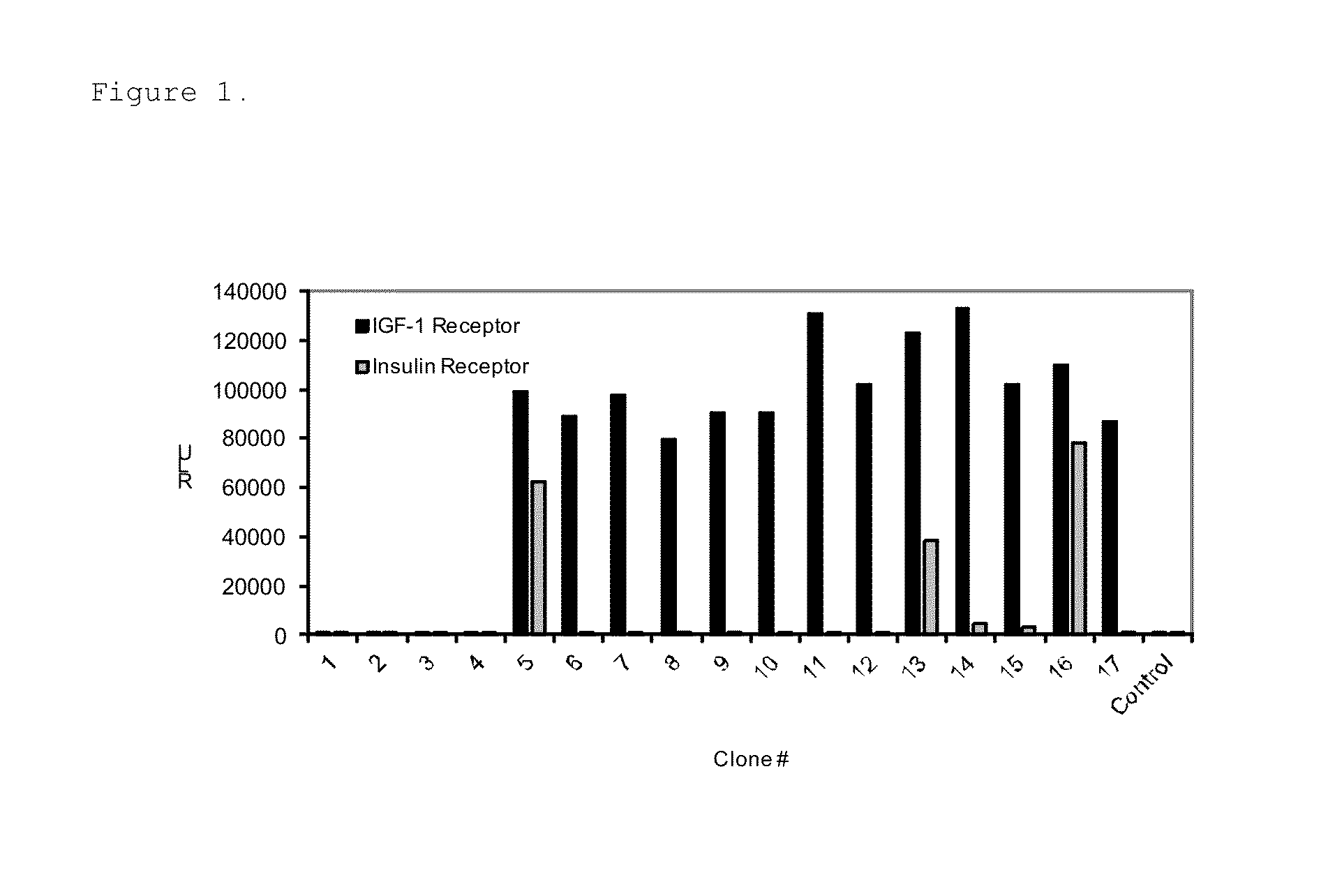
[0063]Total of 384 individual phage lysates from the panning were tested for binding specificity towards sIGF1R in a solid phase phage ELISA. Briefly, 100 μl / well of 5 μg / ml sIGF1R (R&D Systems, Minneapolis, Minn.) was bound to Black Maxisorp Plates (Nunc, Roche...
example 2
Characterization of the IGF1R Binding Peptides
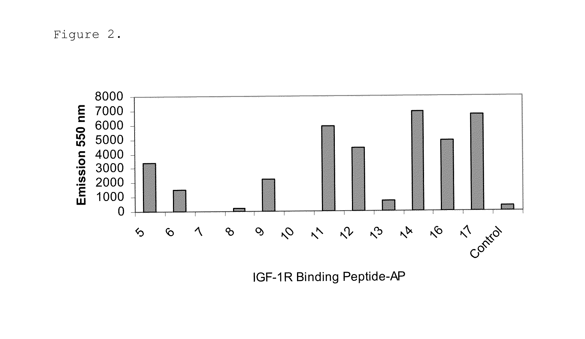
[0068]The identified IFG1R binding peptides were cloned in-frame to the C-terminus of the protein G IgG domain (PG) in a modified pET17b vector (EMD Chemicals, Gibbstown, N.J.) having a ligation independent cloning site (LIC) to generate PG-peptide fusions. The IgG binding domain of protein G is stable and thus enabled easy purification of the fusion protein from bacterial lysates. The amino acid sequence of the protein G IgG domain used is shown in SEQ ID NO: 31. The PG-peptide fusions were expressed in bacteria upon 1 mM IPTG induction and purified using IgG Sepharose beads (GE Healthcare Life Sciences, Piscataway, N.J.) from the bacterial lysates cleared by centrifugation at 16,000 g, 4° C. for 20 min.
[0069]Relative binding affinities of the PG-peptide fusions for sIGF1R were measured using ELISA. The Kds were determined using GraphPad Prism 4 software with a one site binding equation. 100 μl of 2 μg / ml sIGF1R(R&D Systems, Minneapolis...
example 3
In Vitro BBB Model
[0073]Select sIGF1R binding peptides were further characterized in the in vitro blood-brain barrier model, rat brain microvascular endothelial cell model.
[0074]Rat Brain capillary endothelial cells were prepared as described (Perriere et al., J. Neurochem. 93:279-289, 2005). Briefly, brains from 6-8 week old male Sprague Dawley rats were rolled on 3 MM chromatography paper to remove the meniges, cut sagitally, and the white matter dissected leaving the cortices, which were then minced thoroughly. The minced cortices were transferred to a 50 ml polypropylene conical tube with 20 ml DMEM supplemented with 39 units / ml DNase I (Worthington, Lakewood, N.J.) and 0.7 mg / ml Collagenase type 2 (Worthington, Lakewood, N.J.) at final concentration and incubated at 37° C. with gentle mixing for 1.25 hrs. After a brief centrifugation the resultant pellet was resuspended in 20 ml 20% BSA (Sigma, St. Louis, Mo.) in DMEM, centrifuged and the microvessel enriched pellet was isolate...
PUM
| Property | Measurement | Unit |
|---|---|---|
| molecular weight | aaaaa | aaaaa |
| temperature | aaaaa | aaaaa |
| volume | aaaaa | aaaaa |
Abstract
Description
Claims
Application Information
 - R&D
- Intellectual Property
- Life Sciences
- Materials
- Tech Scout
- Unparalleled Data Quality
- Higher Quality Content
- 60% Fewer Hallucinations
Browse by: Latest US Patents, China's latest patents, Technical Efficacy Thesaurus, Application Domain, Technology Topic, Popular Technical Reports.
© 2025 PatSnap. All rights reserved.Legal|Privacy policy|Modern Slavery Act Transparency Statement|Sitemap|About US| Contact US: help@patsnap.com