Stand-alone mini gamma camera including a localization system for intrasurgical use

a technology of localization system and gamma camera, which is applied in the field of medical nuclear physics and radioguided surgery, can solve the problems of no mini gamma camera that can be plugged in and unplugged, and no mini gamma camera that can solve the serious edge and compression problems affecting the image,

Active Publication Date: 2013-05-28
GENERAL EQUIP FOR MEDICAL IMAGING SL +2
View PDF21 Cites 128 Cited by
  • Summary
  • Abstract
  • Description
  • Claims
  • Application Information

AI Technical Summary

Benefits of technology

[0041]This invention also presents a special treatment for the surfaces of the crystal, if the scintillation crystal of the mini gamma camera is of the continuous type, and a method that makes it possible to use almost the entire area thereof without the serious disadvantage of image compression and edge effects that occur with the use of continuous crystals. It also introduces the innovation of using rare-earth-based scintillation crystals.
[0119]An additional object of the present invention is a device that makes it possible, through the use of light emitters, to generate a singular light form with a fixed position on a surface, even if the distance from the emitters to the surface can vary along an axis that runs from the device to the surface, with the particularity that said axis does not cross the spatial position of the light emitters, but can be aligned or made to coincide with the characteristic planes of emission of the light sources.

Problems solved by technology

Meanwhile, new fields of application that are almost impossible for large equipment are being discovered for mini cameras, such as intra-operative use.
Likewise, there are no mini gamma cameras that can be plugged in and unplugged without having to switch the computer off and in which all the necessary voltages for their operation, including high voltage, are generated inside the camera and not on a card or other device that is connected to the computer.
However, none of said patents explain how to overcome the serious edge and compression problems affecting the image that occur when the detection surface is small in relation to the width of the crystal, in the direction in which the gamma rays enter, as is the case, by definition, with mini gamma cameras.
With all the IFIC's aforementioned prototypes and publications there is the problem that in practice the limited broadband (1.2 Mbps) of the USB port used (1.0) only makes it possible to reach a low rate of event acquisition (maximum 1100 counts per second) in relation to the normal rate used in nuclear medicine, even though the physical detector was capable of acquiring data at a greater speed.
Moreover, all these prototypes developed by the IFIC presented the problem that they were not suitable for medical use, making their practical application in humans unviable for both diagnostic and intra-operative use.
Lastly, the painting technique used to treat the surfaces of the crystals used in the IFIC's prototypes and publications was practically unworkable, as there were many defects at the edges and there were inhomogeneities throughout the sensitive area, which gave the sensor systems thus created a very short useful life and caused losses of uniformity that were unacceptable according to medical standards.
Likewise, there are no known cameras or mini gamma cameras that produce real time video gamma images, or still images in a few seconds, using the low doses of radiation permitted in nuclear medicine.
The difficulties found with the intra-surgical use of mini gamma cameras include viewing low-activity regions (such as lymph nodes) close to high-activity points (such as peritumoural injection sites).
As has just been mentioned in the previous paragraph, another of the problems presented by mini gamma cameras is that of locating objects that appear in the images that they capture in the patient's body, i.e. the correspondence between image and organism.
The localisation method using a bright pointer presents the disadvantage that it leaves a static trail behind as the pointer passes over an area of the image.
“Functional Navigator”, P200202220 and PCT ES 03-00497), based on an infrared or radiofrequency localiser, is of the transparent type, although it differs completely in that it requires (and this is a disadvantage) two detection systems, which are furthermore based on different technologies.

Method used


Image

  • Stand-alone mini gamma camera including a localization system for intrasurgical use
  • Stand-alone mini gamma camera including a localization system for intrasurgical use
  • Stand-alone mini gamma camera including a localization system for intrasurgical use

Examples


example 1

Embodiment of a Compact Stand-Alone Mini Gamma Camera with Localisation System for Intra-Surgical Use

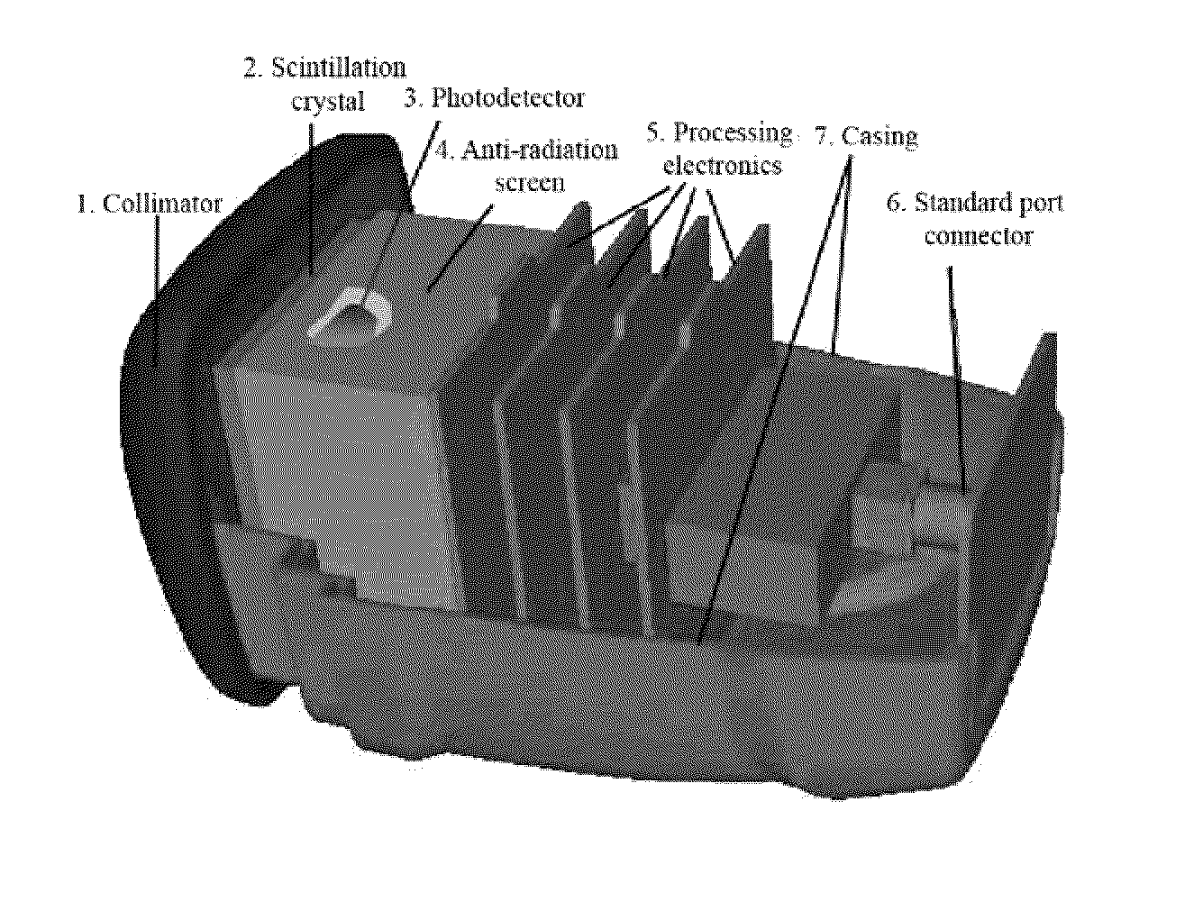
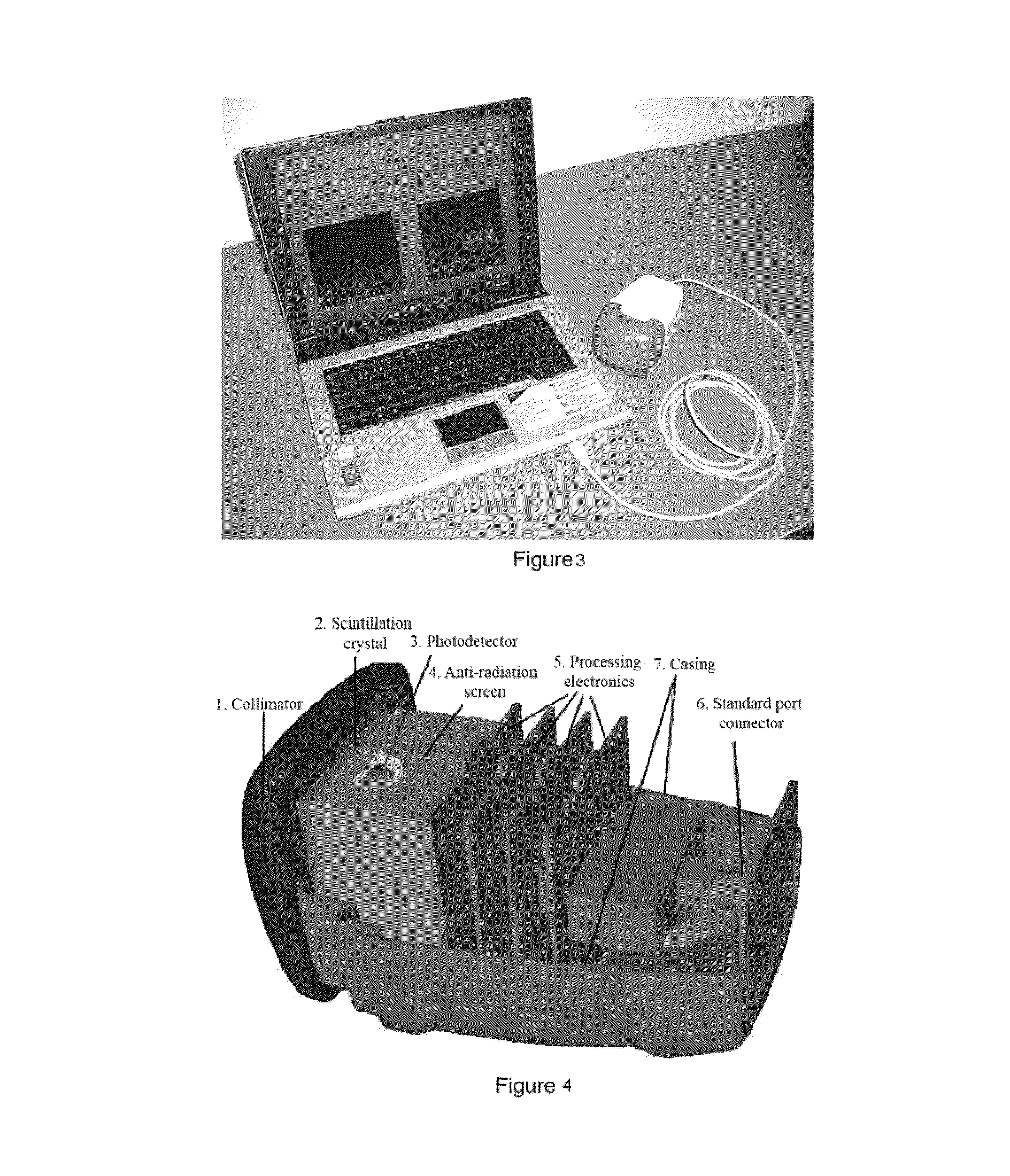
[0144]The mini gamma camera that is shown (FIGS. 3 and 4) consists only of the sensor head, a USB cable and a standard computer. It has an irregular, ergonomic, rectangular prismatic shape with the following dimensions: 70 mm high, 90 mm wide and 140 mm long. It weighs just under 1 kg, including the collimator. The system's small dimensions, ergonomic shape and light weight make this a portable, hand-held device. This mini gamma camera is used to view small organs such as the sentinel lymph node in an intra-operative situation with a positional resolution of less than 2 mm.

[0145]This portable mini gamma camera can “see” an energy range from 15 keV to 250 keV, although its design has been optimised for the 50 keV to 200 keV region, in which the most widely used radioactive sources in medical imaging are found (e.g. Tc-99m, which emits 140 KeV gamma rays). This camera has a single posi...

example 2

Use of a Compact Stand-Alone Mini Gamma Camera with Gamma Navigation System for Intra-Surgical Use

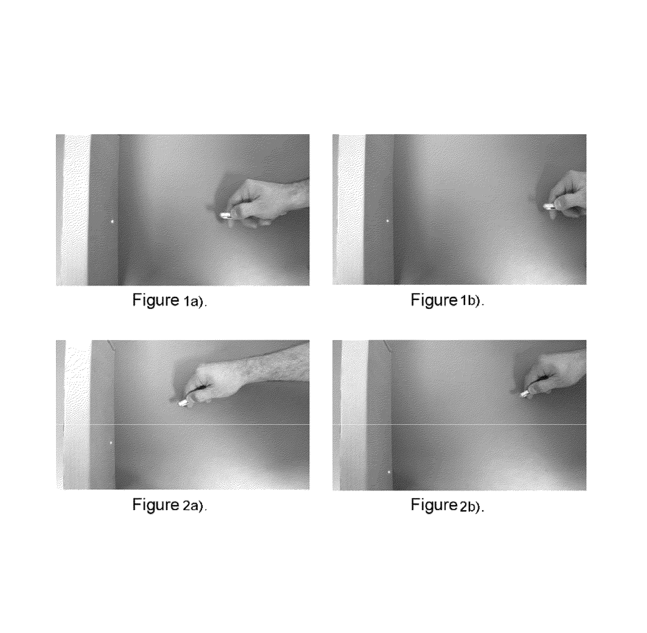
[0148]FIGS. 7 and 8 show the mini gamma camera that is the object of the present invention being used with radioactive navigation systems.

[0149]FIGS. 7a and 7b show two images captured by the mini gamma camera, at the moments when a 1251 pointer was positioned above and below one of two cobalt sources that were used as sentinel lymph node phantoms.

[0150]FIGS. 8a, 8b and 8c show three photos of the mini camera gamma and the computer connected thereto, at the three moments when the opaque pointer (lead screen) was a) absent, b) positioned over the right-hand source and c) positioned over the left-hand source, whilst the computer shows the shadow effect left by said pointer on the scintigraphic image, showing how the method can be used to locate “hotspots” such as sentinel lymph nodes.


PUM


Abstract

The invention relates to a portable mini gamma camera for intrasurgical use. The inventive camera is based on scintillation crystals and comprises a stand-alone device, i.e. all of the necessary systems have been integrated next to the sensor head and no other system is required. The camera can be hot-swapped to any computer using different types of interface, such as to meet medical grade specifications. The camera can be self-powered, can save energy and enables software and firmware to be updated from the Internet and images to be formed in real time. Any gamma ray detector based on continuous scintillation crystals can be provided with a system for focusing the scintillation light emitted by the gamma ray in order to improve spatial resolution. The invention also relates to novel methods for locating radiation-emitting objects and for measuring physical variables, based on radioactive and laser emission pointers.

Description

[0001]This application is a U.S. national phase application under 35 U.S.C. §371 of International Patent Application No. PCT / ES2006 / 070200 filed Dec. 22, 2006, which claims the benefit of priority to Spanish Patent Application No. P200503198 filed Dec. 26, 2005, the disclosures of all of which are hereby incorporated by reference in their entireties. The International Application was published in Spanish on Jul. 5, 2007 as WO 2007 / 074201.FIELD OF THE INVENTION[0002]The present invention belongs to the field of Medical Nuclear Physics and radioguided surgery. It is a device that makes it possible to produce in vivo images of small human and animal organs using radioisotopes that are traditionally used in nuclear medicine, with a high resolution and in real time. The device is designed for functional studies and detection and / or precise localisation of cancer in organs such as the thyroid gland, kidneys, prostate, breasts and lymph nodes. It is applicable to preliminary, intra-operati...

Claims


Application Information

Patent Timeline
Patent Type & Authority Patents(United States)
IPC IPC(8): A61B19/00G01T1/29
CPCA61B19/52A61B6/4258G01T1/161G01T1/1642A61B19/5212A61B19/5244A61B2019/5268A61B2019/5287A61B2019/542A61B6/037A61B2019/5293A61B34/20A61B90/36A61B90/361A61B2034/2068A61B2090/363A61B2090/366A61B2090/392A61B6/03G01T1/1644G01T1/1647
Inventor BAVIERA, JOSE MARIA BENLLOCHMARTINEZ, FILOMENO SANCHEZLERCHE, CHRISTOPHHERNANDEZ, NORIEL PAVONLEIVA, JESUS MODIA
Owner GENERAL EQUIP FOR MEDICAL IMAGING SL
Why Eureka
Try Eureka