Targeted therapeutics based on engineered proteins for tyrosine kinases receptors, including IGF-IR

a technology of tyrosine kinases and engineered proteins, which is applied in the direction of fusion polypeptides, peptide/protein ingredients, unknown materials, etc., can solve the problems of slow attempts to make antagonists or other types of inhibitors, inability to make selective antagonists, and difficulty in making therapeutics, so as to improve the improve the effect of delivery or therapeutic properties, and improve the effect of production efficiency

Active Publication Date: 2013-06-25
BRISTOL MYERS SQUIBB CO
View PDF49 Cites 39 Cited by
  • Summary
  • Abstract
  • Description
  • Claims
  • Application Information

AI Technical Summary

Benefits of technology

[0029]In some embodiments, the polypeptide comprises a fibronectin-based scaffold protein. In some embodiments, the polypeptide comprising a tenth fibronectin type III (10Fn3) domain, wherein the 10Fn3 domain (i) comprises a loop, AB; a loop, BC; a loop, CD; a loop, DE; a loop EF; and a loop FG; (ii) has at least one loop selected from loop BC, DE, and FG with an altered amino acid sequence relative to the sequence of the corresponding loop of the human 10Fn3 domain, and (iii) binds human IGF-IR with a disassociation constant of about 1 μM or less. In some embodiments, at least 1, 2, 3, 4, 5, 6, 7, 8, 9, or 10 amino acids in at least one loop selected from loop BC, DE, and FG are substituted with an amino acid that differs from the wild-type sequence. In some embodiments, at least 1, 2, 3, 4, 5, 6, 7, 8, 9, or 10 amino acids are deleted or added to at least one loop selected from loop BC, DE, and FG. In some embodiments, loop BC; and loop FG have an altered amino acid sequence relative to the sequence of the corresponding loop of the human 10Fn3 domain. In some embodiments, the polypeptide comprises the amino acid sequence of any of one of SEQ ID NOS: 2-125, 184-203, or 226. Substantially monovalent binding to IGF-IR via fibronectin based scaffolds is helpful in reducing IGF-IR activation in this and other embodiments of the invention.
[0030]In some embodiments, the polypeptides of the invention further comprise one or more pharmacokinetic (PK) moieties. PK moieties improve one or more pharmacokinetic properties of the polypeptides, e.g., bioavailability, serum half-life, in vivo stability, and drug distribution. In some embodiments, the PK moiety is selected from: a polyoxyalkylene moiety, a human serum albumin binding protein, sialic acid, human serum albumin, transferrin, and Fc or an Fc fragment. In some embodiments, the PK moiety is polyethylene glycol (PEG). In some embodiments, the PEG moiety is covalently linked to the polypeptide of the invention via a Cys or Lys amino acid. In some embodiments, a polypeptide of the invention is linked by at least one peptide bond to a second protein that binds human serum albumin (HSA) with a binding affinity of about 300 nM or less.
[0034]In some embodiments of the invention, polypeptides of the invention comprise a first and second protein linked via a polypeptide, wherein the first or second protein or polypeptide linker has a protease site that is cleavable by a protease in the blood or target tissue. Such embodiments can be used to release two or more therapeutic proteins for better delivery or therapeutic properties or more efficient production compared to separately producing such proteins.
[0035]In some embodiments, a first and second protein are linked using a biocompatible polymer such as a polymeric sugar. Such polymeric sugar can include an enzymatic cleavage site that is cleavable by an enzyme in the blood or target tissue. Such embodiments can be used to release two or more therapeutic proteins for better delivery or therapeutic properties or more efficient production compared to separately producing such proteins.
[0038]PEG may be used in many aspects of the invention and different sizes may be used as described herein for the desired therapeutic or other in vivo effect, such as imaging. Larger PEGs are preferred to increase half life in the body, blood, non-blood extracellular fluids or tissues. For in vivo cellular activity, PEGs of the range of about 10 to 60 kDa are preferred, as well as PEGs less than about 100 kDa and more preferably less than about 60 kDa, though sizes greater than about 100 kDa can be used as well. For in vivo imaging application, smaller PEGs, generally less than about 20 kDa, may be used that do not increase half life as much as larger PEGs so as to permit quicker distribution and less half life.
[0051]Stably linked proteins of the invention may be of use for therapeutic treatment of cancer. Multispecific proteins of the invention have the advantage of modulating, blocking or inhibiting more than one therapeutic target when directed to 2, 3, 4 or more therapeutic targets or epitopes.

Problems solved by technology

Difficulty of Making a Therapeutic Based on IGF-IR Antagonism or Inhibition
While IGF-IR has attractive biology as a therapeutic target, attempts to make antagonists or other types of inhibitors have been slow, largely due to one or more of the following factors depending on the therapeutic paradigm: difficulty or inability to antagonize the target without agonism; difficulty or inability to make selective antagonists or inhibitors due to undesired cross reactivity with other tyrosine kinase targets or receptors, such as the insulin receptor; difficulty in effective administration due to short half life of the potential therapeutic; difficulty in effective administration clue to low solubility of the potential therapeutic; and difficulty in effective administration due to aggregation of the potential therapeutic.
However, both antisense and siRNA therapeutic strategies suffer from administration challenges and potential cost of goods obstacles.
In addition, antibodies that are shown to “inhibit” may not have been carefully characterized and may actually activate IGF-IR under certain conditions, such as lower antibody concentrations.
Although IR3 antibody is the most commonly used inhibitory antibody for IGF-IR studies in vitro, it suffers from the drawback that it exhibits agonistic activity in transfected 3T3 and CHO cells expressing human IGF-IR (Kato, H. et al., 1993, J. Biol. Chem., 268, 2655-2661; Steele-Perkins, G. and Roth, R. A., 1990, Biochem. Biophys. Res. Commun., 171, 1244-1251).
Although anti-IGF-IR antibodies have been reported present in certain patients with autoimmune diseases, none of these antibodies has been purified and none has been shown to be suitable for inhibiting IGF-I activity for diagnostic or clinical procedures.
Additionally, it has been historically difficult to make antibodies and other therapeutics due to specificity, agonism, aggregation and solubility, as well as bi-specific therapeutics that include IGF-IR as a target, particularly with using antibodies.
Small molecule chemistry programs have largely not been successful in creating a therapeutic to IGF-IR.
The druggability of this target by proteins and small molecules has been difficult for a number of reasons including the ability to obtain selective chemistry for IGF-IR while not inhibiting or binding to related tyrosine kinases or receptors, such as the insulin receptor.
In addition, the small molecules are often of low solubility making bioavailability, administration or formulation challenging.
Current potential therapeutic strategies, although capable of inducing apoptosis in vitro or for inhibiting in vitro cell proliferation associated with increased IGF-I, increased IGF-II, and / or increased IGF-IR receptor levels include suppressing IGF-I levels or IGF-II levels or preventing the binding of IGF-I to the IGF-IR, remain unsuitable for better therapeutics.
However, none of these agents has been suitable for longer-term administration to human patients.
In addition, although IGF-I has been administered to patients for treatment of short stature, osteoporosis, decreased muscle mass, neuropathy or diabetes, the binding of IGF-I to IGFBPs has often made treatment with IGF-I difficult or ineffective.

Method used


Image

  • Targeted therapeutics based on engineered proteins for tyrosine kinases receptors, including IGF-IR
  • Targeted therapeutics based on engineered proteins for tyrosine kinases receptors, including IGF-IR
  • Targeted therapeutics based on engineered proteins for tyrosine kinases receptors, including IGF-IR

Examples


example 1

Initial Identification of IGF-IR Binding Molecules

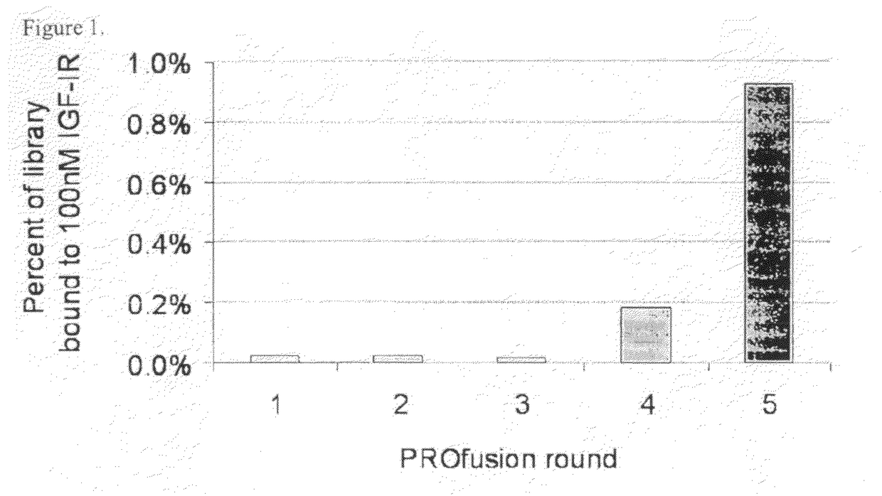
[0280]A library of approximately 1013 RNA-protein fusion variants was constructed based on the scaffold of the tenth type 3 domain of human fibronectin with three randomized regions at positions 23-29, 52-55 and 77-86 (amino acid nos. are referenced to SEQ ID NO:1) (three loop library; Xu et al, Chemistry & Biology 9:933-942, 2002). After conversion to mRNA / cDNA heteroduplexes, the library of a trillion mRNA / cDNA-protein fusions was bound to 100 nM biotinylated IGF-IR in solution, and captured on streptavidin-coated magnetic beads. The cDNA was eluted, amplified by PCR, and used to generate a new library of mRNA / cDNA-protein fusions. Five rounds of selection were carried out in this manner and the percentage of library binding to IGF-IR was monitored by quantitative PCR after every round to identify target binding enrichment (FIG. 1).

example 2

Identification of Competitive IGF-IR Clones

[0281]Proteins encoded by independent clones were analyzed for inhibition of target binding in the presence of IGF-IR using a competitive binding assay. Over one hundred-clones were identified that inhibited the binding of IGF-I to IGF-IR, suggesting that these clones bound IGF-IR at or near the natural ligand (IGF-I) binding site. The SEQ ID NOs (see FIG. 30) of these one hundred clones and their percent inhibition in the competitive binding assay are presented in Table 1.

[0282]

TABLE 1SEQ ID NOs of IGF-IR Competitive Clones and % InhibitionCloneSEQ ID NO:% inhibition223B02289.1%223A04387.0%223F03486.8%214D04586.5%225E09684.6%224A02784.6%223A07883.8%223E11982.2%223H041082.2%215G061180.5%219F081280.0%223B011379.7%225B011479.4%214C111578.9%219C071678.8%218C041777.0%215A071876.1%223D011976.0%225D032074.8%225A022174.6%223A102273.7%225B032373.7%218C052473.6%225A052572.1%224A012672.0%214F032772.0%214A082870.6%223H112970.5%218B033070.4%215E093170....

example 3

Examination of the Electrophoretic Properties of IGF-IR Competitive Clones

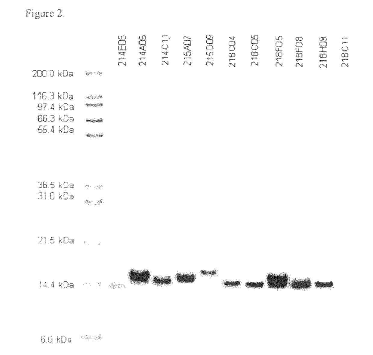
[0283]The purity and electrophoretic properties of selected clones were examined by SDS-PAGE (FIG. 2). These were found to be reasonably pure considering their single-column purification and had apparent molecular weights consistent with their theoretical masses (taking into account the additional mass associated with C-terminal tags).


PUM

PropertyMeasurementUnit
Tmaaaaaaaaaa
concentrationsaaaaaaaaaa

Abstract

The present invention provides innovative proteins that bind to insulin-like growth factor-I receptor (IGF-IR), as well as other important proteins. The invention also provides innovative proteins in pharmaceutical preparations and derivatives of such proteins and the uses of same in diagnostic, research and therapeutic applications. The invention further provides cells comprising such proteins, polynucleotide encoding such proteins or fragments thereof, and vectors comprising the polynucleotides encoding the innovative proteins.

Description

RELATED APPLICATIONS[0001]This application is a U.S. national stage application filed under 35 U.S.C. §371 of International Application PCT / US2007 / 024316, filed Nov. 21, 2007, which claims the benefit of U.S. Provisional Patent Application Nos. 60 / 860,605 filed Nov. 22, 2006 and 60 / 879,666 filed Jan. 9, 2007, which applications are hereby incorporated by reference in their entirety. International Application No. PCT / US2007 / 024316 was published under PCT Article 21(2) in English.FIELD OF THE INVENTION[0002]The present invention relates to innovative proteins that bind to insulin-like growth factor-I receptor (IGF-IR), as well as other important proteins. The invention also relates to innovative proteins in pharmaceutical preparations and derivatives of such proteins and the uses of same in diagnostic, research and therapeutic applications. The invention further relates to cells comprising such proteins, polynucleotide encoding such proteins or fragments thereof, and to vectors compri...

Claims


Application Information

Patent Timeline
Patent Type & Authority Patents(United States)
IPC IPC(8): A61K38/17A61K38/39A61K47/48C07K14/435C07K14/71
CPCC07K14/78C12N15/1062C40B30/04C40B40/08C07K16/00A61K38/18C07K14/00C07K14/765C07K14/79C07K2319/74C40B40/10A61K47/60A61K47/643A61K47/644A61K47/68A61K38/39A61P35/00A61P43/00C07K14/47C12N15/10C07K14/71C07K16/2863C07K2317/92C07K2319/30A61K39/3955A61K2039/505C07K19/00A61K38/179A61K47/10
Inventor CAMPHAUSEN, RAYFABRIZIO, DAVIDWRIGHT, MARTIN C.GAGE, PATRICKMENDLEIN, JOHN
Owner BRISTOL MYERS SQUIBB CO
Why Eureka
Try Eureka