
Insulinotropic complex using an immunoglobulin fragment

a technology of immunoglobulin and complex, which is applied in the field of insulinotropic peptide conjugate, can solve the problems of easy denature, loss of activity, and small siz

Active Publication Date: 2013-07-02
HANMI SCI CO LTD
View PDF30 Cites 19 Cited by
  • Summary
  • Abstract
  • Description
  • Claims
  • Application Information

AI Technical Summary

Benefits of technology

The present invention provides a long-acting preparation of insulinotropic peptide that maintains its in-vivo activity and increases its blood half-life. This is achieved by linking an immunoglobulin Fc, and a non-peptidyl polymer, to an insulinotropic peptide site-specifically at an amino acid residue other than the N-terminus by a covalent bond. The resulting conjugate exhibits a remarkably increased in-vivo efficacy and half life compared to other insulinotropic peptide conjugates.

Problems solved by technology

Peptides tend to be easily denatured due to their low stability, degraded by in-vivo proteolytic enzymes, thus losing the activity, and have a relatively small size, thereby easily passing through the kidney.
However, the peptide drugs are usually administered in the form of injectable preparations, and such frequent administration cause severe pain for the patients.
However, this approach is still difficult in maintaining the in-vivo activity of the peptide drug due to the low in-vivo transfer efficiency, as compared to the injection.
However, with the method for increasing the resistance to DPP IV only, the physiological activity is not sufficiently sustained, and for example, in the case of a commercially available exendin-4 (exenatide), it needs to be injected to a patient twice a day, which is still difficult for patients.
These insulinotropic peptides have a problem, usually in that the size of the peptide is small.
Thus, they cannot be recovered in the kidney, and are then extracorporeally discharged.
However, this method increases the molecular weight of PEG, thereby increasing the in-vivo residence time of the peptide drug, while as the molecular weight is increased, the titer of the peptide drug is remarkably reduced, and the reactivity with the peptide is also reduced.
Accordingly, it undesirably lowers the yield.
These methods can address the problems such as low pegylation yield and non-specificity, but they still have a problem in that the effect of increasing the blood half-life is not noticeable as expected, and sometimes the titers are also low.
Further, if a peptide having disulfide bonds, such as BNP is used, there is a high probability of misfolding.
As a result, such peptide can hardly be used.

Method used


Image

  • Insulinotropic complex using an immunoglobulin fragment
  • Insulinotropic complex using an immunoglobulin fragment
  • Insulinotropic complex using an immunoglobulin fragment

Examples


example 1

Pegylation of Exendin-4 and Isolation of Positional Isomer

[0087]3.4K PropionALD(2) PEG (PEG having two propionaldehyde groups, IDB Inc., South Korea) and the N-terminus of the exendin-4 (AP, USA) were subjected to pegylation by reacting the peptide and the PEG at 4° C. for 90 min at a molar ratio of 1:15, with a peptide concentration of 3 mg / ml. At this time, the reaction was performed in a NaOAc buffer at pH 4.0 at a concentration of 100 mM, and 20 mM SCB (NaCNBH3) as a reducing agent was added thereto to perform the reaction. 3.4K PropionALD(2) PEG and the lysine (Lys) residue of the exendin-4 were subjected to pegylation by reacting the peptide and the PEG at 4° C. for 3 hours at a molar ratio of 1:30, with a peptide concentration of 3 mg / ml. At this time, the reaction was performed in a Na-Phosphate buffer at pH 9.0 at a concentration of 100 mM, and 20 mM SCB as a reducing agent was added thereto to perform the reaction. A mono-pegylated peptide was purified from each of the rea...

example 2

Preparation of Exendin-4(N)-PEG-Immunoglobulin Fc Conjugate

[0088]Using the same method as described in EXAMPLE 1, 3.4K PropionALD(2) PEG and the N-terminus of the exendin-4 were reacted, and only the N-terminal isomers were purified, and then coupled with immunoglobulin Fc. The reaction was performed at a ratio of peptide:immunoglobulin Fc of 1:8, and a total concentration of proteins of 50 mg / ml at 4° C. for 17 hours. The reaction was performed in a solution of 100 mM K—P (pH 6.0), and 20 mM SCB as a reducing agent was added thereto. The coupling reaction solution was purified using two purification columns. First, SOURCE Q (XK 16 ml, Amersham Biosciences) was used to remove a large amount of immunoglobulin Fc which had not participated in the coupling reaction. Using 20 mM Tris (pH 7.5) and 1 M NaCl with salt gradients, the immunoglobulin Fc having relatively weak binding power was eluted earlier, and then the exendin-4-immunoglobulin Fc was eluted. Through this first purification...

example 3

Preparation of Exendin-4(Lys27)-PEG-Immunoglobulin Fc Conjugate

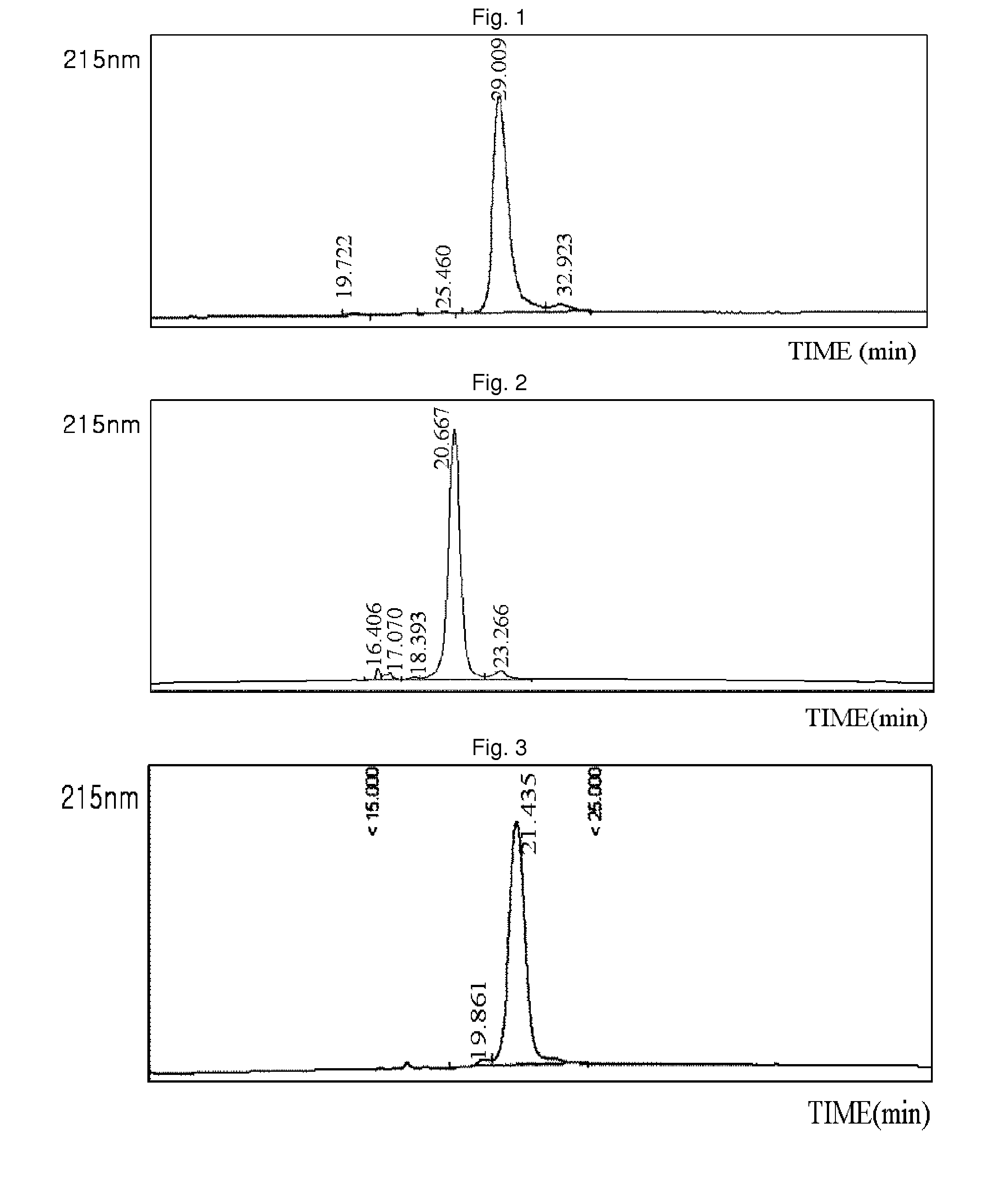
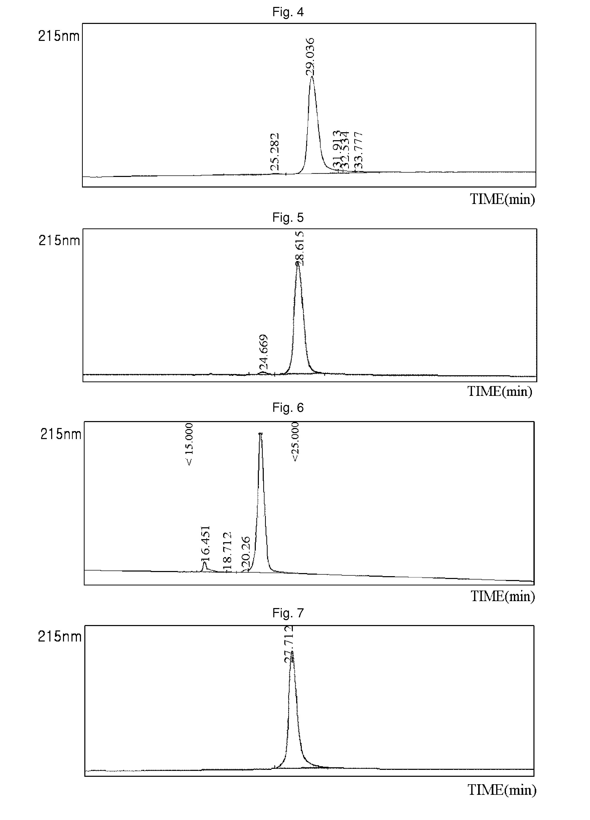
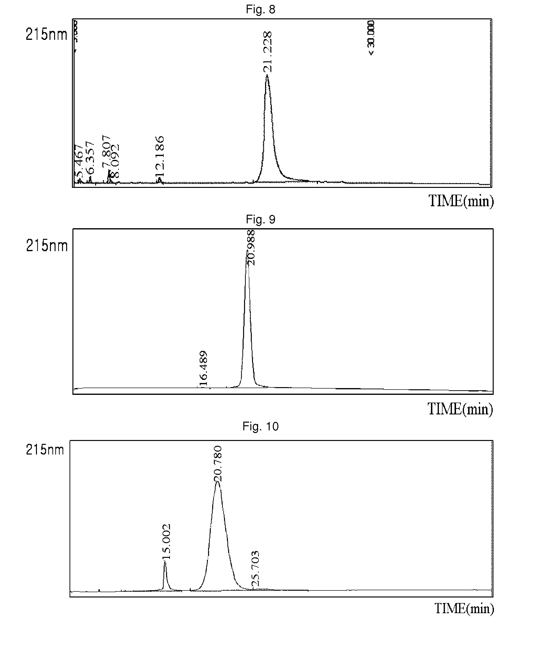
[0089]Using the same method as described in EXAMPLE 1, 3.4K PropionALD(2) PEG and the lysine (Lys) of the exendin-4 were reacted, and only the Lys isomers were purified, and then coupled with immunoglobulin Fc. Among the two isomer peaks, the last isomer peak (positional isomer of Lys27), which has more reaction and which is easily distinguishable from the N-terminal isomer peaks, was used for the coupling reaction. The reaction was performed at a ratio of peptide:immunoglobulin Fc of 1:8, and a total concentration of proteins of 50 mg / ml at 4° C. for 16 hours. The reaction was performed in a solution of 100 mM K—P (pH 6.0), and 20 mM SCB was added as a reducing agent. After the coupling reaction, the two-step purification process using SOURCE Q 16 ml and SOURCE ISO 16 ml was the same as in EXAMPLE 2. The purity measured by reverse phase HPLC was 91.7%. (FIG. 2)


PUM

PropertyMeasurementUnit
pHaaaaaaaaaa
pHaaaaaaaaaa
molecular weightaaaaaaaaaa

Abstract

The present invention relates to an insulinotropic peptide conjugate having improved in-vivo duration of efficacy and stability, comprising an insulinotropic peptide, a non-peptide polymer and an immunoglobulin Fc region, which are covalently linked to each other, and a use of the same. The insulinotropic peptide conjugate of the present invention has the in-vivo activity which is maintained relatively high, and has remarkably increased blood half-life, and thus it can be desirably employed in the development of long acting formulations of various peptide drugs.

Description

[0001]This is a national stage application of PCT / KR2008 / 000061 filed on Jan. 4, 2008, which claims priority from Korean patent application 10-2007-0001662 filed Jan. 5, 2007, contents of which are incorporated herein by reference.TECHNICAL FIELD[0002]The present invention relates to an insulinotropic peptide conjugate for long acting formulation of an insulinotropic peptide. Specifically, the present invention relates to a modified insulinotropic peptide conjugate having a remarkably improved in-vivo duration of efficacy generated by covalently linking the insulinotropic peptide with a non-peptidyl polymer and an immunoglobulin Fc, and a method for preparation thereof.BACKGROUND ART[0003]Peptides tend to be easily denatured due to their low stability, degraded by in-vivo proteolytic enzymes, thus losing the activity, and have a relatively small size, thereby easily passing through the kidney. Accordingly, in order to maintain the blood levels and the titers of a medicament comprisi...

Claims


Application Information

Patent Timeline
Patent Type & Authority Patents(United States)
IPC IPC(8): A61K38/00A61K39/395A61P5/50
CPCC07K14/605C07K14/57563A61K47/48507A61K38/00A61K47/6835A61P15/00A61P15/08A61P3/10A61P3/04A61P3/08A61P5/50A61P9/00A61P9/10A61K47/50A61K38/16A61K38/28
Inventor SONG, DAE HAELIM, CHANG KISONG, TAE HUNKIM, YOUNG HOONKWON, SE CHANGLEE, GWAN SUNJUNG, SUNG YOUBCHOI, IN YOUNG
Owner HANMI SCI CO LTD
Learn More