Tissue-mimicking phantom for prostate cancer brachytherapy

a brachytherapy and tissue-mimicking technology, applied in the field of phantoms for prostate cancer brachytherapy, can solve the problems of incontinence and/or sexual dysfunction, inadequate and inaccurate placement of seeds, and no suitable animal model that would permit the evaluation of both needle insertion and ultrasound guidan

Active Publication Date: 2013-07-09
NAT RES COUNCIL OF CANADA
View PDF27 Cites 62 Cited by
  • Summary
  • Abstract
  • Description
  • Claims
  • Application Information

AI Technical Summary

Benefits of technology

[0018]A tissue-mimicking phantom for prostate cancer brachytherapy of the present invention mimics the complex 3D morphology of the prostate gland as well as the imaging and physical properties of soft tissue structures (e.g. prostate tissue, perineal tissue, skin tissue). The tissue-mimicking phantom of the present invention advantageously mimics not only the imaging properties of the prostate and surrounding tissues, but also their mechanical properties, thereby providing a realistic phantom for prostate cancer brachytherapy. The tissue-mimicking phantom of the present invention is especially useful for mimicking human prostate glands and surrounding tissues.

Problems solved by technology

For some men, cancerous cells may form within the prostate but grow too slowly to cause problems.
Although the procedure is safe and effective it is still fraught with inadequate and inaccurate placement of the seeds.
The consequences are zones of diseased tissue that are not destroyed resulting in re-growth of the cancer (requiring subsequent brachytherapy (ies)) and / or destruction of adjacent healthy cells that control the bladder sphincter muscle and / or penile erector muscles, which can result in incontinence and / or sexual dysfunction.
There are no suitable animal models that would permit the evaluation of both the needle insertion and ultrasound guidance.
They acknowledged that they do not know if the mechanical properties represent the mechanical forces of tissues.
Since a training simulator requires a phantom with both mechanical and ultrasound imaging properties, this commercial product is not appropriate and cannot be modified to suit this application.
However, some authors have indicated that PVA is not stable enough and too stiff to be suitable for application to phantoms [41].

Method used


Image

  • Tissue-mimicking phantom for prostate cancer brachytherapy
  • Tissue-mimicking phantom for prostate cancer brachytherapy
  • Tissue-mimicking phantom for prostate cancer brachytherapy

Examples


example 1

Preparation of Polyvinyl Alcohol Cryogels

[0053]Into a 1000 ml three-necked flat-bottomed flask at room temperature is charged polyvinyl alcohol (PVA) powder (Mw=124,000-164,000; Aldrich 36, 316-2) and distilled or deionized water in amounts as shown in Table 1 depending on the desired amount and PVA concentration. At this stage, the PVA solution is abbreviated “PVA-S”.

[0054]The solution is mixed with a stirring rod and the flask is placed on a warm heating mantle. The necks of the flask are outfitted with a condenser, a thermometer and a stirrer (Heidoph type RZR.1). The solution is heated with stirring to a temperature of 80° C., being careful not to allow the temperature to exceed 95° C. (for higher proportions of PVA (>25%) the temperature is allowed to reach 80° C. before stirring is commenced). The solution is cooked at 80° C. for 1-2 hours (1 hour for 300 ml batches, 2 hours for 1000 ml batches) to produce a gooey PVA solution abbreviated as “PVA-H”, which is allowed to cool. ...

example 2

Preparation of Skin Tissue Phantom

[0057]A 3.5 mm thick PVA-C skin tissue phantom is created as follows.

[0058]A 300 ml batch of aqueous PVA-H solution comprising 20% w / w PVA is prepared in accordance with the procedure in Example 1. When the heating / stirring is finished, the temperature of the solution is allowed to drop to 70° C. A solution of 0.2% w / w Germall™ Plus (a biocide from ISP Sutton Laboratories) and approximately 2 ml distilled water is formed and gently stirred into the cooked PVA, being careful not to introduce air bubbles into the mixture. The resulting PVA solution is injected into a plate mould having dimensions suitable for the skin tissue phantom until the plate mould is full and allowed to cool for 1 hour.

[0059]Six freeze-thaw cycles are then performed on the PVA solution in the mould. The freeze-thaw cycles are performed in a heated / refrigerated circulator bath (VWR model 1187P) or an Environmental Chamber (Cincinnati Sub-Zero model ZH-8-1-H / AC). The temperature ...

example 3

Preparation of Urethra Tissue Phantom

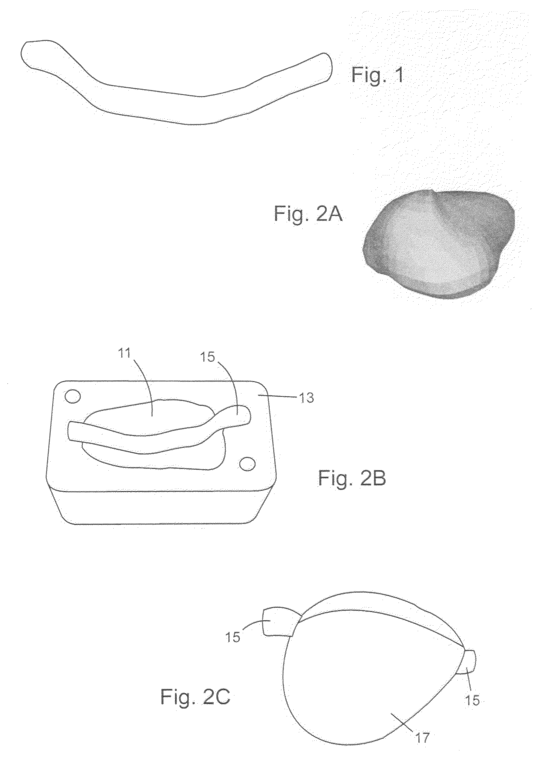
[0061]An anatomically shaped PVA-C urethra tissue phantom is created as follows.

[0062]A mould in the shape of a urethra having the proper size and shape is created by using ultrasound diagnostic imaging to define tissue morphology by 3D reconstruction of ultrasound images. The mould is then constructed using fused deposition modelling (FDM).

[0063]A 300 ml batch of aqueous PVA-H solution comprising 15% w / w PVA is prepared in accordance with the procedure in Example 1. When the heating / stirring is finished, the temperature of the solution is allowed to drop to less than 55° C. A 20 ml aliquot of the PVA-H is placed in a beaker and 9% w / w (1.8 g) Sigmacell™ Type 50 (cellulose particles from Sigma-Aldrich) is gently stirred into the 20 ml aliquot of cooked PVA in the beaker, being careful not to introduce air bubbles into the mixture. The resulting PVA solution from the beaker is injected into the urethra-shaped mould until the mould is full and allo...


PUM


Abstract

A phantom for prostate cancer brachytherapy has a prostate tissue phantom shaped like a real prostate gland, a perineal tissue phantom surrounding the prostate tissue phantom, a skin tissue phantom separating the perineal tissue phantom from an outside environment, and an enclosure. The prostate tissue phantom is a polyvinyl alcohol cryogel (PVA-C) having undergone 3-5 freeze-thaw cycles and having 10-20% w / w PVA in a solvent (e.g. water), 4-8% w / w oil and an amount of acoustic scattering particles to ultrasonically distinguish the prostate-tissue phantom from its surroundings. The perineal tissue phantom is a PVA-C having undergone 1-2 freeze-thaw cycles and having 10-20% w / w PVA in a solvent, 4-8% w / w oil and an amount of acoustic scattering particles to ultrasonically distinguish the perineal tissue phantom from its surroundings. The skin tissue phantom is a PVA-C having undergone at least 6 freeze-thaw cycles and having 15-25% w / w PVA in a solvent. The phantom mimics the imaging and mechanical properties of the prostate and surrounding tissues, providing a realistic phantom for prostate cancer brachytherapy.

Description

CROSS-REFERENCE TO RELATED APPLICATIONS[0001]This application claims the benefit of U.S. Provisional patent application Ser. No. 61 / 136,128 filed Aug. 13, 2008, the entire contents of which is herein incorporated by reference.FIELD OF THE INVENTION[0002]The present invention relates to a phantom for prostate cancer brachytherapy.BACKGROUND OF THE INVENTION[0003]The prostate is a gland about the size and shape of a walnut. The prostate is located between the pubic bone and rectum. It surrounds the upper part of the urethra, the tube that carries urine from the bladder. As a man ages, his prostate may change. Non-cancerous (benign) growths may form. Or some cells may change into precancerous cells and cancerous cells may form a malignant tumor. For some men, cancerous cells may form within the prostate but grow too slowly to cause problems. In other cases, cancerous tumors may grow inside the prostate, then spread.[0004]Prostate cancer is the most frequently diagnosed cancer and the s...

Claims


Application Information

Patent Timeline
Patent Type & Authority Patents(United States)
IPC IPC(8): G09B23/28
CPCG09B23/28B33Y80/00
Inventor CAMPBELL, GORDONHOMBLOWER, VICTORIA
Owner NAT RES COUNCIL OF CANADA
Why Eureka
Try Eureka