Methods for approximation and fastening of soft tissue

a soft tissue and approximation technology, applied in the field of soft tissue approximation and fastening, can solve the problems of large skin incisions, tedious and time-consuming process of approximating individual layers of tissue and securely fastening, and the method used in these devices, so as to achieve less variation in tissue reconfiguration and greater freedom for surgeons

Inactive Publication Date: 2013-08-06
LONGEVITY SURGICAL
View PDF84 Cites 7 Cited by
  • Summary
  • Abstract
  • Description
  • Claims
  • Application Information

AI Technical Summary

Benefits of technology

[0011]The distal tool assembly includes mechanisms for engaging tissue at two or more spaced-apart tissue locations on a tissue surface, and is further configured to allow the operator to reposition and / or move at least one of the spaced-apart tissue locations toward another spaced-apart tissue location thereby approximating the engaged tissue locations near the distal end of the device. In certain embodiments, the device may be designed and configured to allow the tissue engagement at two or more locations to be performed sequentially, whereas in other embodiments the device may be designed and configured to the allow tissue engagement at two or more locations to be performed simultaneously. A wide variety of tissue engagement mechanisms are possible within the scope of the present invention, including needles, hooks, barbs, clamps, grippers, forceps, jaws, teeth, vacuum ports, and the like. In certain embodiments, the distal tool assembly further incorporates mechanisms for deploying one or more individual tissue fasteners into the approximated tissue, to securely hold the tissue in the approximated configuration after the device is removed from the treatment site. A wide variety of fasteners may be used within the scope of the present invention, including sutures, staples, screws, tacks, clips, hooks, clamps, rivets, t-tags, expandable anchors, and the like. The devices may be configured to deliver a single fastener, requiring reloading after each tissue engagement, approximation and fastening cycle, or alternatively, in other embodiments, the device may be configured as a multi-fire instrument in which a plurality of fasteners are pre-loaded into the device, allowing successive tissue engagement, approximation and fastening cycles to be completed without reloading or removing the device from the patient. In some embodiments the device is configured having separate and independently operable tissue approximation and fastening mechanisms, whereas in other embodiments multi-functional components provided within the distal tool assembly are designed and configured to provide both the tissue approximation and fastening functions. This can simplify the mechanisms required, lowering cost and increasing reliability, as well as reducing the device profile.
[0016]In other embodiments, two or more such moveable members are provided, each having an associated tissue engagement mechanism positioned near its distal end. In this example, the step of engaging tissue at two locations occurs after initially deploying the distal end of said moveable members away from the longitudinal axis of the device. Upon actuated retraction of said moveable members, the engaged tissue locations are both moved toward to the longitudinal axis of the device, being moved toward one another and approximated near the distal end of the device. Some advantages of this type of device configuration over the previously described embodiments are that the distal end of the device itself need not become attached to the target tissue, nor does it need to be significantly repositioned to effect the approximation. This results in greater freedom and ease of use for the surgeon, and less variation in the tissue reconfiguration which may be attributable to surgeon technique. As described previously, once the tissues have been approximated near the distal end of the device in this manner, the fastener may be deployed to securely hold tissues in the approximated configuration after the device has been removed from the treatment site.

Problems solved by technology

The process of approximating the individual layers of tissue and fastening them securely is tedious and time consuming.
The disadvantages and limitations of the methods used in these devices include relatively large skin incisions and resultant scars from providing the surgeon access to the abdominal cavity.
Extended time under anesthesia due to the time consuming nature of suturing the wound closed, and lengthy recovery times for patients who undergo these invasive procedures resulting in high costs.
These are significant drawbacks for this type of intervention.
However these devices are complicated to manufacture, expensive and typically require the tissue being fastened must also be transected.
Additionally the approximation possible with these types of devices is limited by the size of the aperture of the open jaws of the instrument, often being less than the outside diameter of the instrument shaft itself.
Another disadvantage of these devices occurs when the tissue to be fastened does not completely fill the stapler jaws; staples not in contact with tissue fall loosely into the patient's abdomen.
These are significant disadvantages for this type of device.
While tedious, time consuming, and technique sensitive, laparoscopic suturing is a method used for fastening tissues to this day.
In this disclosure, the device fixates portions of tissue together so that the tissue can fuse or scar over, however Gannoe specifically discusses the need to apply a clamping force that does not clamp too tightly, thus leading to complications such as pressure necrosis, or too lightly, which may result in an incomplete tissue union.
Thus, inconsistent securement is a problem that requires precise application of force.

Method used


Image

  • Methods for approximation and fastening of soft tissue
  • Methods for approximation and fastening of soft tissue
  • Methods for approximation and fastening of soft tissue

Examples


Embodiment Construction

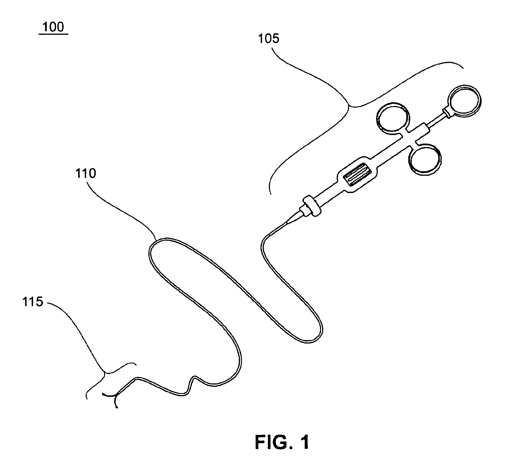
[0035]According to one embodiment of the present invention, illustrated in FIG. 1, tissue approximation device 100 is configured for laparoscopic or endoscopic use, consisting of a proximal handle assembly 105, longitudinal tube assembly 110 and distal tool assembly 115. Longitudinal tube assembly 110 is typically produced from flexible biocompatible materials and is configured to be inserted into the body via a laparoscopic access port (e.g. a small incision or trocar) or flexible endoscope. It is preferably between 0.5 mm and 20 mm in diameter, more preferably between 1 mm and 15 mm in diameter, and most preferably between 1.5 mm and 10 mm in diameter. It may consist of a single tube, multiple concentric tubes, and combinations thereof.

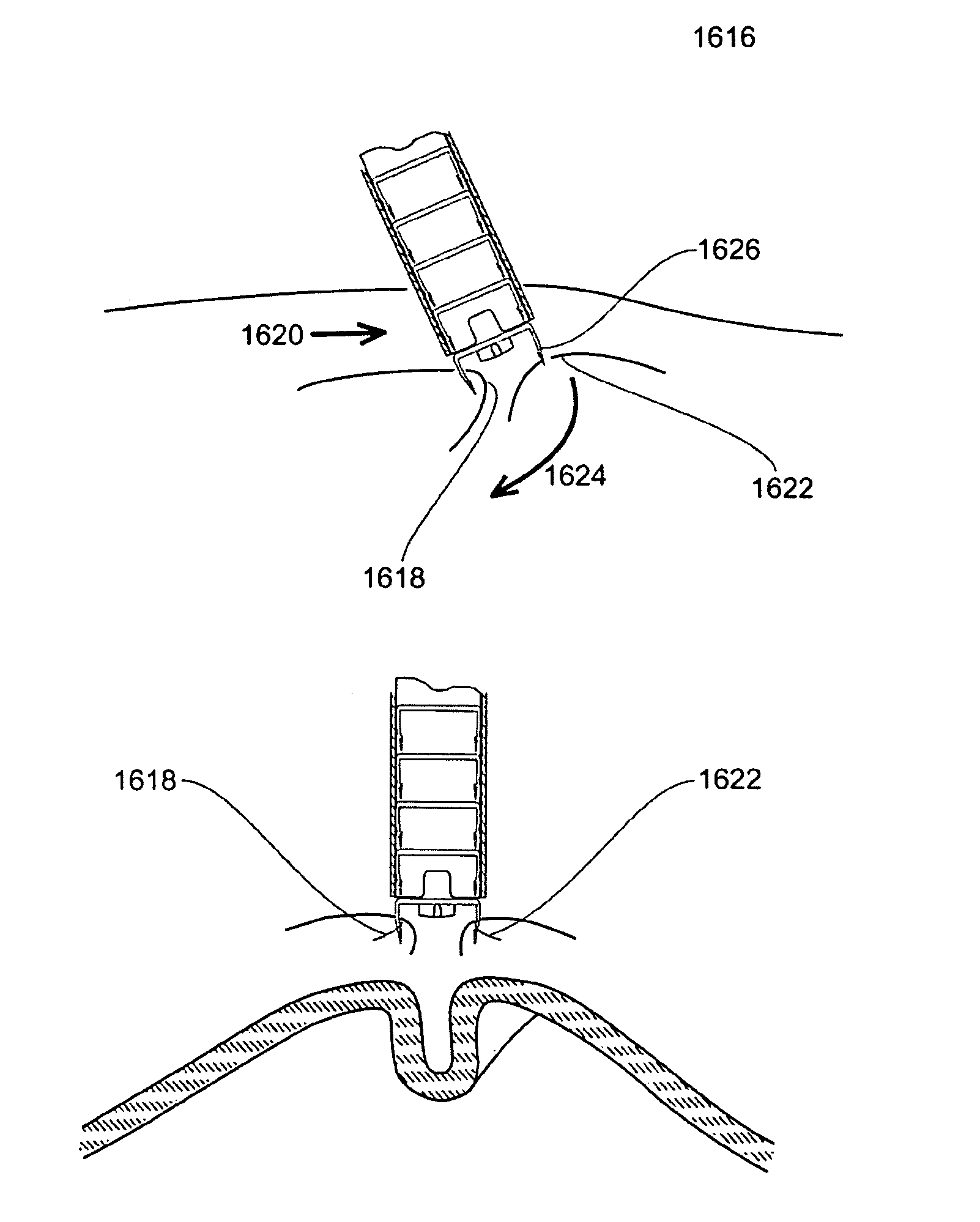
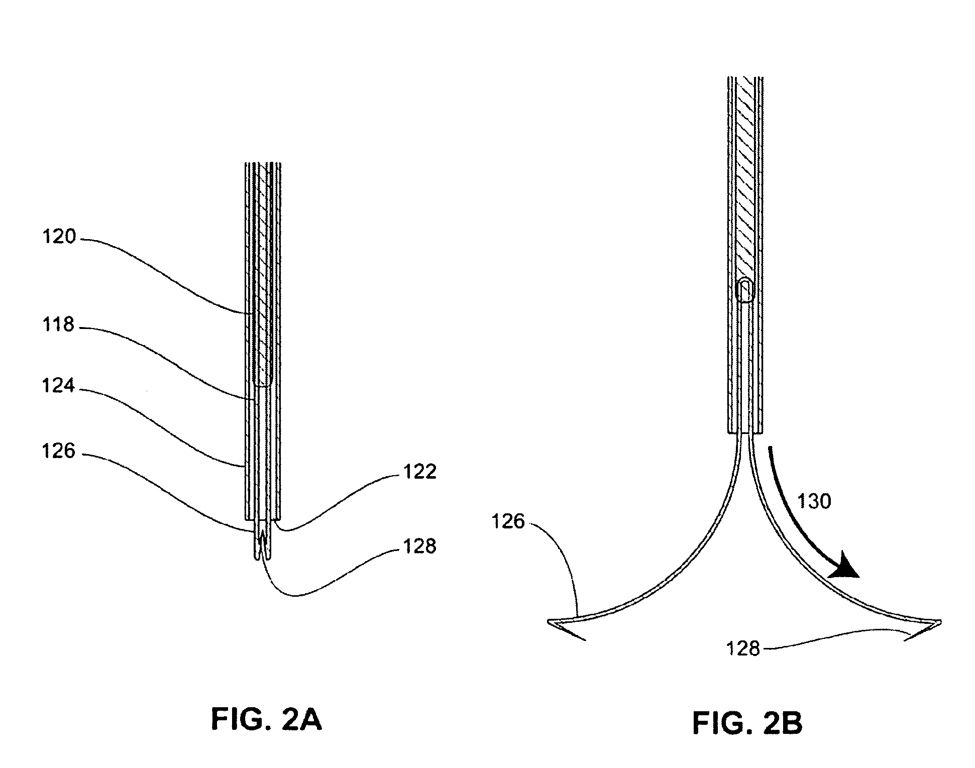
[0036]FIG. 2 shows close up details of distal tool assembly 115. In the pre-deployed configuration (FIG. 2A) two moveable arms 118 are configured as individual longitudinal components that are operatively connected to the distal end of internal shaf...


PUM


Abstract

Methods for approximating and fastening tissue by application of one or more tissue fasteners are provided. In one embodiment, spaced apart tissue locations are engaged by tissue penetrating members of a deformable fastener, one of more of the engaged tissue locations is moved toward another engaged tissue location to approximate the spaced apart locations, and the deformable fastener is deployed to secure the approximated tissue locations. These methods may be used in laparoscopic plication gastroplasty procedures for forming an invaginated tissue fold, to close holes in the gastrointestinal lumen, and in a variety of interventional procedures.

Description

REFERENCE TO PRIORITY APPLICATIONS[0001]This application claims priority to U.S. International Patent Application No. PCT / US2008 / 56921 filed Mar. 13, 2008 and is a continuation-in-part of U.S. patent application Ser. No. 12 / 048,206, filed Mar. 13, 2008 and issued as U.S. Pat. No. 8.142.450 on Mar. 27, 2012, which claims priority to U.S. Provisional Patent Application No. 60 / 894,626 filed Mar. 13, 2007. This application also claims priority under 35 U.S.C. §119(e) to U.S. Provisional Patent Application No. 61 / 031,124 filed Feb. 25, 2008. These patent applications are incorporated herein by reference in their entireties.FIELD OF THE INVENTION[0002]The present invention relates generally to methods and devices for approximating and fastening of soft tissues within the body. Uses for the methods and devices of the present invention include laparoscopic and endoscopic interventions involving reconfiguration or repair of soft tissues, such as reconfiguration or repair of gastrointestinal ...

Claims


Application Information

Patent Timeline
Patent Type & Authority Patents(United States)
IPC IPC(8): A61B17/08A61D1/00
CPCA61B17/0057A61B17/0684A61B17/10A61B17/083A61B17/0643A61B2017/00668A61B2017/00349A61B2017/00637
Inventor HARRIS, PETER S.RABIN, BARRY HAL
Owner LONGEVITY SURGICAL
Why Eureka
Try Eureka