
Pharmaceutical compositions and methods to vaccinate against candidiasis

a composition and composition technology, applied in the field of candidiasis surface adhesion proteins, can solve the problems of unfavorable candidiasis survival rate, unfavorable candidiasis treatment effect, so as to prevent, treat, or alleviate the effect of disseminated candidiasis

Inactive Publication Date: 2013-09-24
LOS ANGELES BIOMEDICAL RES INST AT UCLA HARBOR MEDICAL CENT +1
View PDF7 Cites 11 Cited by
  • Summary
  • Abstract
  • Description
  • Claims
  • Application Information

AI Technical Summary

Benefits of technology

The patent describes a new way to fight fungal infections using a combination of traditional anti-fungal agents and immunotherapeutics. Immunotherapy involves administering a vaccine to stimulate the body's immune system, which then generates antibodies to fight the fungal infection. The antibodies can be administered directly to patients, or they can be generated in a laboratory and given to patients. This approach offers the advantage of using highly specific antibodies that are typically less toxic than regular anti-fungal agents. The patent also describes a specific protein called ALS, which is involved in adhesion to endothelial cells. A null mutant of this protein would not adhere to cells as well, while over-expression would increase adhesion. Antisera to the protein would cross-react with anti-ALS antibodies and provide increased survival times in animal models of fungal infections.

Problems solved by technology

C. albicans infections are difficult to diagnose and the organism can survive in vivo without causing overtly detectable disease symptoms and can cause overt disease symptoms that vary in site and in severity.
While potent antifungal agents exist that are microbicidal for Candida, the attributable mortality of candidemia is approximately 38%, even with treatment with potent anti-fungal agents such as amphotericin B. Also, existing agents such as amphotericin B tend to exhibit undesirable toxicity.

Method used


Image

  • Pharmaceutical compositions and methods to vaccinate against candidiasis
  • Pharmaceutical compositions and methods to vaccinate against candidiasis
  • Pharmaceutical compositions and methods to vaccinate against candidiasis

Examples


example 1

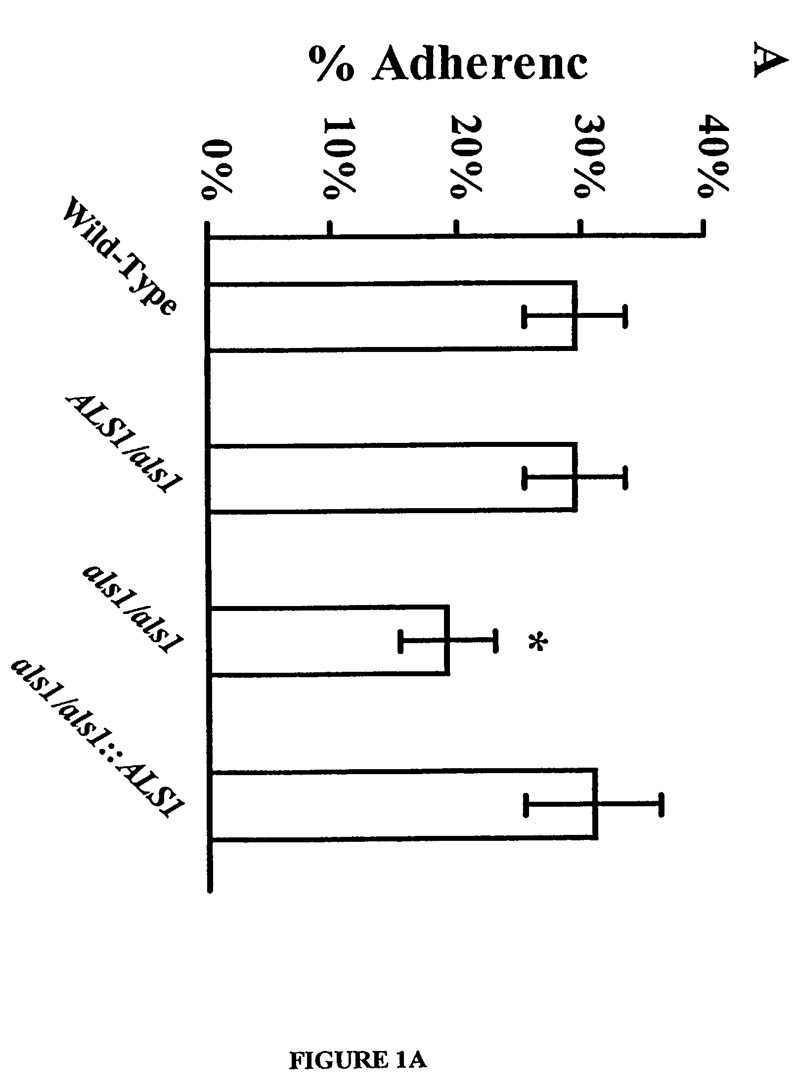
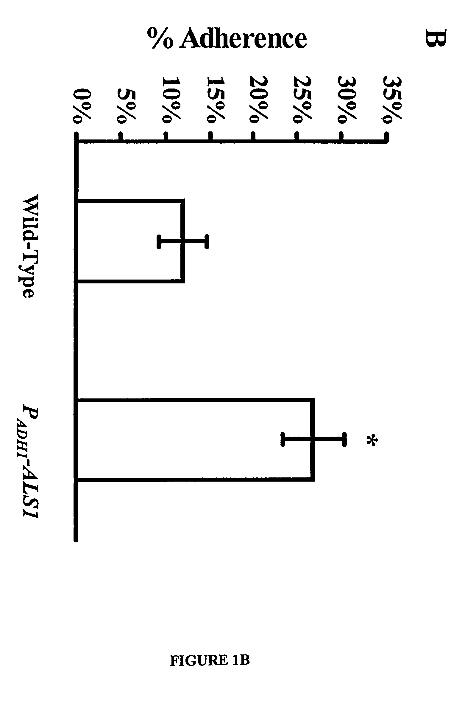
Als1 Mediates Adherence of C. albicans to Endothelial Cells

[0050]The URA blaster technique was used to construct a null mutant of C albicans that lacks expression of the Als1p. The als1 / als1 mutant was constructed in C. albicans strain CAI4 using a modification of the Ura-blaster methodology [W. A. Fonzi and M. Y. Irwin, Genetics 134, 717 (1993)] as follows: Two separate als1-hisG-IRA3-hisG-als1 constructs were utilized to disrupt the two different alleles of the gene. A 4.9 kb ALS1 coding sequence was generated with high fidelity PCR (Boehringer Mannheim, Indianapolis, Ind.) using the primers: 5′-CCGCTCGAGATGCTTCAACAATTTACATTGTTA-3′ (SEQ ID NO.1) and 5′-CCGCTCGAGTCACTAAATGAACAAGGACAATA3′ (SEQ ID NO. 2). Next, the PCR fragment was cloned into pGEM-T vector (Promega, Madison, Wis.), thus obtaining pGEM-T-ALS1. The hisG-URA3-hisG construct was released from pMG-7 by digestion with KpnI and Hind3 and used to replace the portion of ALS1 released by Kpn1 and Hind3 digestion of pGEM-T-ALS...

example 2

Localization of Als1p

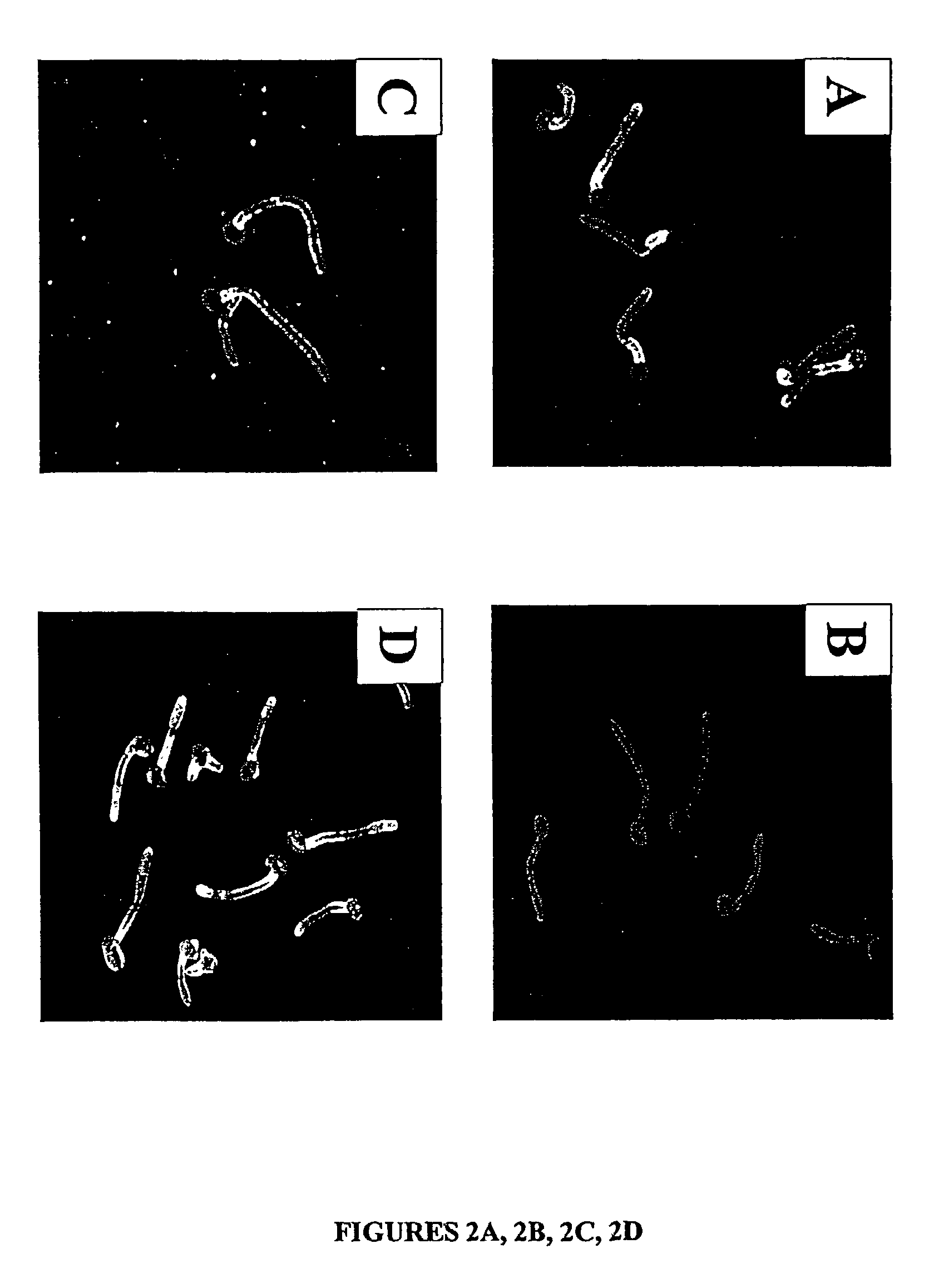
[0056]For a number of the ALS family to function as an adhesin protein, it must be located on the cell surface. The cell surface localization of Als1p, for example, was verified using indirect immunofluorescence with the anti-Als1p monoclonal antibody. Diffuse staining was detected on the surface of blastospores during exponential growth. This staining was undetectable on blastospores in the stationary phase. Referring to FIG. 2A, when blastospores were induced to produce filaments, intense staining was observed that localized exclusively to the base of the emerging filament. No immunofluorescence was observed with the als1 / als1 mutant, confirming the specificity of this antibody for Als1p. See FIG. 2B. These results establish that Als1p is a cell surface protein.

[0057]The specific localization of Als1p to the blastospore-filament junction implicates Als1p in the filamentation process. To determine the mechanism, the filamentation phenotype of the C. albicans AL...

example 3

Purification of ALS1 Adhesin Protein, Truncated N-Terminal Protein

[0063]For use as an immunogen, an ALS protein synthesized by E. coli is adequate when vaccination with a traditional protocol yield an immune response generating B cells expressing measurable anti-ALS anti-sera or levels of serum Ig from which polyclonals may be obtained. However, eukaryotic proteins synthesized by E. coli may not be functional due to improper folding or lack of glycosylation. Therefore, to determine if the ALS1 protein can block the adherence of C. albicans to endothelial cells, the protein is, preferably, purified from genetically engineered C. albicans, and formulated into a substantially pure pharmaceutical composition that is pharmacologically effective for prophylactic or therapeutic treatment of disseminated candidiasis.

[0064]PCR was used to amplify a fragment of ALS1 (SEQ ID NO 7), from nucleotides 52 to 1296.

[0065]This 1246 bp fragment encompassed the N-terminus of the predicted ALS1 protein ...


PUM

PropertyMeasurementUnit
adhesionaaaaaaaaaa
sizeaaaaaaaaaa
morphologiesaaaaaaaaaa

Abstract

A Candida albicans bloodstream infections cause significant morbidity and mortality in hospitalized patients. Filament formation and adherence to host cells are critical virulence factors of C. albicans. Multiple filamentation regulatory pathways have been discovered, however the downstream effectors of these regulatory pathways remain unknown. The cell surface proteins in the ALS group are downstream effectors of the filamentation regulatory pathway. Particularly, Als1p mediates adherence to endothelial cells in vitro and is required for virulence. The blocking of adherence by the organism is described resulting from the use of a composition and method disclosed herein. Specifically, a pharmaceutical composition comprised of a gene, gene product, or specific antibody to the ALS gene family is administered as a vaccine to generate an immune response capable of blocking adherence of the organism.

Description

SEQUENCE LISTING[0001]The present application contains a Sequence Listing which has been submitted in ASCII format via EFS-Web and is hereby incorporated by reference in its entirety. Said ASCII copy, created on Sep. 23, 2011, is named 50665—006001_Sequence_Listing09—23—2011_ST25.txt and is 154 KB in size.[0002]This application is a continuation-in-part of Ser. No. 09 / 715,876 filed on Nov. 18, 2000, which issued as U.S. Pat. No. 7,067,138, which claims priority from Provisional Application Ser. No.: 60 / 166,663 filed Nov. 19,1999. This invention was made with Government support under Public Health Service grants PO-1Al-37194, RO1Al-19990, and MO1 RR0425.The Government has certain rights in this invention. The priority of the prior applications are expressly claimed, and the disclosure of each of these prior applications are hereby incorporated by reference in their entirety.FIELD OF INVENTION[0003]This invention relates to Candida albicans surface adhesin proteins, to antibodies resu...

Claims


Application Information

Patent Timeline
Patent Type & Authority Patents(United States)
IPC IPC(8): A61K39/00A61K38/00A61K49/00C07K1/00C07K16/14
CPCA61K39/0002C07K14/40A61K39/39575A61K45/06A61K38/16C07K16/14A61K2039/505A61K2039/53
Inventor EDWARDS, JR., JOHN E.FILLER, SCOTT G.SHEPPARD, DONALD C.IBRAHIM, ASHRAF S.FU, YUESPELLBERG, BRADLEY J.
Owner LOS ANGELES BIOMEDICAL RES INST AT UCLA HARBOR MEDICAL CENT
Learn More