Analyte-releasing beads and use thereof in quantitative elispot or fluorispot assay
a technology of analyte-releasing beads and quantitative elispot or fluorispot assay, which is applied in the field of reage, can solve the problem that assays can only provide limited data
- Summary
- Abstract
- Description
- Claims
- Application Information
AI Technical Summary
Benefits of technology
Problems solved by technology
Method used
Image
Examples
example 1
Preparation of Immunoglobulin-Releasing Reagent
[0062]Construction of Beads:
[0063]Beads were obtained from commercial bead makers such as Bangs Laboratories or Micromod. Materials used to make such beads include latex, polymer, and silica, and the beads may contain iron oxide to give the beads a paramagnetic nature. Bead sizes included 8 microns, 10 microns, and 12 microns (polystyrene, catalog #UMC3N Compel™ Magnetic COOH-modified, polystyrene, catalog #CP01N SuperAvidin Coated Microspheres, Bangs Laboratories), 10 microns (latex, #08-19-104 Micromod, GmbH) and 12 microns (latex, #08-19-124, Micromod, GmbH) in diameter. They are permanently coated with Streptavidin protein, which can bind biotin protein.
[0064]DSB-X-Biotin-Antigen Conjugation:
[0065]A DSB-X-biotin Protein Labeling Kit (D20655, Molecular Probes, Inc.) was used to conjugate human IgG (Sigma-Aldrich, Inc) to DSB-X biotin. DSB-X biotin is a derivative of desthiobiotin, which is a stable biotin pre-cursor, and has a lower ...
example 2
Preparation of Immunoglobulin-Releasing Magnetic Microparticles
[0068]Wash:
[0069]For magnetic beads, a slightly different protocol was used. Example 1 is modified as follows. 10 μl aliquots of beads were washed in 500 μl PBS and then placed in a magnet-rack and allowed to settle against the tube. Then the supernatant was carefully removed. The beads were resuspended in 500 μl PBS and allowed to settle. This step was repeated. Antigen solutions were prepared as described in Example 1.
[0070]Label:
[0071]The supernatant was carefully removed, and then the tubes were carefully removed from the magnet-rack. The beads were resuspended in antigen solution and incubated for 30 minutes.
[0072]Wash:
[0073]The beads were placed in the magnet-rack and allowed to settle against the tube, and then the supernatant was carefully removed. The beads were resuspended in 500 μl PBS and allowed to settle. This step was repeated. The beads were then ready to be counted and used in an ELISpot assay.
example 3
ELISpot Assay for Immunoglobulin Secreting Cells
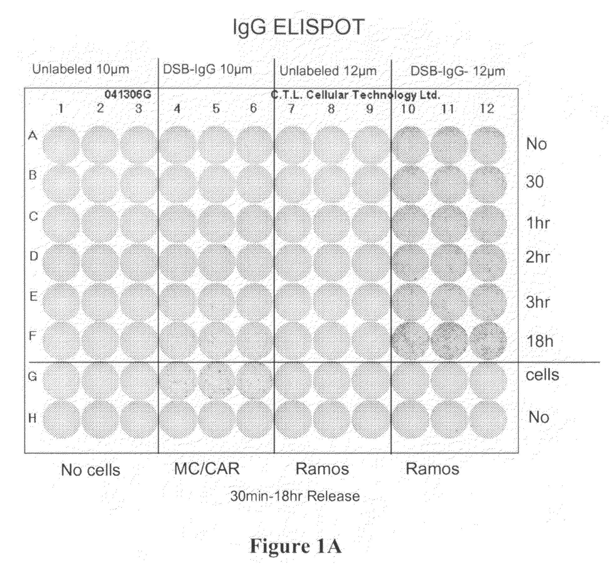
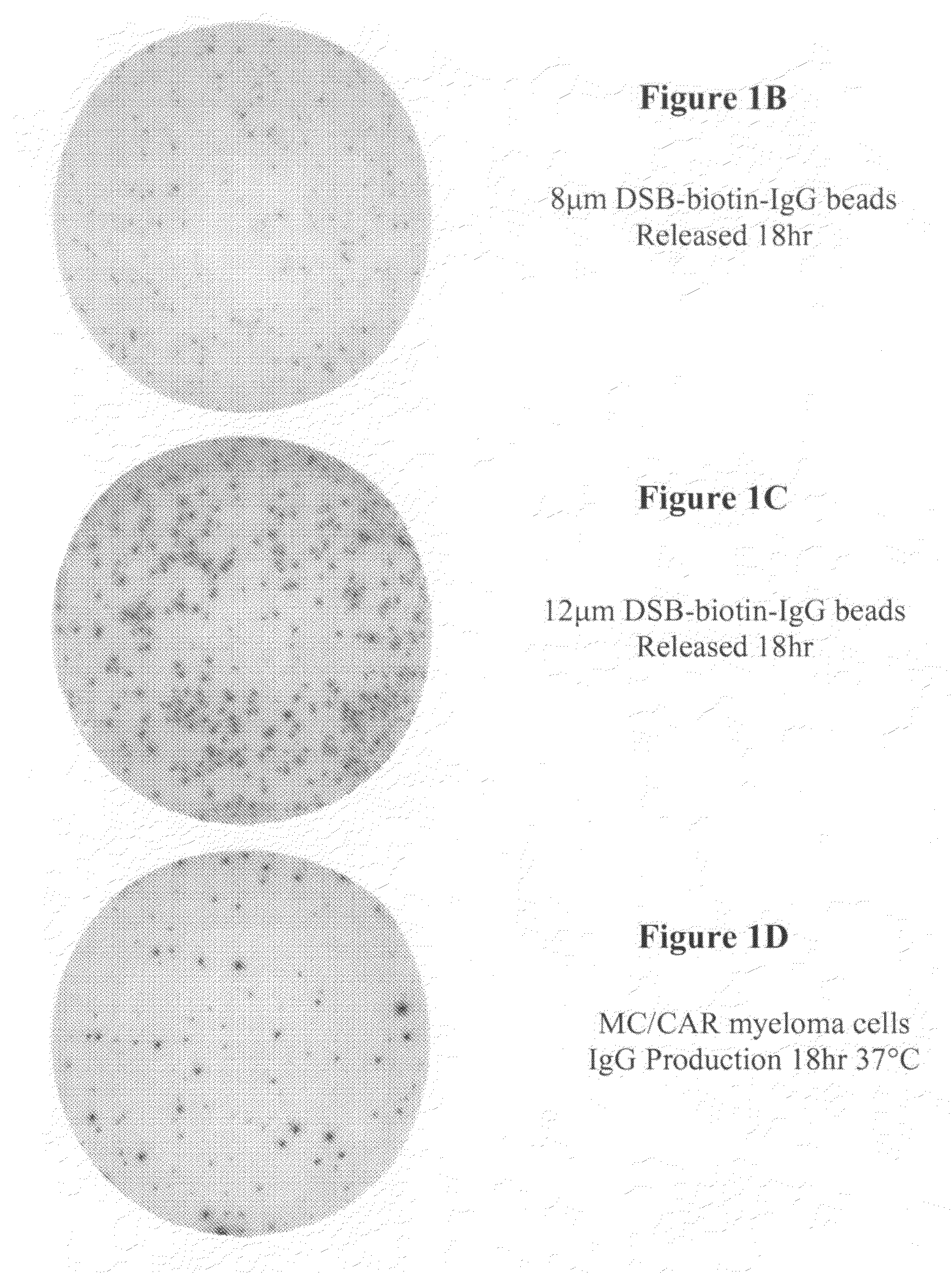
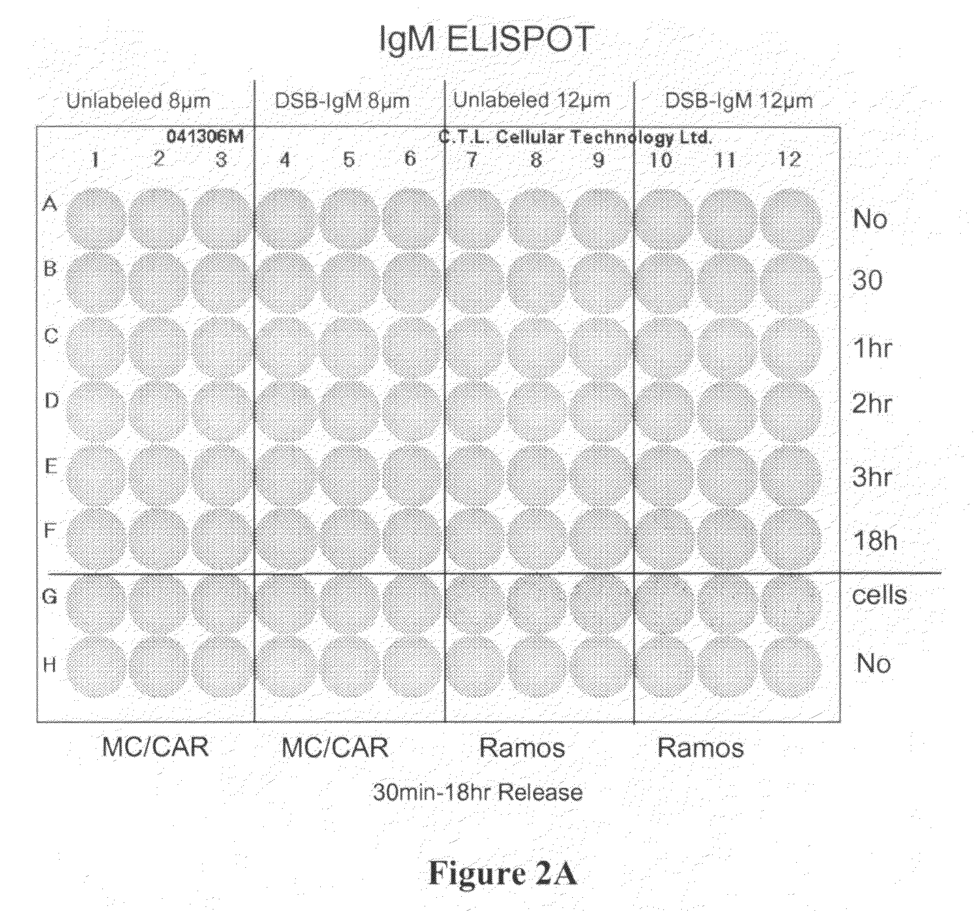
[0074]ELISpot Assay was carried out according to the procedure described below. Materials and reagents that were used included Millipore PVDF 96-well multiscreen ELISpot plates, RPMI Cell Culture Medium (Gibco, Inc.), Fetal Bovine Serum (FBS), Pen / Strep, Bovine Serum Albumin (BSA) (powder), Fraction V Sigma A4059-1006, 1× Phosphate Buffered Saline (PBS), Strepavidin-Alkaline Phosphatase (Southern Biotech #7100-04), Vector Alkaline Phosphatase substrate kit III, 70% ethyl alcohol, and Immunospot ELISpot Analyzer (CTL Corp.).
[0075]The reagents were prepared as follows. 1×PBS composed of 40 g NaCl, 1 g KCl, 5.75 g Na2HPO4.7H2O, 1 g KH2PO4, 5 L ddH2O was mixed thoroughly. The capture antibody was prepared at 10 μg / ml in PBS (see Table 1 below). The cell culture medium was prepared using sterile technique, RPMI plus 10% Fetal Bovine Serum with penicillin / streptomycin. PBS and 2% BSA (100 ml+2 gm BSA) was prepared. The detecting antibody was...
PUM
| Property | Measurement | Unit |
|---|---|---|
| bead sizes | aaaaa | aaaaa |
| bead sizes | aaaaa | aaaaa |
| diameter | aaaaa | aaaaa |
Abstract
Description
Claims
Application Information
 - R&D
- Intellectual Property
- Life Sciences
- Materials
- Tech Scout
- Unparalleled Data Quality
- Higher Quality Content
- 60% Fewer Hallucinations
Browse by: Latest US Patents, China's latest patents, Technical Efficacy Thesaurus, Application Domain, Technology Topic, Popular Technical Reports.
© 2025 PatSnap. All rights reserved.Legal|Privacy policy|Modern Slavery Act Transparency Statement|Sitemap|About US| Contact US: help@patsnap.com


