Methods of detecting antibodies specific for denatured HLA antigens

a technology of hla antibodies and antibodies, applied in the field of detecting antibodies specific for denatured hla antibodies, can solve the problems of increasing the risk of rejection of the transplanted organ, cell-based screening assays are unlikely to detect the presence of antibodies, and the available screening methods do not focus on selective detection of these antibodies

Active Publication Date: 2014-01-14
ONE LAMBDA INC
View PDF8 Cites 2 Cited by
  • Summary
  • Abstract
  • Description
  • Claims
  • Application Information

AI Technical Summary

Benefits of technology

[0014]It is important to screen for antibodies that specifically bind to denatured antigens, because there is a chance that a transplant recipient may have a high reactivity to a denatured HLA antigen and may not have the same reactivity for the antigen in its native conformation. Cell-based screening assays are unlikely to detect the presence of antibodies that specifically bind to denatured HLA antigens because there is minimal presentation of denatured antigens on the cell surface. Failure to detect a high reactivity to a denatured HLA antigen increases the risk for rejection of the transplanted organ. Therefore, screening for denatured antigens is an important step in fully analyzing a transplant recipient for HLA antigen reactivity prior to transplant.
[0030]A solid-phase substrate that presents a HLA antigen in its native conformation may also present a HLA antigen in its denatured conformation as an artifact of sample preparation. However, the number of denatured antigens on the solid-phase substrate in this situation should be much lower than the number of native HLA antigens. Thus, when carrying out the absorption step of any of the preceding screening methods, one of skill in the art will appreciate that any effect of underlying denatured HLA antigens may be effectively removed by titrating the volume of test sera and the immunosorbents.

Problems solved by technology

The clinical significance of antibodies specific for denatured HLA antigens was unknown and therefore the available screening methods did not focus on selectively detecting these antibodies.
Cell-based screening assays are unlikely to detect the presence of antibodies that specifically bind to denatured HLA antigens because there is minimal presentation of denatured antigens on the cell surface.
Failure to detect a high reactivity to a denatured HLA antigen increases the risk for rejection of the transplanted organ.
On the other hand, detection of serum reactivity due to antibodies that specifically bind denatured HLA antigens rather than native antigens in a virtual cross-matching assay may be a false positive result that would not be detected in a cell-based assay.
These false positive results may negatively affect transplant organ allocation by labeling a transplant patient ineligible due to an inaccurate HLA antibody profile.

Method used


Image

  • Methods of detecting antibodies specific for denatured HLA antigens
  • Methods of detecting antibodies specific for denatured HLA antigens
  • Methods of detecting antibodies specific for denatured HLA antigens

Examples


example 1

Method of Denaturing HLA Class I Antigens on Beads

[0086]To investigate the presence of antibodies specific for denatured HLA antigens, beads known to present native HLA antigens were subjected to denaturing conditions. FlowPRA Class I beads (FL1-30, FL1SP, One Lambda Inc.), Flow Single antigen beads (FL1HD, One Lambda Inc.), LABCreen class I PRA (LS1PRA, One Lambda Inc.) or LABCreen Single antigen beads (LS1A01, LS1A02, LS1A03, or LS1A04, One Lambda Inc.) were incubated with a pH 3 buffer containing 0.1 M sodium acetate for 30 minutes at room temperature. The beads were subsequently washed twice with PBS and resuspended in PBS.

[0087]Monoclonal antibodies BIH I (One Lambda, Inc.) and W6 / 32 (ATCC) are known to specifically bind to β2m-associated class I antigens and not denatured HLA antigens. Monoclonal antibodies HC10 (ATCC) or HB-296 (ATCC) are known to specifically bind β2m-free class I antigens (denatured HLA antigens) (Perosa et al., J. Immunol. 171: 1918-1926, 2003).

[0088]To co...

example 2

Separation of β2m-Free (Denatured) and β2m-Associated (Native) HLA Class I Antigens

[0090]It was of interest to develop techniques for separating denatured and native HLA antigens. A panel of untreated HLA antigen-presenting beads was incubated with an antibody specific for native HLA antigens (W6 / 32) and an antibody specific for denatured HLA antigens (HB-296; ATCC). The antigens on the beads were purified from human cell lines, and the beads contain both β2 m+ and β2m-free antigens because β2m may be disassociated from class I HLA antigens in vitro during antigen purification or other manipulation.

[0091]The denatured HLA antigens of the above-identified panel were separated from the native HLA antigens using HC10 affinity column absorption. HC10 is an antibody that is specific for denatured HLA antibodies. Mixture of native and denatured HLA class I antigens were diluted to about 10-50 μg / ml and run through an HC10 affinity column at a flow rate of 0.5 ml / min. The native antigen re...

example 3

Screening HLA Antibodies with Antigen Beads

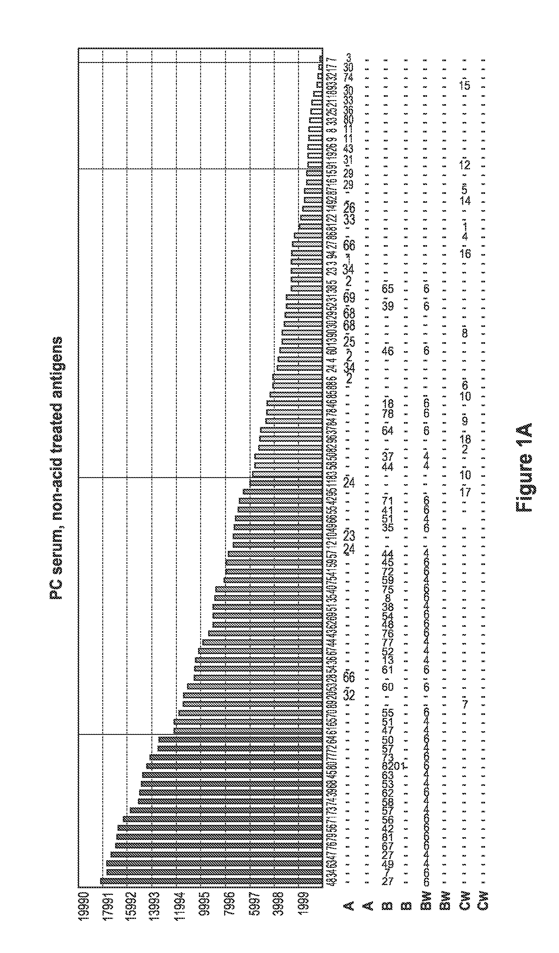
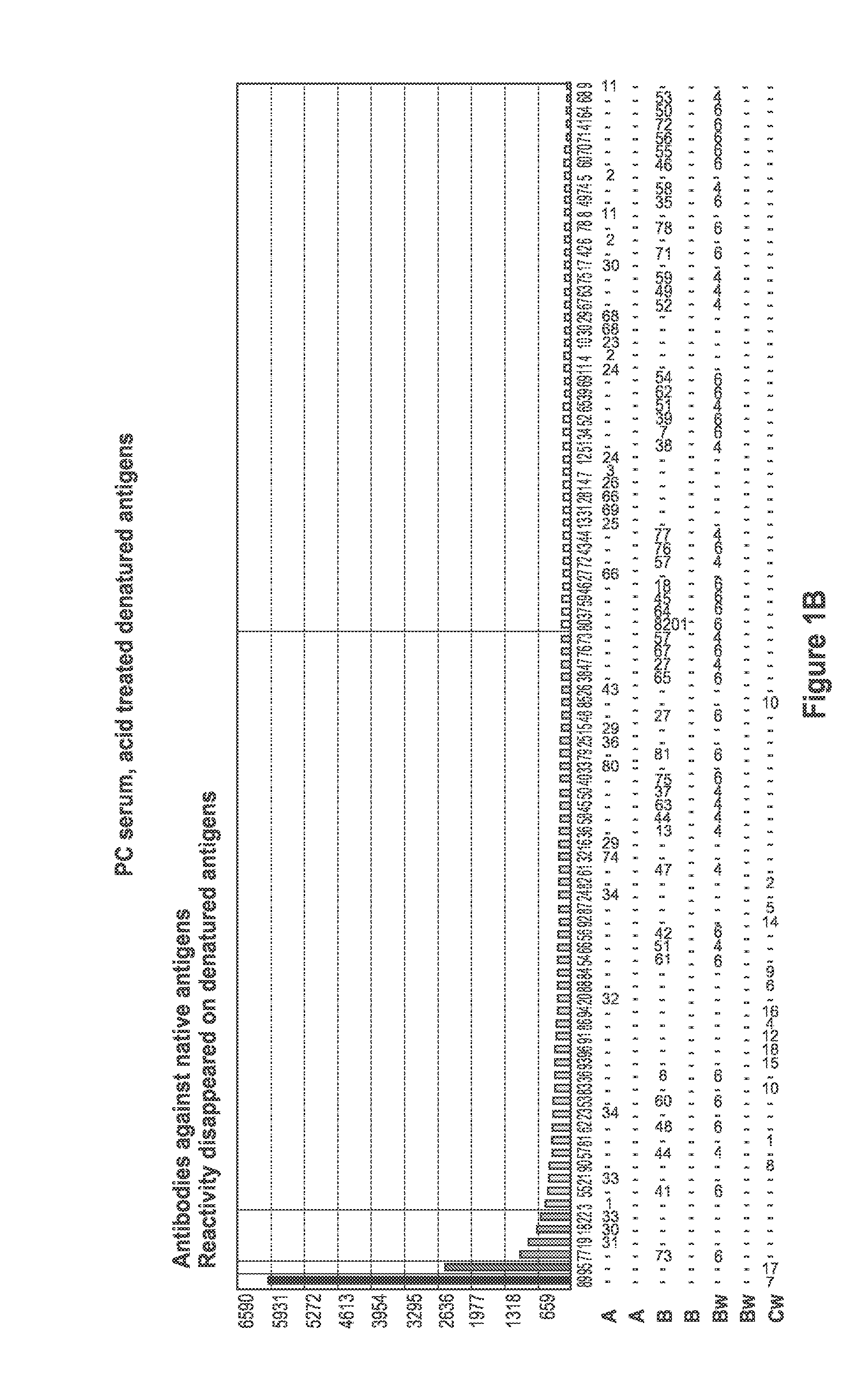
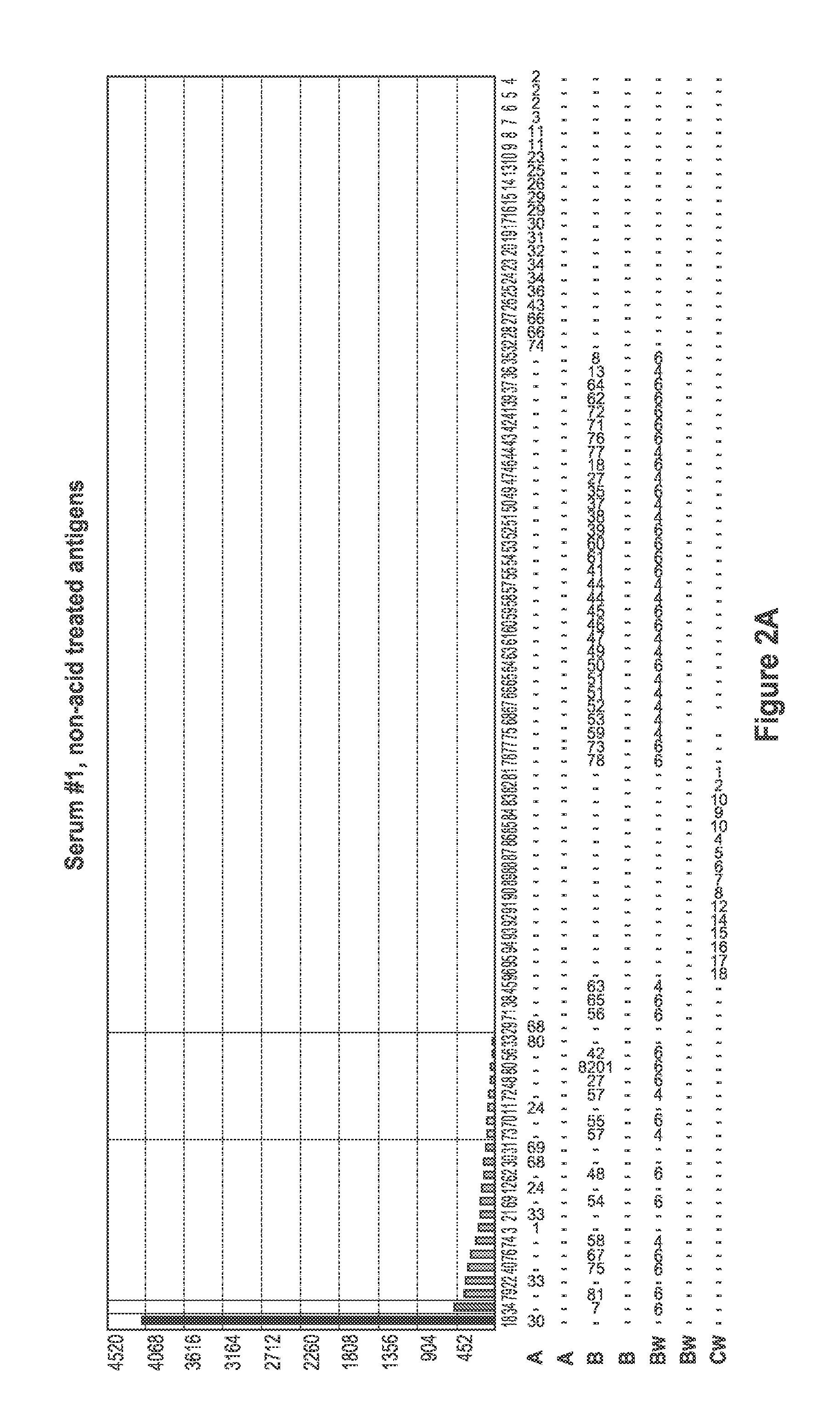
[0093]Sera samples from patients waiting for kidney transplant were screened for the presence of antibodies specific for denatured HLA antigens using panels of beads which present both native HLA antigens and denatured HLA antigens. Twenty microliters of serum were incubated with 5 μl of each of the antigen beads with or without acid treatment (as described in Example 1) for 30 minutes at 20-25° C. with gentle rotation. The microbeads were washed 3 times with 1 ml wash buffer (One Lambda., Inc.) by centrifugation at 9000 g for 2 minutes and then incubated with 100 FITC or PE-conjugated F(ab)2 fragment of goat anti-human IgG (Jackson ImmunoResearch). The microbeads were then washed twice with 1 ml wash buffer, and then 0.5 ml fixing solution (PBS with 0.5% formaldehyde) was added to the beads. The beads were then analyzed by a flow cytometer or a Luminex machine.

[0094]FIG. 1 depicts a comparison of positive control (PC) serum reaction patter...


PUM

PropertyMeasurementUnit
pHaaaaaaaaaa
flow rateaaaaaaaaaa
pHaaaaaaaaaa

Abstract

The invention is directed to methods of screening for HLA antibodies comprising detecting antibodies specific for native HLA antigens and denatured HLA antigens. The invention also provides for methods of removing antibodies specific for denatured HLA antigens or antibodies specific for native HLA antigens from a serum sample. In addition, the invention also provides for method of predicting whether a transplant recipient has an increased risk for rejecting the transplanted organ.

Description

[0001]This application is a continuation-in-part application of U.S. patent application Ser. No. 12 / 163,524, filed Jun. 27, 2008 (now U.S. Pat. No. 7,972,804), which claims priority benefit of U.S. Provisional Patent Application No. 60 / 947,872, filed Jul. 3, 2007 and U.S. Provisional Patent Application No. 60 / 949,806, filed Jul. 13, 2007, all of which are incorporated by reference herein in their entirety.FIELD OF INVENTION[0002]The invention is directed to methods of screening for HLA antibodies comprising detecting antibodies specific for native HLA antigens and denatured HLA antigens. Native class I HLA antigens are non-covalently associated with J32-microglobulin and denatured HLA class I antigens are disassociated from J32-microglobulin. Native HLA class II antigens are non-covalently bound hetero-dimers and denatured HLA class II antigens are monomers. The invention also provides for methods of removing antibodies specific for denatured HLA antigens or antibodies specific for ...

Claims


Application Information

Patent Timeline
Patent Type & Authority Patents(United States)
IPC IPC(8): G01N33/564G01N33/543G01N1/00
CPCG01N33/6854G01N2800/245C07K16/2833G01N33/505G01N33/543G01N2333/70539Y10T436/25125
Inventor LEE, JAR-HOWPEI, RUINONG, THOA
Owner ONE LAMBDA INC
Why Eureka
Try Eureka