Method for detection of liver cancer cell using anti-glypican-3 antibody

a technology of antiglypican and liver cancer, which is applied in the field of in vitro immunoassay methods for detecting the presence of liver cancer cells, can solve the problems of false positive or false negative, misinterpretation of staining, and inability to achieve ideal fixatives, and achieve the effect of accurate detection of the expression pattern of glypican 3

Active Publication Date: 2014-03-04
UNIVERSITY OF MIYAZAKI +1
View PDF6 Cites 9 Cited by
  • Summary
  • Abstract
  • Description
  • Claims
  • Application Information

AI Technical Summary

Problems solved by technology

However, unless sufficient care is taken to, for example, possible artifacts derived from formalin-fixed tissues themselves, not only does staining end unsuccessfully, but also such artifacts sometimes bring about alteration of antigens attributed to formalin fixation and embedding or alteration of antibody penetration or reactivity, resulting in false positive or false negative.
Such false positive or false negative may end in misinterpretation of staining results.
Formalin fixation used in usual histological search is useful for maintaining morphology but is not an ideal fixative from the viewpoint of maintaining affinity for antibodies.
However, a fixation method has not yet been established which is excellent in maintaining morphology and can preserve the affinity of every antigen for antibodies.
Accordingly, the formalin fixation is a practical fixation method widely used, although it has a problem in maintaining affinity for antibodies.
However, glypican 3 undergoes post-translational modification by protease or the like after being expressed on cell surface and therefore has limitations on epitopes capable of binding to antibodies.
Thus, this approach lacks epitope diversity for selecting a suitable epitope.
However, this method cannot be expected to significantly increase sensitivity.
However, it has been confirmed that as the number of reactions increases, non-specific background staining also tends to increase.
However, in the detection of antigens in formalin-fixed tissues, the use of the conventional method such as ABC method allows diagnosis of tumor tissues as being positive and normal or non-tumor tissues as being negative, on the basis of which tumor tissues can be differentiated from non-tumor tissues, whereas the use of the highly sensitive method provides detection of even a trace amount of antigens in non-tumor tissues and may therefore fail to make such differentiation depending on the types of antigens.
Moreover, the highly sensitive method used also had the problem of increased background staining, because of having high sensitivity.
This method had such problems as peel-off of sections from glass attributed to the digestion of the sections themselves, and unstable staining results.
However, since anti-glypican 3 antibodies exhibit cross-reactivity with the epithelial cells of blood vessels or hepatic sinusoids, this approach requires such complicated treatment that blocking reaction with a normal liver cell-derived protein lysate is performed in advance to exclude such cross-linking (Non-Patent Document 4).
Thus, the HIER method conventionally used cannot accurately detect glypican 3 in liver cancer tissues in such a way that glypican 3 originally expressed on the cell membrane is observed as if this antigen is cytoplasmically expressed (Non-Patent Document 2).

Method used


Image

  • Method for detection of liver cancer cell using anti-glypican-3 antibody
  • Method for detection of liver cancer cell using anti-glypican-3 antibody
  • Method for detection of liver cancer cell using anti-glypican-3 antibody

Examples


example 1

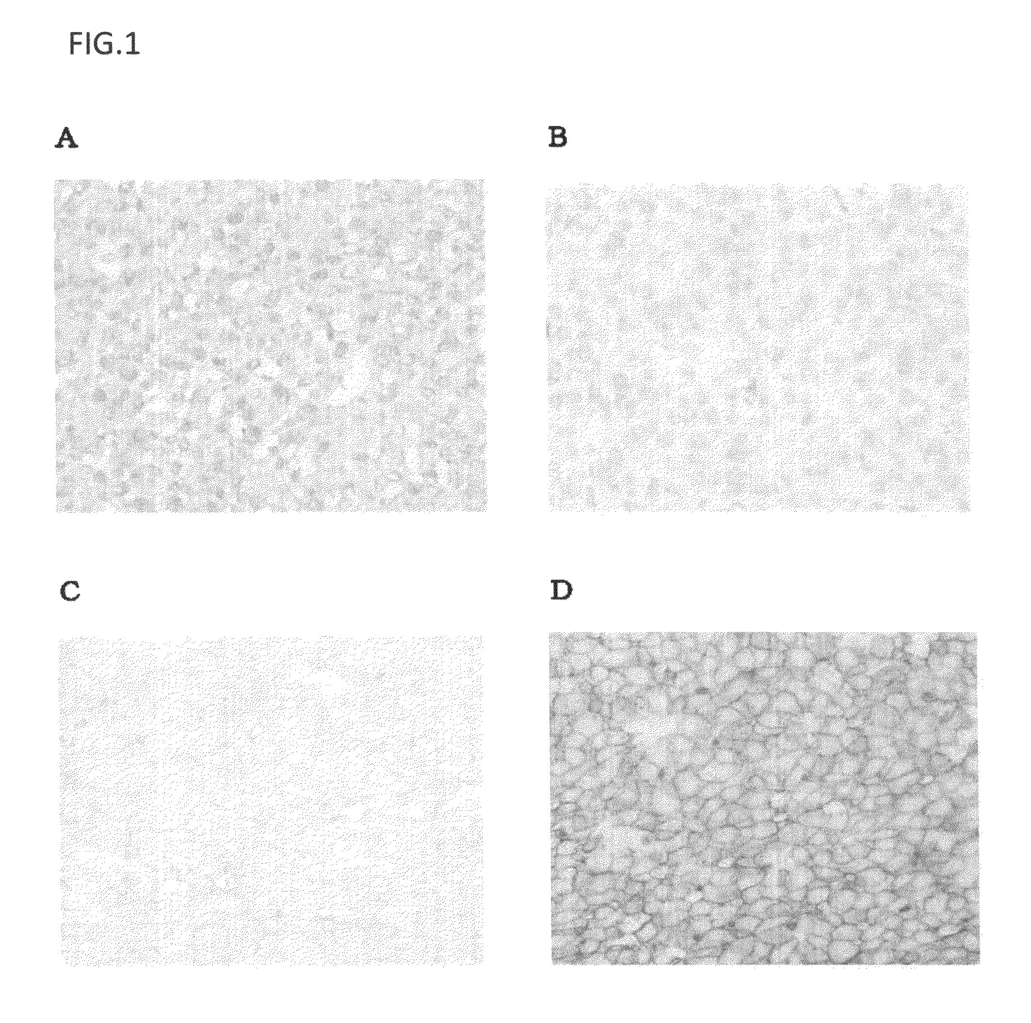
Immunostaining of Glypican 3 Using Mouse Models in which Human Liver Cancer Cell Strain Expressing Glypican 3 was Subcutaneously Transplanted into the Abdominal Region

(1) Cell Strain

[0129]The liver cancer cell strains used were HuH-7 cells (Health Science Research Resources Bank, HSRRB) and HepG2 cells (ATCC). HuH-7 was maintained and subcultured in Dulbecco's Modified Eagle's Medium (SIGMA-ALDRICH CO.) containing 10% FBS (BIONET). HepG2 was maintained and subcultured in Minimum Essential Medium Eagle medium (SIGMA-ALDRICH CO.) containing 10% FBS, 1 mmol / L MEM Sodium Pyruvate (Invitrogen Corp.), and 1 mmol / L MEM Non-Essential Amino Acid (Invitrogen Corp.).

(2) Measurement of GPC3 Expression Level

(2-1) Measurement Method

[0130]The GPC3 expression levels of the HuH-7 and HepG2 cells were measured using a mouse anti-human GPC3 monoclonal antibody (clone name: GC33, described in WO2006 / 006693) and QIFI-Kit (DakoCytomation). The measurement method was conducted according to a method descri...

example 2

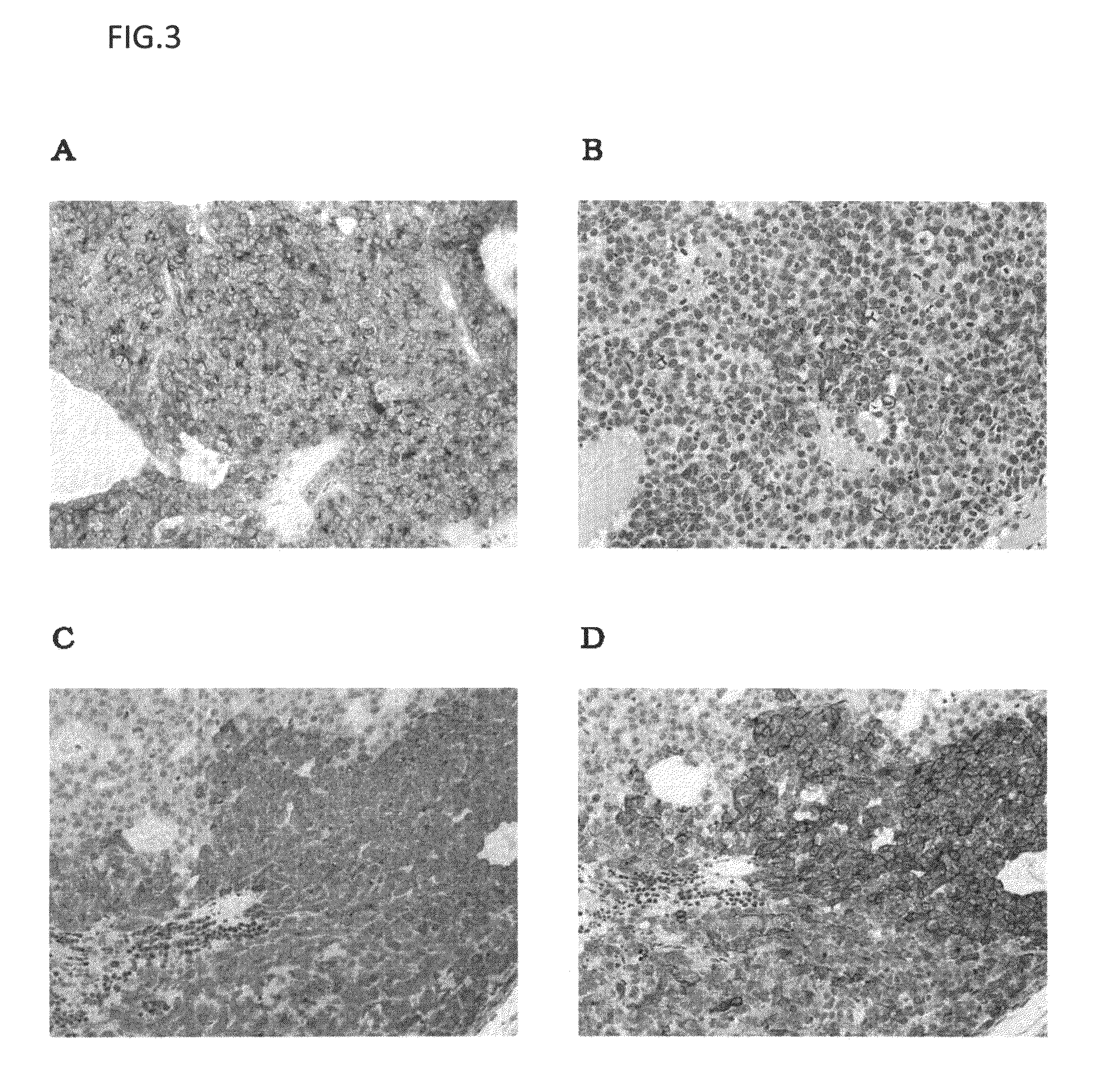
Antigen Retrieval Effect of Protease in GPC3 Immunostaining Using Human Hepatocellular Carcinoma Tissue Preparation

(1) Immunohistochemical Staining of Human Hepatocellular Carcinoma Samples

(1-1) Methods for Preparing and Staining Preparations

[0187]Human hepatocellular carcinoma samples were dipped in 10% neutral buffered formalin for the predetermined time or longer for fixation. Next, paraffin-embedded block preparations were prepared according to a standard method. The block preparations were cut into thin slices, and these tissue sections were used in immunohistochemical staining. The immunohistochemical staining was carried out by the same procedures as in Example 1.

(1-2) Method for Evaluating Staining Results

[0188]On the basis of the description of the paragraph (6-2) of Example 1, stainability in the immunostaining using the mouse anti-human GPC3 antibody was evaluated according to three parameters (rate of positive cells: PR, staining intensity: SI, and cell membrane staining...

example 3

Drug Efficacy of Anti-GPC3 Antibody on GPC3-Expressing Human Liver Cancer Cell Strain-Transplanted Mouse Models

(1) Cell Strain

[0214]Cells used in transplantation were HuH-7 cells and HepG2 cells. The HuH-7 cells were maintained and subcultured in Dulbecco's Modified Eagle's Medium (SIGMA-ALDRICH CO.) containing 10% FBS (BIONET). The HepG2 cells were maintained and subcultured in Minimum Essential Medium Eagle medium (SIGMA-ALDRICH CO.) containing 10% FBS, 1 mmol / l MEM Sodium Pyruvate (Invitrogen Corp.), and 1 mmol / l MEM Non-Essential Amino Acid (Invitrogen Corp.).

(2) Preparation of Human Liver Cancer Cell Strain-Transplanted Mouse Models

[0215]The cells of these strains were separately adjusted to 5×10⁷ cells per ml with a solution containing equal amounts of the medium for maintenance and subculture described above and Matrigel Matrix (BD Biosciences). One day before cell transplantation, 100 μl of an anti-asialo GM1 antibody (Wako Pure Chemical Industries, Ltd.; 1 vial was dissolve...


PUM

PropertyMeasurementUnit
timeaaaaaaaaaa
timeaaaaaaaaaa
timeaaaaaaaaaa

Abstract

The present invention relates to an in-vitro immunoassay method for detecting the presence of liver cancer cells in a subject. In the method of the present invention, antigen retrieval treatment based on heat-induced epitope retrieval method and antigen retrieval treatment based on protease-induced epitope retrieval method can be combined in the detection of glypican 3 antigen expression in liver cancer tissues to thereby detect the difference in the expression level or expression pattern of the glypican 3 antigen by immunohistochemical staining method. This enables samples, which has been determined by the conventional HIER method as highly expressing glypican 3, to be graded according to the expression level of glypican 3.

Description

TECHNICAL FIELD[0001]The present invention relates to an in-vitro immunoassay method for detecting the presence of liver cancer cells in a subject.BACKGROUND ART[0002]Since glypican 3 is highly expressed in liver cancer with frequency, the expression profile analysis of glypican 3 in liver cancer is thought to be probably useful for the functional identification of glypican 3 in liver cancer, the treatment or diagnosis of liver cancer, and the prognostic prediction of liver cancer. For the general expression analysis of proteins, immunohistochemistry, particularly, enzyme antibody technique, is widely used in pathological diagnosis. The immunohistochemistry is a method for detecting, highly sensitively and specifically, the presence and distribution of a substance (antigen) in vivo using biological reagents such as antibodies or enzymes. Examples of the features of the immunohistochemistry include: 1. its procedures are convenient; 2. the method is widely applicable in such a way th...

Claims


Application Information

Patent Timeline
Patent Type & Authority Patents(United States)
IPC IPC(8): G01N33/53
CPCC07K16/303G01N33/57438A61K2039/505
Inventor KATAOKA, HIROAKITAKAI, HIROTAKEKATO, ATSUHIKOSUZUKI, MASAMISUGIMOTO, MASAMICHI
Owner UNIVERSITY OF MIYAZAKI
Why Eureka
Try Eureka