Gene expression signature of genomic instability in breast cancer
a genomic instability and gene expression technology, applied in the field of gene expression signature of genomic instability in breast cancer, can solve the problem of tissue cell diversity and genetic instability
- Summary
- Abstract
- Description
- Claims
- Application Information
AI Technical Summary
Benefits of technology
Problems solved by technology
Method used
Image
Examples
example 1
[0057]The following example demonstrates the correlation of gene expression with genomic instability.
[0058]Tumor samples were collected from 48 patients diagnosed with breast cancer. Clinical material was collected from surgically removed tumors, diagnosed on H&E-stained tissue sections, and graded according to Elston et al., Histopathology, 19(5), 403-10 (1991). The clinical data are summarized in Table 1. After surgery, clinical tissue was used for touch preparation slides for quantitative measurement of the nuclear DNA content. The tissue samples were then snap frozen until further processing with TRIzol reagent (Invitrogen, Carlsbad, Calif.) for DNA and RNA extraction. In addition, paraffin-embedded specimens of the same tumors were used for histopathology and immunohistochemistry.
[0059]Image cytometry was performed on Feulgen-stained touch preparation slides. The staining procedure, internal standardization, and tumor cell selection were based on methods described previously (A...
example 2
[0065]The following example demonstrates the correlation between genomic instability and the expression of a 12-gene signature.
[0066]A supervised machine learning approach was applied in two sample settings to identify a genomic instability signature from the expression profiles of 7,657 genes in 44 of the primary breast cancer samples described in Example 1. Using random forests, the gene expression data from the three groups of tumors (dGS, aGS, and aGU) was compared. This comparison and classification generated a list of 70 genes. The top 10 genes of this set were selected with Relief, and then used to individually classify the three groups. This classification achieved an accuracy of 80%.
[0067]Random forests was then used to establish a gene list that discerns the genomically stable (dGS and aGS) tumors from the genomically unstable ones (aGU). Seven genes were identified from the analysis. In leave-one-out cross-validation, this 7-gene signature allowed classification of genomi...
example 3
[0071]The following example demonstrates the use of the 12-gene signature to classify tumors in an independent validation set.
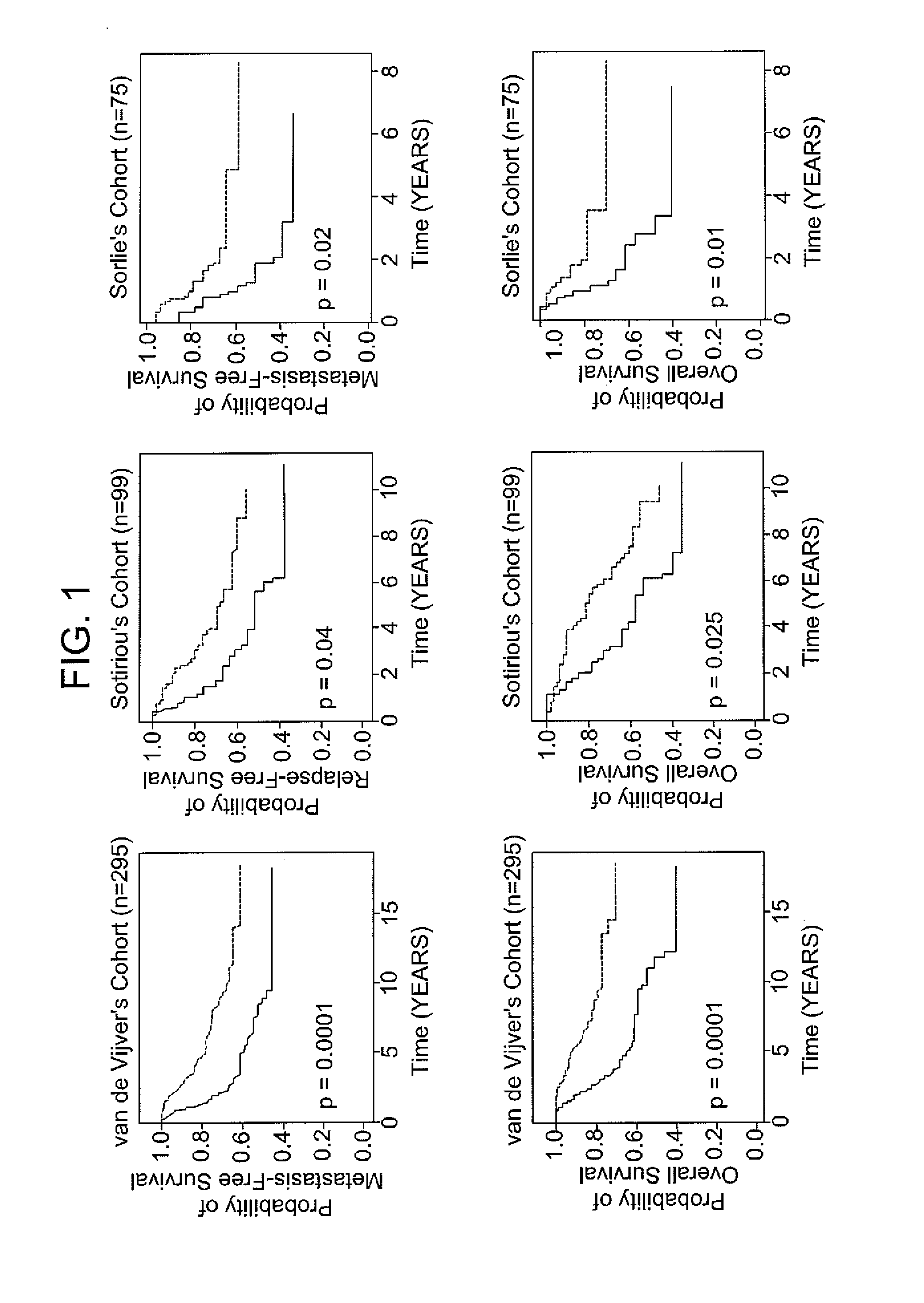
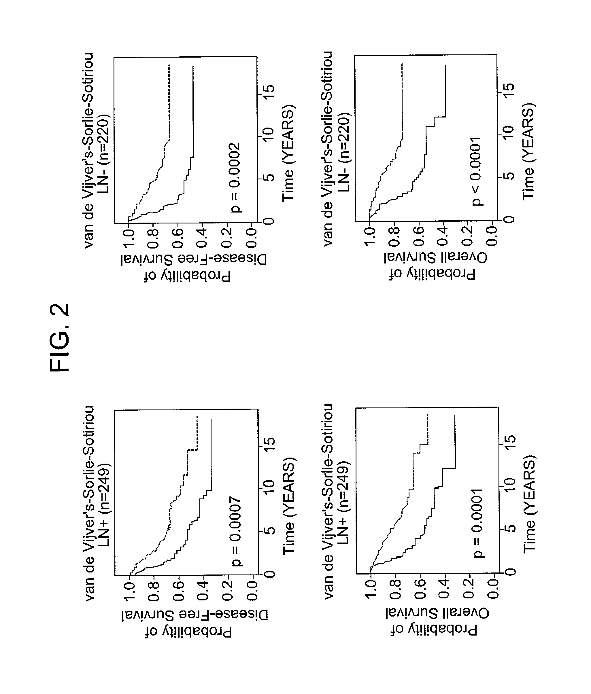
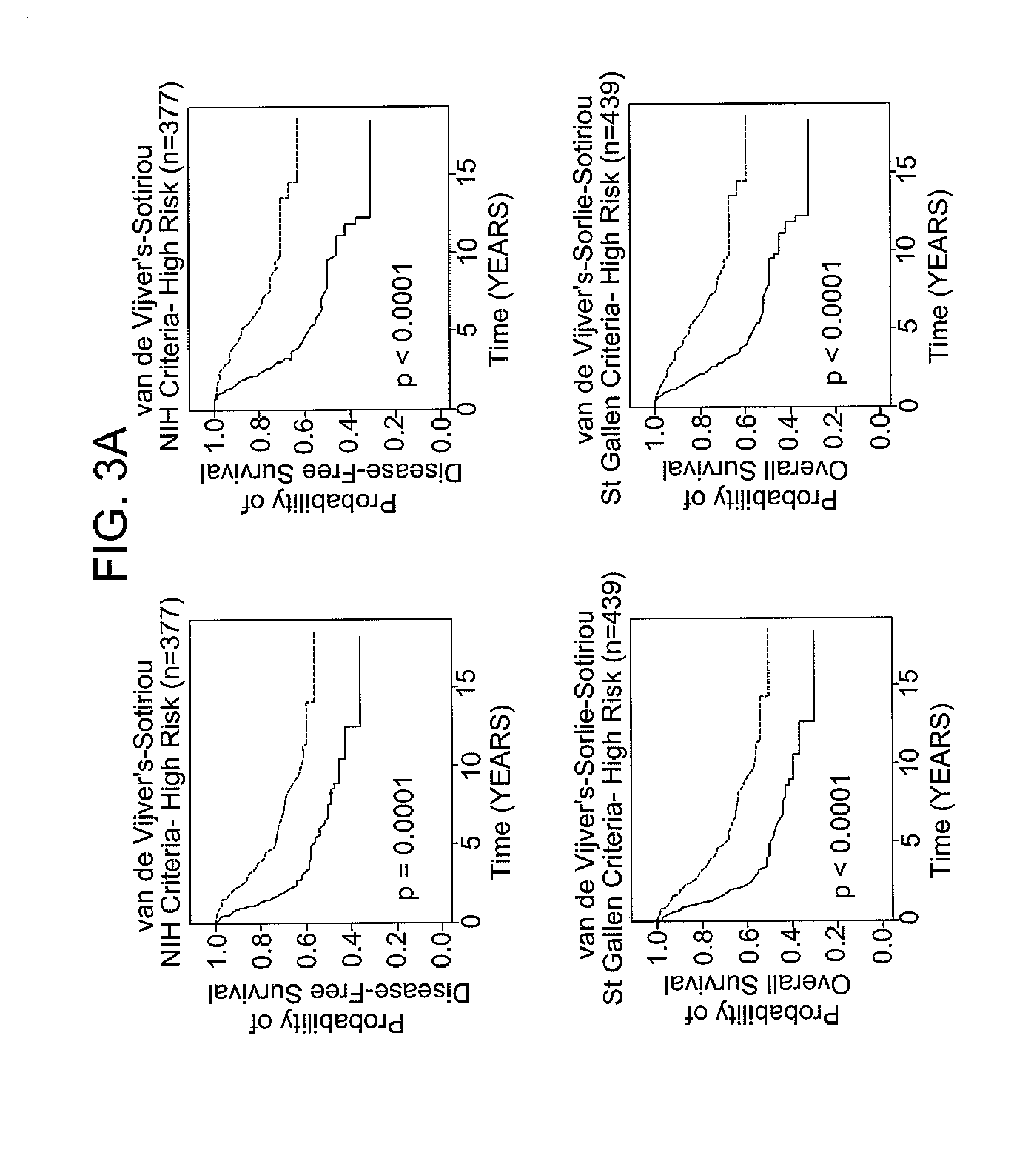
[0072]Patients with breast cancer from three data sets (n=469), for which different clinical endpoints were available, were used to validate the 12-gene signature. The three data sets are described in van de Vijver et al., N Engl J Med, 347 (25), 1999-2009 (2002), Sotiriou et al., Proc Natl Acad Sci USA, 100 (18), 10393-8 (2003), and Sorlie et al., Proc Natl Acad Sci USA, 100 (14), 8418-23 (2003). These datasets comprise gene expression profiles from 469 patients with heterogeneous histology and different disease stages. The clinical parameters included tumor grade, tumor size, lymph node status, estrogen receptor status, progesterone receptor status, age, and histology. The clinical endpoints used for the validation of the classifiers included relapse-free survival, metastasis-free survival, disease-free survival (here a disease event refers to either breast...
PUM
| Property | Measurement | Unit |
|---|---|---|
| size | aaaaa | aaaaa |
| size | aaaaa | aaaaa |
| Northern Blotting | aaaaa | aaaaa |
Abstract
Description
Claims
Application Information
 - R&D
- Intellectual Property
- Life Sciences
- Materials
- Tech Scout
- Unparalleled Data Quality
- Higher Quality Content
- 60% Fewer Hallucinations
Browse by: Latest US Patents, China's latest patents, Technical Efficacy Thesaurus, Application Domain, Technology Topic, Popular Technical Reports.
© 2025 PatSnap. All rights reserved.Legal|Privacy policy|Modern Slavery Act Transparency Statement|Sitemap|About US| Contact US: help@patsnap.com


