NG tube with gastric volume detection

a gastric volume and tube technology, applied in the field of ng tube with gastric volume detection, can solve the problems of patient's bowels eventually shutting down for lack of use, unable to aspirate stomach contents, and unable to meet the needs of patients,

Inactive Publication Date: 2015-03-24
CR BARD INC
View PDF16 Cites 24 Cited by
  • Summary
  • Abstract
  • Description
  • Claims
  • Application Information

AI Technical Summary

Benefits of technology

[0010]The enteral tube with a GRV detection sensor is flexible and may be long enough to be looped around within the stomach to access and conform to various regions within the gastric cavity and access the areas of the stomach at which the gastric contents may pool, regardless of the position of the patient. Weights may be employed within the GRV detection sensor and spaced at intervals to keep the sensor located generally at the lowest portion of the gastric cavity as determined by the position of the patient at all times. The enteral tube may also have a specific geometry (e.g. a flexible yet slightly curved pre-formed shape) that will allow the GRV detection sensor to be directed towards and access particular areas of the stomach when rotated at appropriate times during the insertion process.
[0011]The GRV detection sensor may include a sealed air column terminating with a flexible membrane. When the enteral tube and sealed air column are inserted into the stomach, it generally will come to rest in the lowest portion of the stomach due to the weights in the GRV detection sensor. To the extent that the sealed air column is submerged in gastric contents, pressure is exerted against the flexible membrane, and therefore against the sealed air column. The greater the height of the gastric contents, the greater the pressure. A pressure sensor detects the pressure within the sealed air column and provides a signal or level representative of the amount of gastric contents in the stomach to the volume indication device. The sealed air column may also be terminated with a sealed and flexible bulbous diaphragm, which can provide a larger signal or pressure reading to the pressure sensor representative of the amount of gastric contents in the stomach due to the larger surface area upon which pressure may be applied by the gastric contents.

Problems solved by technology

One disadvantage of IV feeding is that the patient's bowels may eventually shut down for lack of use, especially for patients with extended ICU stays.
However, enteral tube feeding can cause complications.
Some patients receiving enteral tube feeding experience nausea and vomiting (which can lead to aspiration of stomach contents into the lungs and subsequent pneumonia) due to delayed gastric emptying.
When an NG tube is inserted into the stomach, the lower esophageal sphincter at the junction between the esophagus and the stomach may not be able to form an adequate seal, which can again lead to aspiration of stomach contents into the lungs and subsequent pneumonia.
Other possible complications include diarrhea, constipation, and malabsorption / maldigestion (impaired absorption of nutrients).
However, there is currently no direct and relatively immediate confirmation of emptying of the stomach after formula has been forced into the patient's stomach via an NG tube.
Furthermore, even if the stomach empties, it cannot be immediately determined whether the formula is being tolerated (the nutrients are being absorbed), or whether the formula is just passing through the patient's digestive system, resulting in diarrhea, for example.
However, because the stomach contents extracted using this GRV detection procedure have an unpleasant odor and can produce biohazards, contamination, bacteria, and infections, there may be a reluctance on the part of the clinician to regularly perform the procedure.
In addition, because GRV detection results in the removal of the stomach contents, the patient experiences a loss of nutrition if the gastric contents are not returned to the patient's stomach through the NG tube.

Method used


Image

  • NG tube with gastric volume detection
  • NG tube with gastric volume detection
  • NG tube with gastric volume detection

Examples


Embodiment Construction

in conjunction with the accompanying drawings that are first briefly described.

BRIEF DESCRIPTION OF THE DRAWINGS

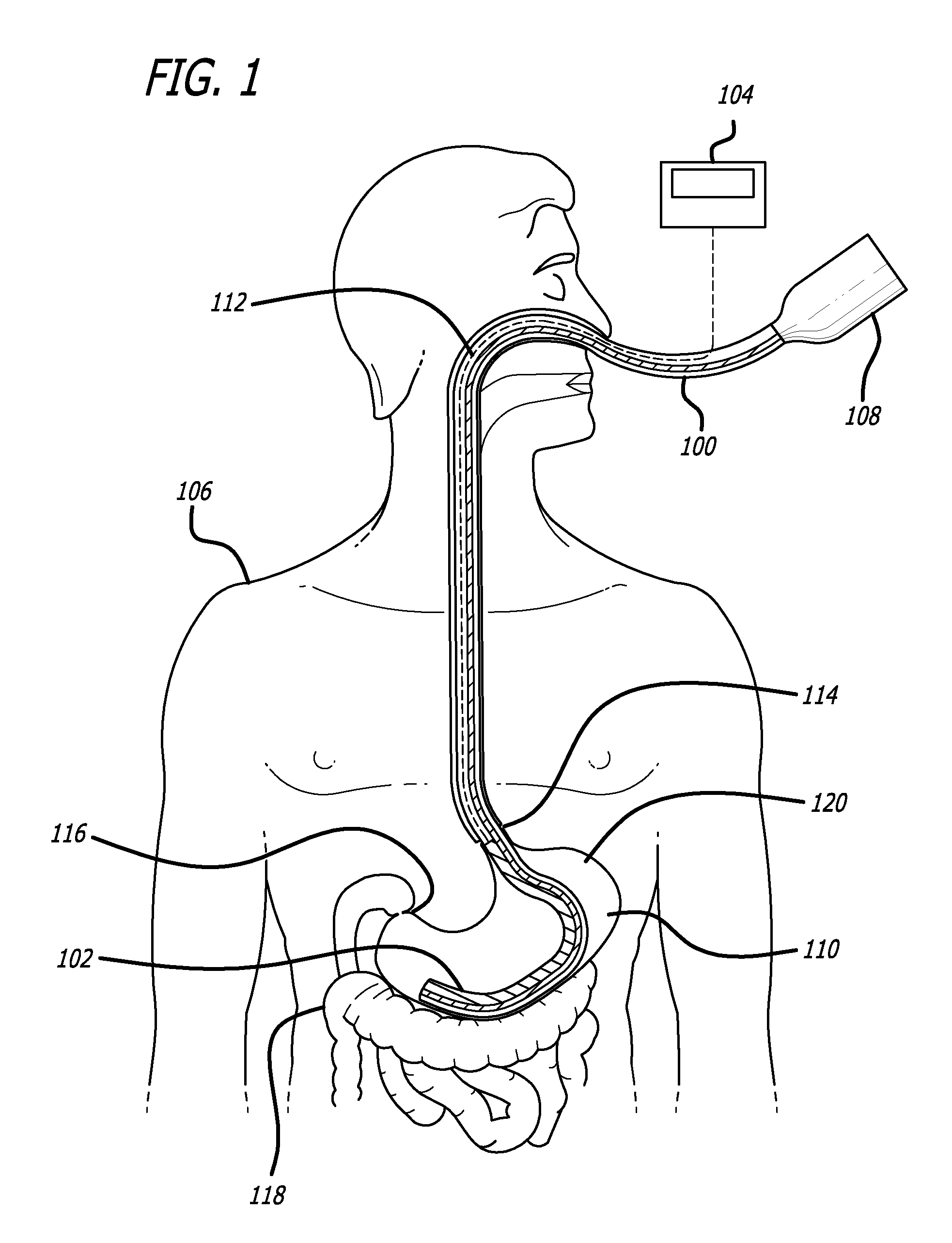
[0015]FIG. 1 illustrates an exemplary enteral tube with an integrated GRV detection sensor inserted into a patient.

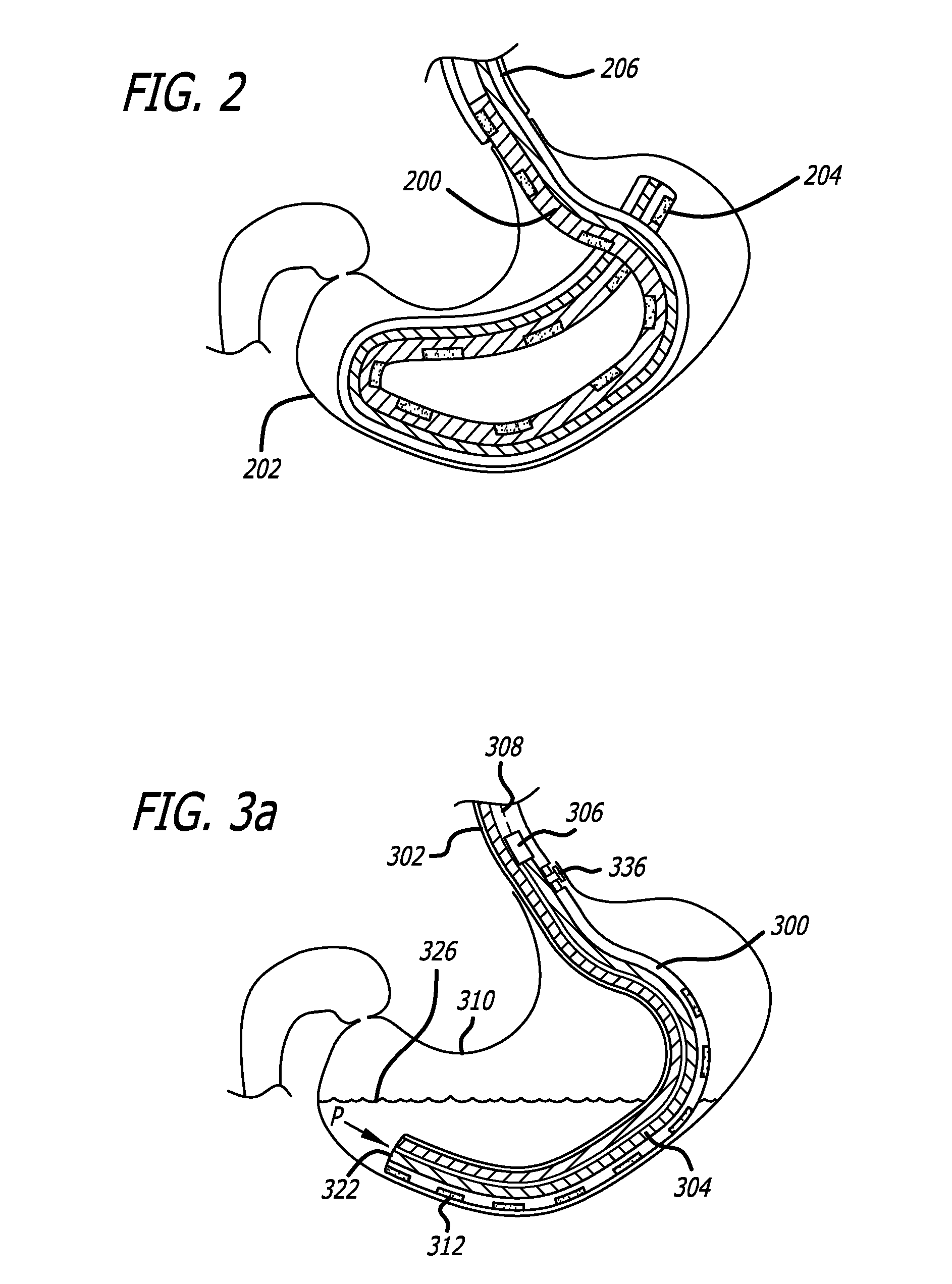
[0016]FIG. 2 is an illustration of an exemplary enteral tube with a GRV detection sensor which is flexible and long enough to be looped around within the stomach.

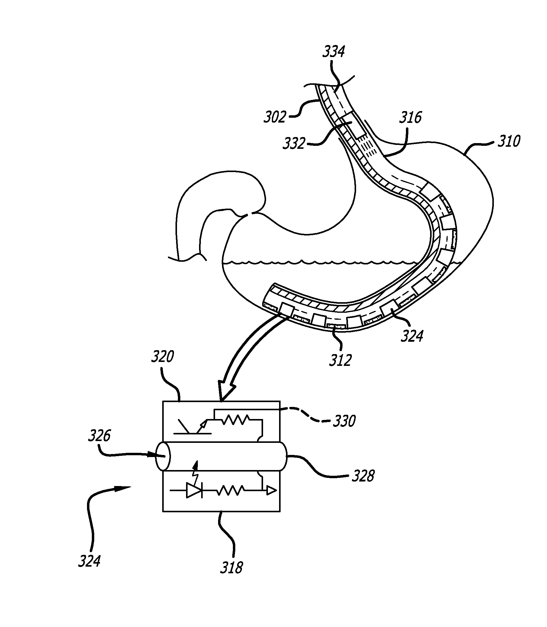
[0017]FIG. 3a is an illustration of an exemplary GRV detection sensor including a sealed air column terminating with a flexible membrane.

[0018]FIG. 3b is an illustration of an exemplary GRV detection sensor including a sealed air column terminating with a flexible and bulbous diaphragm.

[0019]FIG. 3c is an illustration of an exemplary GRV detection sensor including a plurality of fluid detection circuits, each fluid detection circuit including a photodiode and phototransistor pair surrounding a tube or chamber that is open at either end.

[0020]FIG. 4 is an illustration of an exemplary enteral tube inclu...


PUM


Abstract

An enteral tube includes a built-in Gastric Residual Volume (GRV) detection sensor at the distal end and a GRV indicating device at the proximal end for indicating the current GRV without requiring the aspiration of any stomach contents. The enteral tube with integrated GRV detection sensor is flexible and long enough to be looped around within the stomach. Weights may be employed to keep the sensor located generally at the lowest portion of the gastric cavity. The GRV detection sensor may include a sealed air column terminating with a flexible membrane. When the enteral tube and sealed air column are inserted into the stomach and submerged in gastric contents, pressure is exerted against the flexible membrane, and therefore against the sealed air column. A pressure sensor detects the pressure within the sealed air column and provides a signal or level representative of the amount of gastric contents in the stomach.

Description

PRIORITY[0001]This application is a U.S. national stage application under 35 USC§371 of International Application No. PCT / US2008 / 070457, field Jul. 18, 2008, which claims the benefit of priority to U.S. Provisional Patent Application No. 60 / 950,686, filed Jul. 19, 2007, each of which is incorporated by reference into this application as if fully set forth herein.BACKGROUND[0002]There are generally two ways to support patient nutrition for patients who are unable to self-feed orally in the usual manner—parenteral nutrition (by injection, e.g. IntraVenous (IV) feeding of electrolytes), and enteral nutrition (delivery directly into the GastroIntestinal (GI) tract). One disadvantage of IV feeding is that the patient's bowels may eventually shut down for lack of use, especially for patients with extended ICU stays. Enteral nutrition generally provides various physiologic and metabolic benefits as compared to parenteral nutrition. Thus, enteral tube feeding (e.g. NasoGastric (NG) tube fee...

Claims


Application Information

Patent Timeline
Patent Type & Authority Patents(United States)
IPC IPC(8): A61B5/103A61B5/00A61B5/117A61B5/03A61B5/20
CPCA61B5/036A61B5/4238A61B5/4233A61B5/42A61B5/204A61B5/4211A61B5/6852
Inventor NISHTALA, VASU
Owner CR BARD INC
Why Eureka
Try Eureka