Method of guiding prostate biopsy location

a biopsy location and biopsy core technology, applied in the field of biopsy core analysis, can solve the problems of high over-prescription of this highly invasive and costly examination, no above-mentioned screening method provides information on tumor differentiation, location and/or dimensions, and high false-negative rate of trnb

Active Publication Date: 2015-06-09
PROSIGHT
View PDF5 Cites 4 Cited by
  • Summary
  • Abstract
  • Description
  • Claims
  • Application Information

AI Technical Summary

Problems solved by technology

The rate of negative (non-cancerous) findings for TRNB examination is about 75%, which indicates a high over-prescription of this highly invasive and costly examination.
Furthermore, none of the above screening methods provide information on tumor differentiation and / or location and / or dimensions.
The lack of location information, which could serve as guidance during the needle-biopsy examination, leads to high false-negative rates in TRNB (of ˜28% in the first examination and ˜17% in repeated ones).
However, these modalities can not reliably provide information about the pathological stage of the lesions.
Methodologies based on endo-rectal magnetic resonance imaging (ER-MRI) also exist but they are expensive and their use is limited to pre-operative staging.

Method used


Image

  • Method of guiding prostate biopsy location
  • Method of guiding prostate biopsy location
  • Method of guiding prostate biopsy location

Examples


example 1

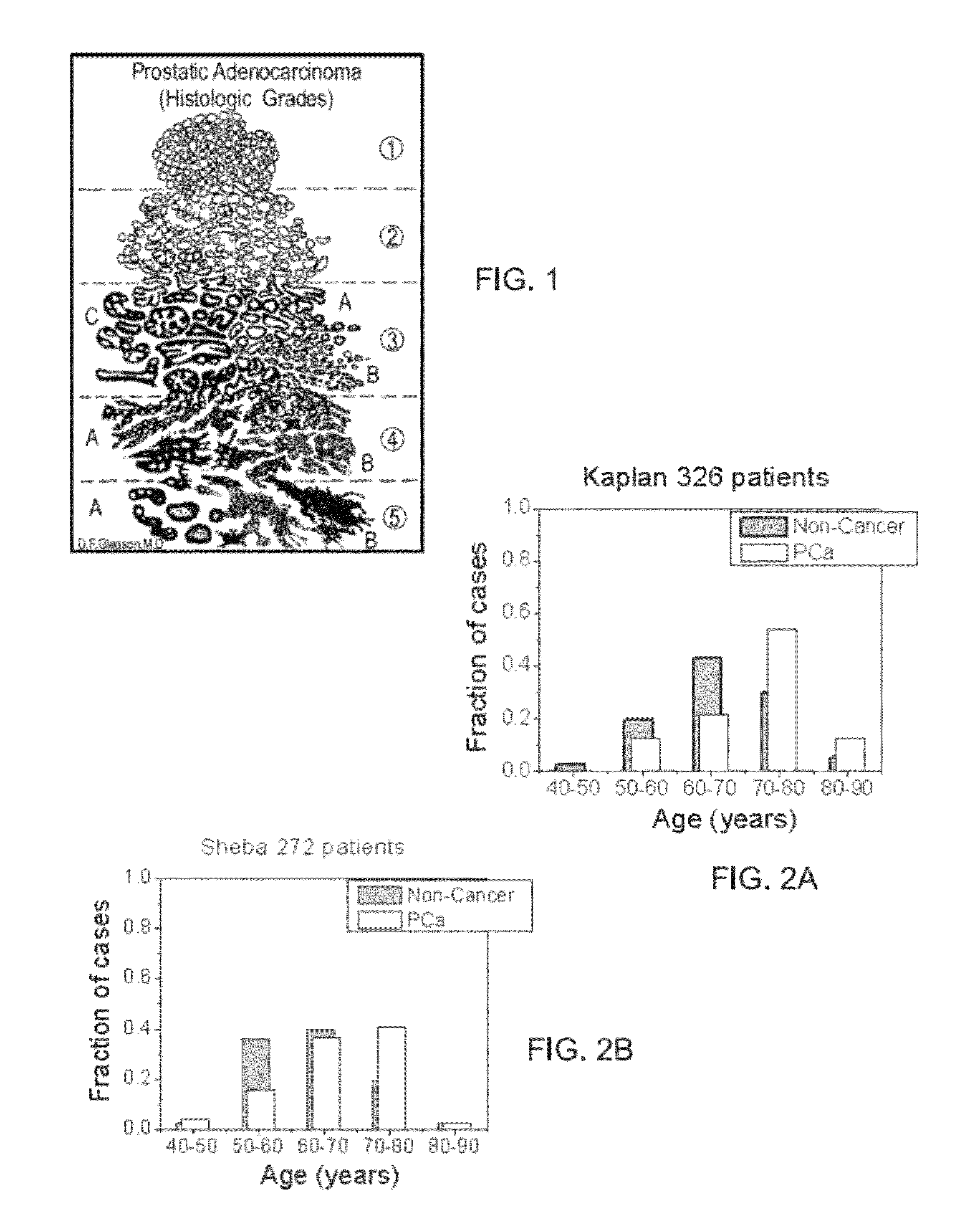
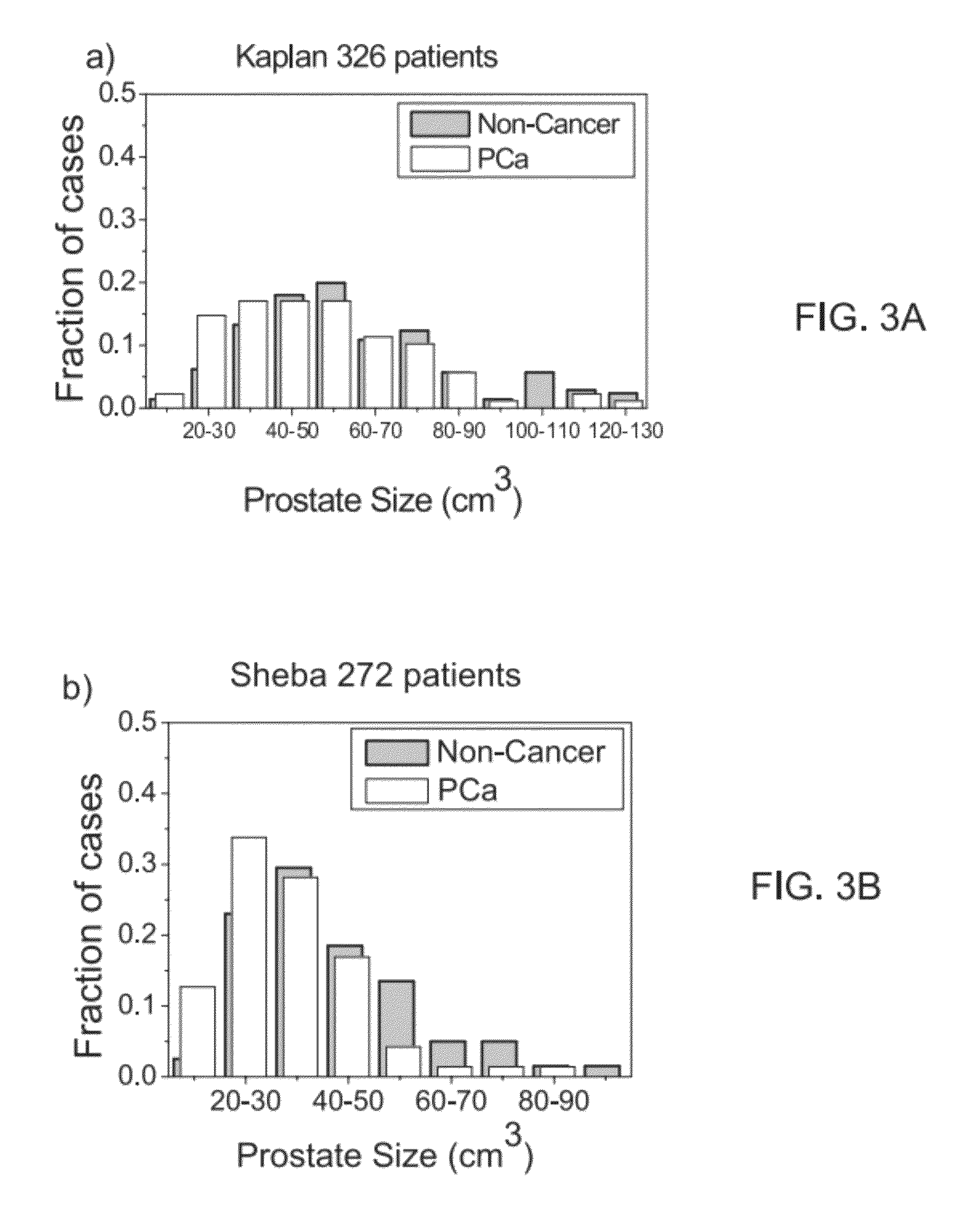
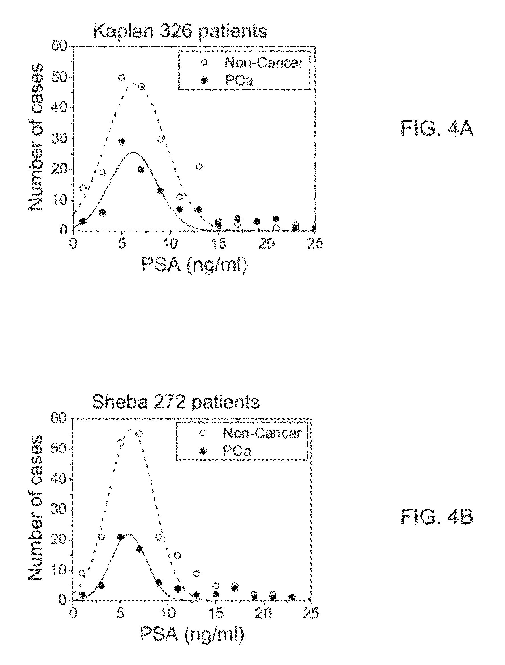
Zinc Depletion, Gleason Scores and Lesion Size

[0212]X-Ray Fluorescence Measurements:

[0213]The methods and sample-handling protocols used in this study were described in details in PCT WO 2004 / 041060 to Breskin et al, filed Nov. 6, 2003, including details on calibration procedures and ways used to avoid sample drying during the measurements. Briefly, zinc concentration in the examined tissue-segments was quantified by X-ray fluorescence (XRF). Under the measuring conditions the sensitivity of both is about 20 counts per ppm, namely zinc concentration sensitivity better than 1 ppm. The calibration of both systems was checked against identical calibration standards, with absolute precision of a few μg / g (˜1%). The reliability was checked by repeated measurements of the same sample, during the entire study, assessed to be within sigma=5%.

[0214]The measurements were carried out, in parallel, in two locations, using two tabletop XRF systems: one a locally-assembled system at the Kaplan Me...

example ii

Zinc Concentration Maps

[0252]In addition to detection and grading of cancerous and pre-cancerous foci, it would be highly desirable to have tools for accurate and non-invasive location and imaging of prostate lesions, in order to detect both location, dimension and grade of lesions. In order to test whether zinc depletion data generated in accordance with some embodiments of the instant invention, can be useful in location and imaging of prostate lesions, zinc-concentration maps were generated from experimental zinc-concentration data. The maps represent prostate-tissue with lesions of different dimensions and histological grades, at various locations within the gland. The maps are then transformed into 8-bit images and processed with a simple image processing algorithm yielding a one-parameter classifier test.

[0253]Generation of Zinc Concentration Maps

[0254]FIG. 16 shows a representative frequency distribution of zinc-concentration for PCa tissue, based on the data presented in FIG...


PUM

PropertyMeasurementUnit
thicknessaaaaaaaaaa
lengthaaaaaaaaaa
threshold L1aaaaaaaaaa

Abstract

A method of analyzing a plurality of biopsy cores extracted from a plurality of respective biopsy locations in a prostate is disclosed. The method comprises: measuring a level of a chemical element in each of the biopsy cores, and generating a chemical element map of at least a portion of the prostate based on the levels and the respective biopsy locations. In some embodiments, the method determines at least one additional biopsy location for a future biopsy in the prostate.

Description

RELATED APPLICATION[0001]This application is a National Phase of PCT Patent Application No. PCT / IL2009 / 001172 having International filing date of Dec. 10, 2009, which claims the benefit of priority of U.S. Provisional Patent Application No. 61 / 121,201, filed Dec. 10, 2008. The contents of the above applications are incorporated herein by reference.FIELD AND BACKGROUND OF THE INVENTION[0002]The present invention, in some embodiments thereof, relates to analysis of biopsy cores, and, more particularly, but not exclusively, to a method and system for automatically analyzing biopsy cores extracted from a plurality of respective biopsy locations in a prostate.[0003]Based on recent statistics, for the general population, a man has about a 17 percent chance (1 in 6) of being diagnosed with prostate cancer at some point during his lifetime (lifetime risk), and a 3 percent chance (1 in 33) of dying from prostate cancer.[0004]Current screening methods for symptomatic prostate-cancer (PCa) det...

Claims


Application Information

Patent Timeline
Patent Type & Authority Patents(United States)
IPC IPC(8): G01N33/574
CPCG01N33/57434
Inventor SIMON, AVIWEKSLER, MEIRBRESKIN, AMOSCHECHIK, RACHELCORTESI, MARCO
Owner PROSIGHT
Why Eureka
Try Eureka