Method for the simultaneous detection of multiple nucleic acid sequences in a sample

a nucleic acid sequence and sample technology, applied in the field of detection of multiple nucleic acid sequences in samples, can solve the problems of limited scope of these assays, limited clinical symptoms, and relatively slow viral culture, so as to reduce time, labor and cost

Active Publication Date: 2015-12-29
PATHOFINDER HLDG BV
View PDF6 Cites 0 Cited by
  • Summary
  • Abstract
  • Description
  • Claims
  • Application Information

AI Technical Summary

Problems solved by technology

This limits the scope of these assays considerably.
However, clinical symptoms are only rarely attributable to a single pathogen.
However, viral culture is relatively slow and therefore routine diagnosis is sub optimal.
Although rapid antigen detection tests are available for some of these viruses, these tests have shown to be less sensitive and less specific than viral culturing.
Disadvantages of gel electrophoresis as a detection technique are that it is laborious and time consuming and therefore rather costly.
Furthermore, the risk of cross contamination is enlarged as each sample has to be opened after the PCR for analysis.
Although the ELISA based assays allow highly multiplex analyses, they posses the same disadvantages as the gel electrophoresis based assays.
The assays are laborious and time consuming and the reaction vessel has to be opened after PCR, thereby increasing the risk of cross contamination.
Real Time PCR capacity to simultaneously detect multiple targets is limited to the number of fluorescent emission peaks that can be unequivocally resolved.
This diminishes the risk of cross contamination and requires no extra handling time.
However, the complexity of the current assays is limited to a maximum of four probes per reaction.
Moreover, complex analyses require more reactions thereby increasing the costs.
However, they require specialized equipment and extensive post PCR handling.
As with the microarray based assays, these assays allow highly complex analyses but require specialized equipment and extensive post PCR handling.
However, this application requires specialized and expensive hardware which, at the moment, is not common on a standard microbiology laboratory.
The above described multiparameter approaches to identify and differentiate the causative agents of a RTI are a great step forward but still have limitations.
They take either too much time (>10 hours), require a lot of hands-on time, have a limited multi-parameter character and / or need expensive equipment or large set-up costs to perform the tests.

Method used


Image

  • Method for the simultaneous detection of multiple nucleic acid sequences in a sample
  • Method for the simultaneous detection of multiple nucleic acid sequences in a sample
  • Method for the simultaneous detection of multiple nucleic acid sequences in a sample

Examples


example 1

Design of Probes and Primers

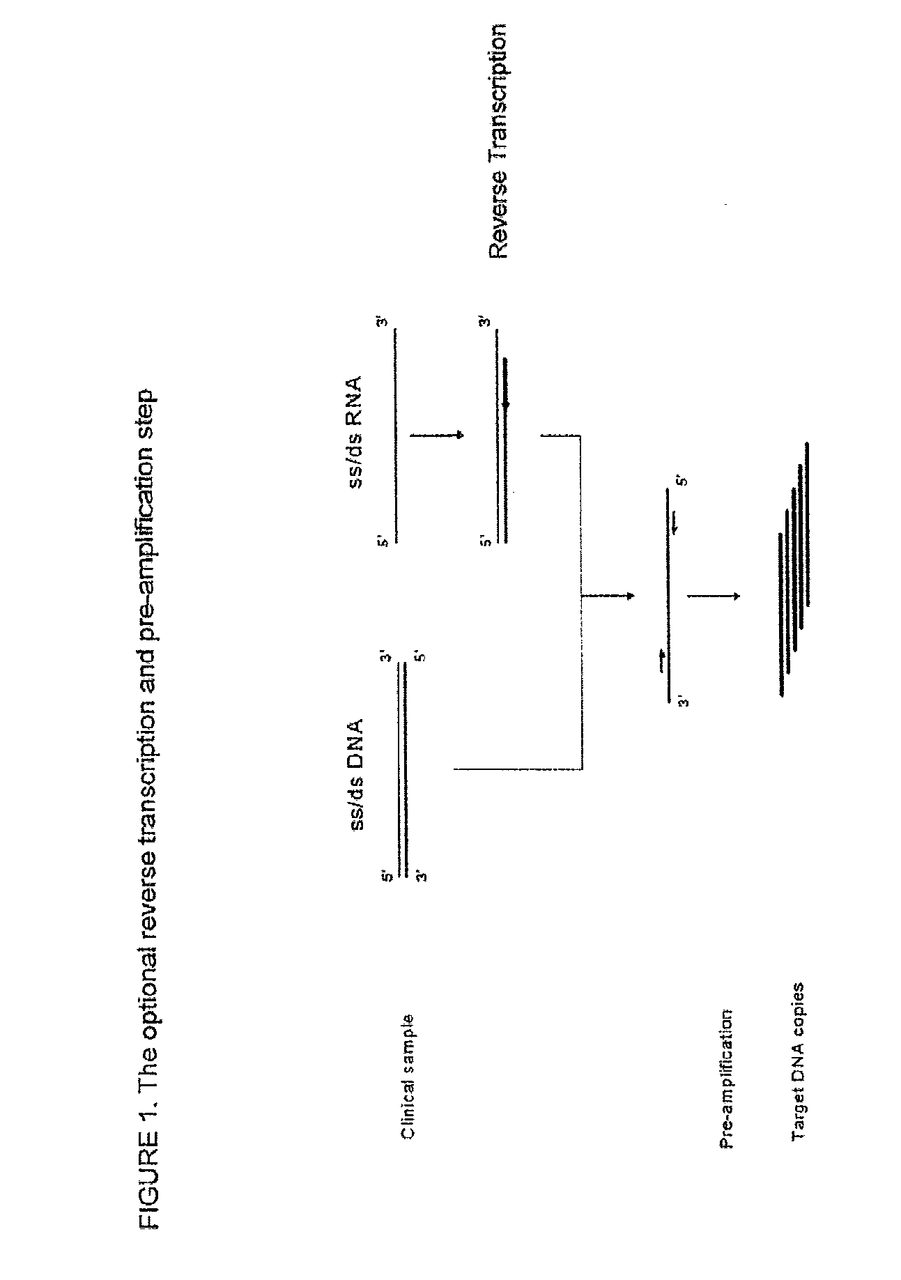
[0187]Viral and bacterial sequences were obtained from GenBank and Los Alamos database. Alignments (Clustal X v. 1.8.1) were performed on a set of sequences for each virus to identify highly conserved regions. Since some of the target pathogens are RNA viruses, a reverse transcriptase step followed by pre-amplification step was performed in order to obtain a plurality of different target DNA templates. Based upon these highly conserved regions, reverse transcriptase (RT)-PCR primers and ligation probes were designed. Table 1 lists the target regions, Gen Bank accession numbers and position for the sequences that serve as a template for the hybridisation of the first and second nucleic acid probes.

[0188]Conserved regions in the matrix protein gene (M1) were selected as the target for amplification and detection of influenza A virus. These primers are suitable for the amplification of a variety of strains, including, but not limited to, H3N2, H2N2, H4N2, H3...

example 2

Sample Preparation

[0206]A nasopharyngeal lavage was used as clinical specimen. The MagnaPure nucleic acid system (Roche Diagnostics, Almere, The Netherlands) was used as extraction method. The Total nucleic acid isolation kit and the Total nucleic acid lysis extraction MagnaPure protocol were applied. Extractions were performed according to the manufacturer's instructions. Briefly, 200 μl of starting material was used and the purified nucleic acid was eluted in a final volume of 100 μl. Before starting the extraction, 5 μl (approximately 150 copies) of an Internal Amplification Control (IAC) was added to the lysed sample.

example 3

Pre-Amplification

[0207]The extracted nucleic acid with IAC was placed in a separate reaction tube and the mix of primers as shown in table 2 was added along with reagents for reverse-transcription followed by pre-amplification (RT-PCR). While any procedure known in the art for RT-PCR may be used, the following procedure was used in this example. OneStep RT-PCR (Qiagen, Hilden, Germany) was performed in 25 μl containing 5 μl OneStep RT-PCR buffer (12.5 mM MgCl2; pH 8.7 (20° C.)), 1 μl deoxy nucleoside triphosphate (dNTP) mix (containing 1.6 μM of each dNTP), 2.5 μl primermix (containing 2 μM of each primer), 1 μl of OneStep RT-PCR Enzyme mix, 5.5 μl RNase free water and 10 μl of the extracted nucleic acid template with IAC. A blank reaction control was prepared by adding RNase free water to one reaction tube in place of nucleic acid template. The reaction tubes were placed in a Biometra T1 Thermocycler (Biometra, Goëttingen, Germany) programmed as follows: 30 minutes at 50° C. revers...


PUM

PropertyMeasurementUnit
melting temperaturesaaaaaaaaaa
timeaaaaaaaaaa
melting temperatureaaaaaaaaaa

Abstract

The invention is in the technical field of detecting nucleic acid sequences in a sample, such as the detection of pathogenic organisms in clinical samples. More specifically, the invention relates to the field of detecting an infection caused by a pathogenic organism such as a virus or a bacterium in a clinical specimen by amplifying and detecting specific nucleic acid sequences from the pathogenic organism. It provides a multiplex assay with the possibility to determine about 30 different target nucleic acid sequences in a single one-tube assay combined with real-time probe detection. The method employs multiplex ligation dependent Probe Amplification (MLPA) employing labelled primers in combination with labelled, probes and detection of the fluorescence.

Description

[0001]This application is the U.S. national phase of International Application No. PCT / EP2008 / 059050 filed 10 Jul. 2008, which designated the U.S. and claims priority to EP Application No. 07112219.6 filed 11 Jul. 2007, the entire contents of each of which are hereby incorporated by reference.FIELD OF THE INVENTION[0002]The invention relates to the technical field of detecting multiple nucleic acid sequences in a sample, such as the detection of pathogenic organisms in clinical samples. More specifically, the invention relates to the field of detecting an infection caused by a pathogenic organism such as a virus or a bacterium in a clinical specimen by means of amplifying and detecting specific nucleic acid sequences from said pathogenic organism.BACKGROUND OF THE INVENTION[0003]Infectious agents such as micro-organisms are typically detected by culturing clinical samples under conditions favourable for the growth of such micro-organisms and monitoring that growth by a number of dif...

Claims


Application Information

Patent Timeline
Patent Type & Authority Patents(United States)
IPC IPC(8): C12P19/34C12Q1/68
CPCC12Q1/6818C12Q1/686C12Q2531/113C12Q2527/107C12Q2533/107C12Q2537/143C12Q2521/107
Inventor REIJANS, MARTINUS GERARDUS CECILIA MARIADINGEMANS, GIJS JOSEPHUS HENRICUSSIMONS, AUGUSTINUS FRANCISCUS MARIA
Owner PATHOFINDER HLDG BV
Why Eureka
Try Eureka