
Imaging of meningiomas using phenylbenzothiazole, stilbene, or biphenylalkyne derivatives

a technology of phenylbenzothiazole and phenylalkyne, which is applied in the field of meningiomas imaging using phenylbenzothiazole derivatives or stilbene derivatives or biphenylalkyne derivatives, can solve the problems of lack of confidence in conventional diagnostic imaging, dramatic and negative effects on patient medical care, and lumbar puncture with cerebrospinal fluid testing is not useful for diagnosing meningiomas

Active Publication Date: 2016-02-16
MAYO FOUND FOR MEDICAL EDUCATION & RES
View PDF13 Cites 2 Cited by
  • Summary
  • Abstract
  • Description
  • Claims
  • Application Information

AI Technical Summary

Benefits of technology

The present invention provides a method for definitively diagnosing meningiomas in patients with intracranial tumors that may be meningioma or metastasis. This method can help differentiate between meningioma and metastasis, which is difficult to do using computed tomography and magnetic resonance imaging alone. This method may also help avoid unnecessary surgeries, therapies, or diagnostic procedures, which can improve patient management and outcomes.

Problems solved by technology

These two factors can lead to a lack of diagnostic confidence with conventional diagnostic imaging.
This lack of confidence can have dramatic and negative effects on a patient's medical care.
Lumbar puncture with cerebrospinal fluid (CSF) testing is not useful for diagnosing meningiomas.
Leptomeningeal invasion can give rise to tumor cell dissemination in the CSF, which can be detected by lumbar puncture with cytology, however this is often falsely negative.
There are no good laboratory tests available for the diagnosis of meningioma.
Thus, there are problems with the current methods for diagnosing meningiomas.
Therefore, the diagnosis of meningioma can not always be made with confidence based on CT and MRI alone, and sometimes when the diagnosis of meningioma is felt to be confident, it is incorrect.
Meningiomas can occasionally occur outside of the cranial vault and outside of the dura, making their correct diagnosis in these locations much more difficult.
The differentiation between meningioma and dural metastasis in particular can be very difficult based on current imaging methods.
Furthermore, metastasis to the dura can occur long after the patient has been in complete remission.
However, diffusion tensor MRI has not been shown to differentiate meningiomas form other types of tumors, such as metastasis.
In addition, intravenous MRI contrast agents are required to perform these studies, therefore patients with renal failure are not able to undergo these exams.
This is because, patients with renal failure are at risk to develop nephrogenic systemic fibrosis if they receive gadolinium containing intravenous MRI contrast agents.
However, since meningiomas and the other tumors that are seen in the same locations can have variable FDG uptake on FDG-PET / CT, there is limited use of FDG-PET / CT for the diagnosis of meningioma.
Thus FDG-PET / CT is only of limited help in the diagnosis of meningioma in these situations.
C11-methionine would not likely be useful for differentiating between aggressive meningiomas and dural metastasis, since both would likely have an increased SUV.
However, like most PET tracers, most single photon emitting tracers are nonspecific for meningiomas.
For example, Thallium-201 SPECT imaging is somewhat useful for predicting histological types of meningiomas, but is nonspecific and is not useful for diagnosing meningiomas.
Octreotide appears to have good sensitivity for meningiomas, but is not perfect.
SPECT imaging, even when combined with CT, lacks the resolution of PET imaging and is generally considered inferior as a modality.
Octreotide studies are time consuming to perform.
However, even 4 hours is a long wait when compared to the more conventional MRI and CT imaging, and is disruptive to patient's schedules.
Thus, patients who have a history of cancer and find a new tumor in the meningeal membranes that envelope the brain are faced with a common diagnostic dilemma.
CT and MRI are clearly inadequate for confident distinction of meningioma from meningeal metastasis in a single imaging evaluation.
Chemotherapy is not currently useful for treating meningiomas.
Unfortunately, because of inadequate diagnostic confidence with current imaging, many patients with probable meningiomas are compelled to have surgery primarily to confirm the diagnosis and only secondarily for the purpose of treating the tumor.

Method used


Image

  • Imaging of meningiomas using phenylbenzothiazole, stilbene, or biphenylalkyne derivatives
  • Imaging of meningiomas using phenylbenzothiazole, stilbene, or biphenylalkyne derivatives
  • Imaging of meningiomas using phenylbenzothiazole, stilbene, or biphenylalkyne derivatives

Examples


example 1

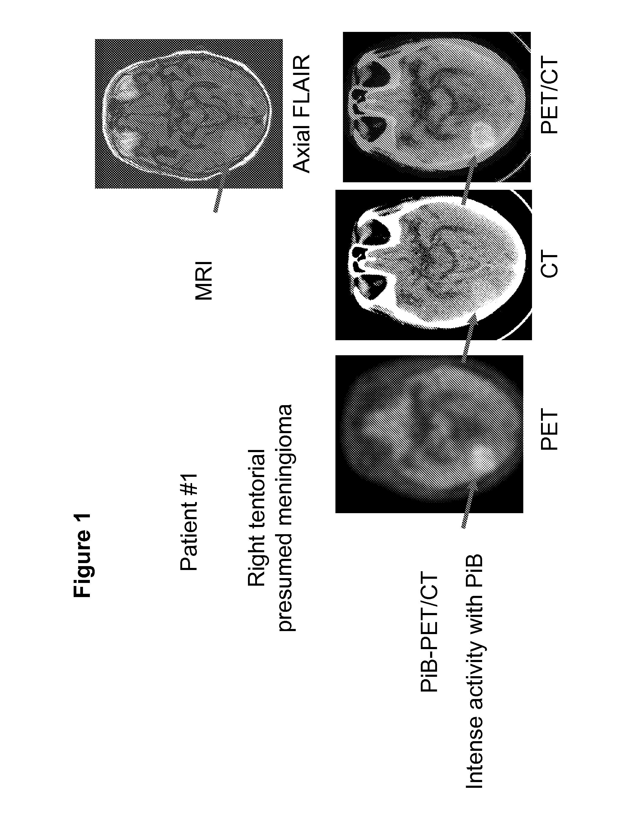
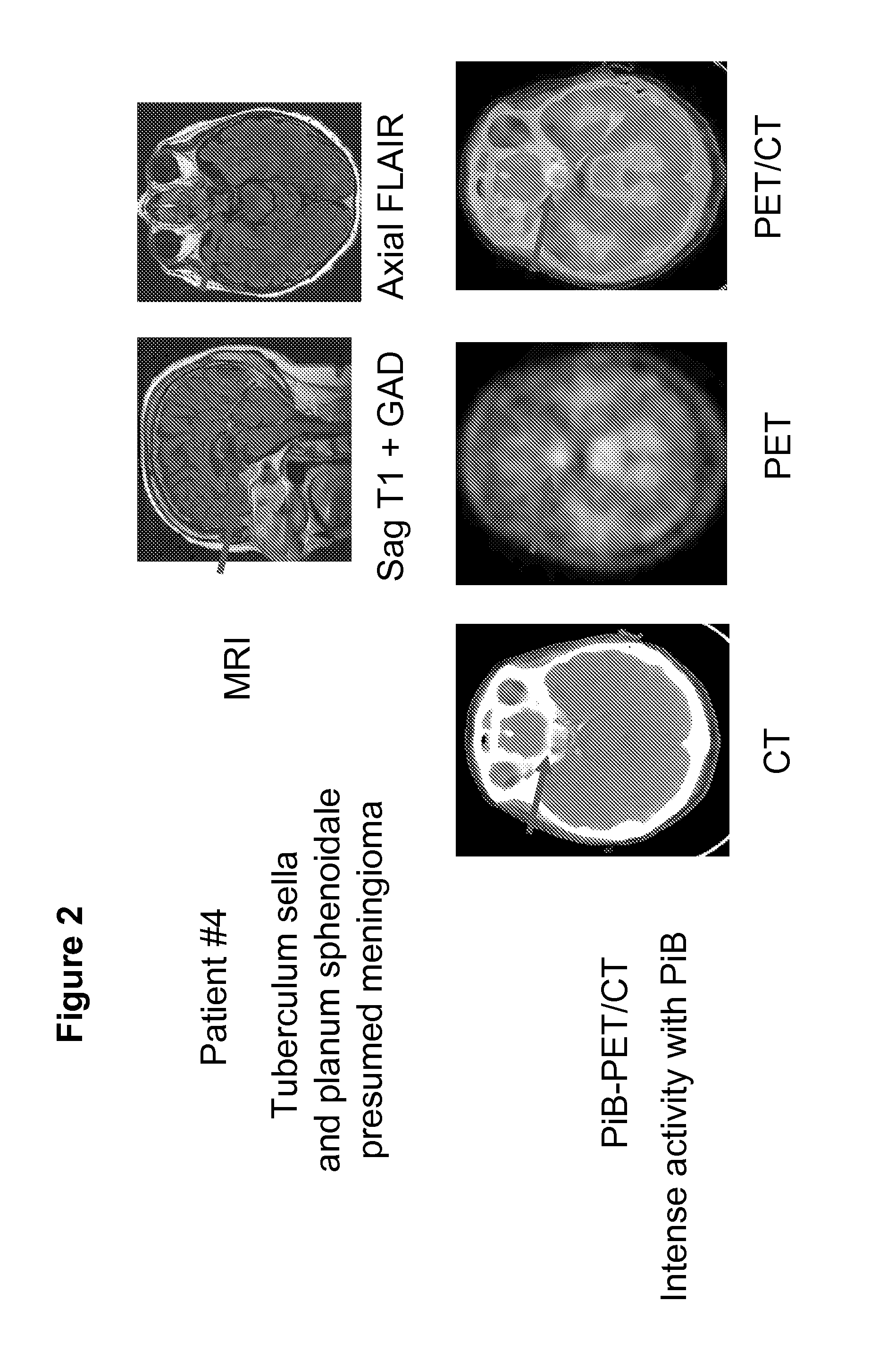
[0080]An Alzheimer's imaging database of two hundred forty-one patients was reviewed. Some of the patients had a history of cognitive impairment that might be due to early Alzheimer's disease and some were normal controls. All patients had been imaged by at least one MRI, as well as by FDG-PET / CT and PET / CT using the compound of formula (V). MRI reports of all patients were reviewed for possible meningiomas and other tumors. Seven patients were found to have the diagnosis of presumed meningioma based on MRI and sometimes also on CT. The diagnostic confidence of the radiologists interpreting the studies varied slightly, depending on factors such as tumor location and previous imaging. Of these seven tumors, six showed intense uptake of the compound of formula (V) on PET / CT. One showed some uptake, but was difficult to evaluate presumably because of its small size (˜4 mm. thick). Tumors smaller than 7 millimeters are generally considered too small to be evaluated by PET or PET / CT. Thi...

example 2

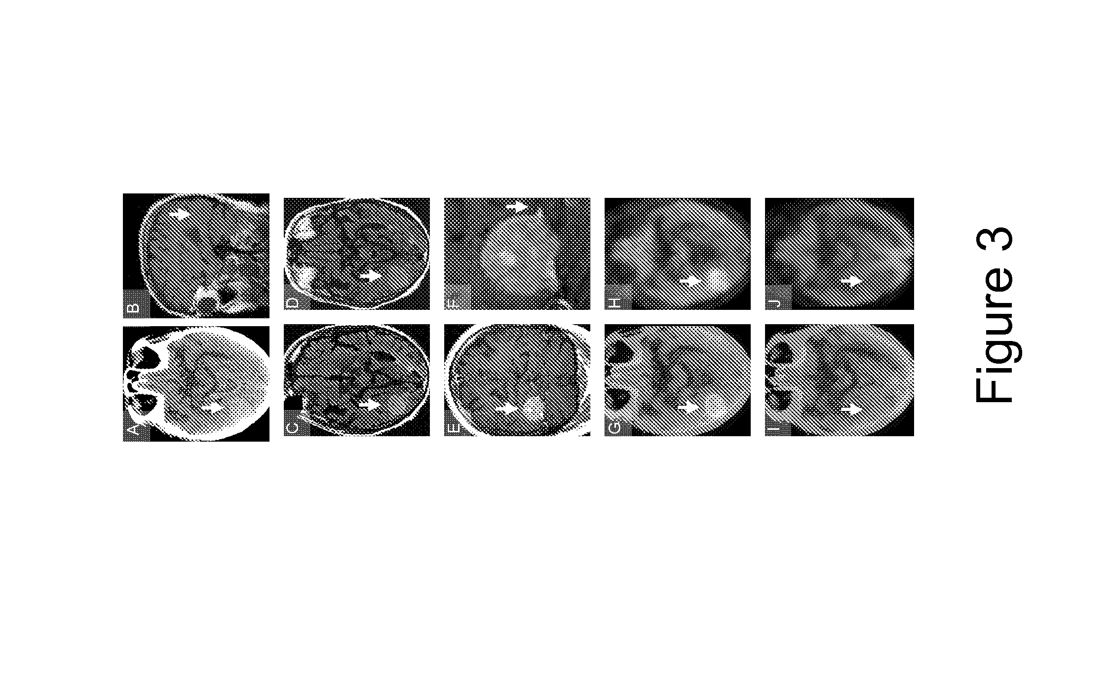
[0083]In Example 1, we identified six patients with meningiomas >5 millimeters in size that showed positive uptake of the compound of formula (V) on PET / CT. We identified three more patients with four presumed meningiomas. All three patients were imaged by PET-CT using the compound of formula (V), FDG PET-CT and MRI within as part of the ongoing amyloid imaging study. In one patient, there are two meningiomas. One is in the orbit, and is therefore extracranial. This orbital meningioma was previously resected and pathologically shown to be a meningioma in 1969, and pathology was confirmed later at our institution. It has now regrown within the orbit. The other meningioma is along the falx. Both show avid uptake of the compound of formula (V). There were two other patients with a single meningioma with uptake of the compound of formula (V). Therefore, there are a total of ten presumed meningiomas that show uptake of the compound of formula (V) on PET / CT.

example 3

[0084]In Examples 1 and 2, we identified ten patients with meningiomas >5 millimeters in size that showed positive uptake of the compound of formula (V) on PET / CT. We identified three more examples of presumed meningiomas that show activity using the compound of formula (V) on PET-CT, for a total of thirteen. All of these formula (V) avid meningiomas are >5 millimeters in size. We also identified three more presumed meningiomas that showed some positive uptake of formula (V), but were non-diagnostic due to common limitations of PET / CT imaging. One of these non-diagnostic presumed meningiomas was too small to evaluate by PET or PET / CT. One of these non-diagnostic presumed meningiomas was necrotic / cystic centrally with a 4 millimeter rim of tumor, and is therefore effectively also too small to evaluate by PET or PET / CT. The other of these non-diagnostic presumed meningiomas was only seen in the last 2 slices of PET data, an area considered to be uninterpretable. In addition, this pati...


PUM

PropertyMeasurementUnit
volumeaaaaaaaaaa
sizeaaaaaaaaaa
sizeaaaaaaaaaa

Abstract

Methods for detecting or ruling out a meningioma in a patient using a phenylbenzothiazole derivative or a stilbene derivative or a biphenylalkyne derivative, and a medical imaging technique such as positron emission tomography / computed tomography are disclosed. In one version of the method, the phenylbenzothiazole derivative is a compound of formula (V):

Description

CROSS-REFERENCES TO RELATED APPLICATIONS[0001]This application is a 371 application of PCT / US2011 / 055753 filed Oct. 11, 2011 which claims priority from U.S. Patent Application No. 61 / 392,282 filed Oct. 12, 2010.STATEMENT REGARDING FEDERALLY SPONSORED RESEARCH[0002]This invention was made with government support under AG016574 and AG011378 awarded by the National Institutes of Health and National Institute on Aging. The government has certain rights in the invention.BACKGROUND OF THE INVENTION[0003]1. Field of the Invention[0004]The invention relates to a method for the imaging of meningiomas using phenylbenzothiazole derivatives or stilbene derivatives or biphenylalkyne derivatives, and using a medical imaging technique such as positron emission tomography imaging.[0005]2. Description of the Related Art[0006]Meningiomas are the second most common tumor inside the skull with an incidence of approximately six per 100,000, and meningiomas account for 13-26 percent of all primary intrac...

Claims


Application Information

Patent Timeline
Patent Type & Authority Patents(United States)
IPC IPC(8): A61K51/00A61M36/14A61K51/04
CPCA61K51/0453A61K51/04A61K51/0446A61K51/0455A61K51/0497
Inventor JOHNSON, GEOFFREY B.LOWE, VAL J.NATHAN, MARK A.PARISI, JOSEPH E.
Owner MAYO FOUND FOR MEDICAL EDUCATION & RES
Learn More