
Methods of decellularizing bone

a bone and bone technology, applied in the field of bone decellularization, can solve the problems of iatrogenic complications, time and expense, and autograft from the iliac crest, proximal tibia or distal femur, and achieve the effect of improving revascularization

Active Publication Date: 2016-03-22
MIROMATRIX MEDICAL
View PDF73 Cites 4 Cited by
  • Summary
  • Abstract
  • Description
  • Claims
  • Application Information

AI Technical Summary

Benefits of technology

The decellularized bone retains mechanical and biochemical properties, enabling effective load-bearing applications and enhanced revascularization, with the potential for recellularization to provide functional and structural support in bone grafting.

Problems solved by technology

At best, the need to harvest the autograft from the iliac crest, proximal tibia or distal femur presents the obvious drawbacks of the discomfort, time and expense of two procedures to accommodate the patient's need for bone grafting.
At worst, the initial harvesting procedure can precipitate chronic pain, significant blood loss, infection and other iatrogenic complications, prolonged hospital stay and recovery time.
The second surgery also adds substantially to the cost of the overall bone graft process.
Additionally, the autograft material is collected in fragments, which is sufficient as a bone filler for small voids, but offers minimal structural stability.
While cadaver-derived allograft, the second most frequent material, precludes the need for a second surgery and can be used for large, load bearing applications, the grafted bone may be incompatible with, and ultimately rejected by, the host bone due to the cellular material still present.
In addition, the effectiveness of allograft material is inconsistent.
The processing of allograft tissue to lower contamination risk also can substantially degrade the biologic and mechanical properties initially present in the donated tissue.
The inherent shortcomings of both autografts and allografts have driven the development of synthetic bone graft substitutes, however, synthetic materials are currently used in only ten percent of orthopedic procedures worldwide.
Many of these materials then dissolve over time, leaving new bone behind.
However, many of them lack the osteoinductive properties of native bone.

Method used


Image

  • Methods of decellularizing bone
  • Methods of decellularizing bone
  • Methods of decellularizing bone

Examples


example 1

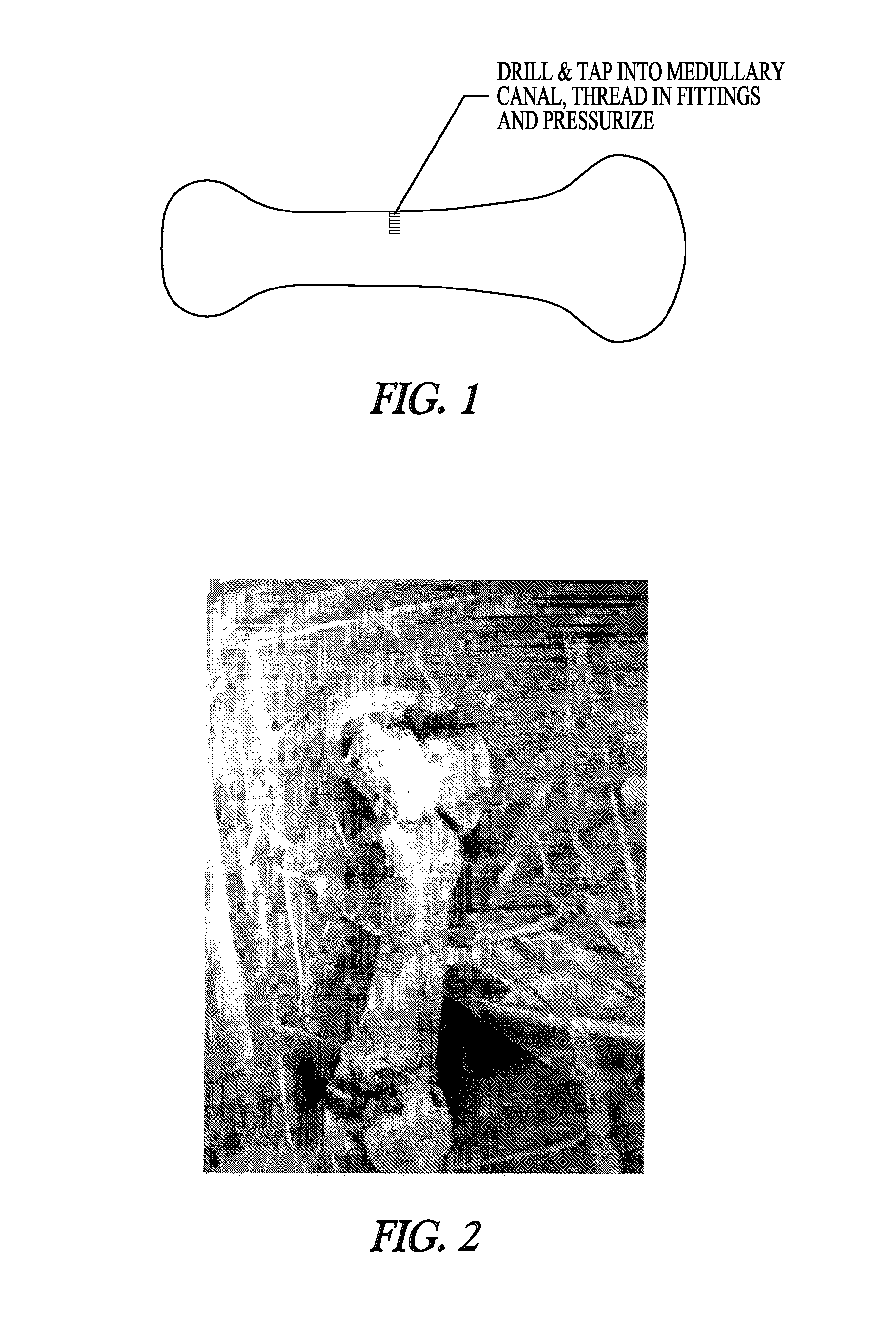
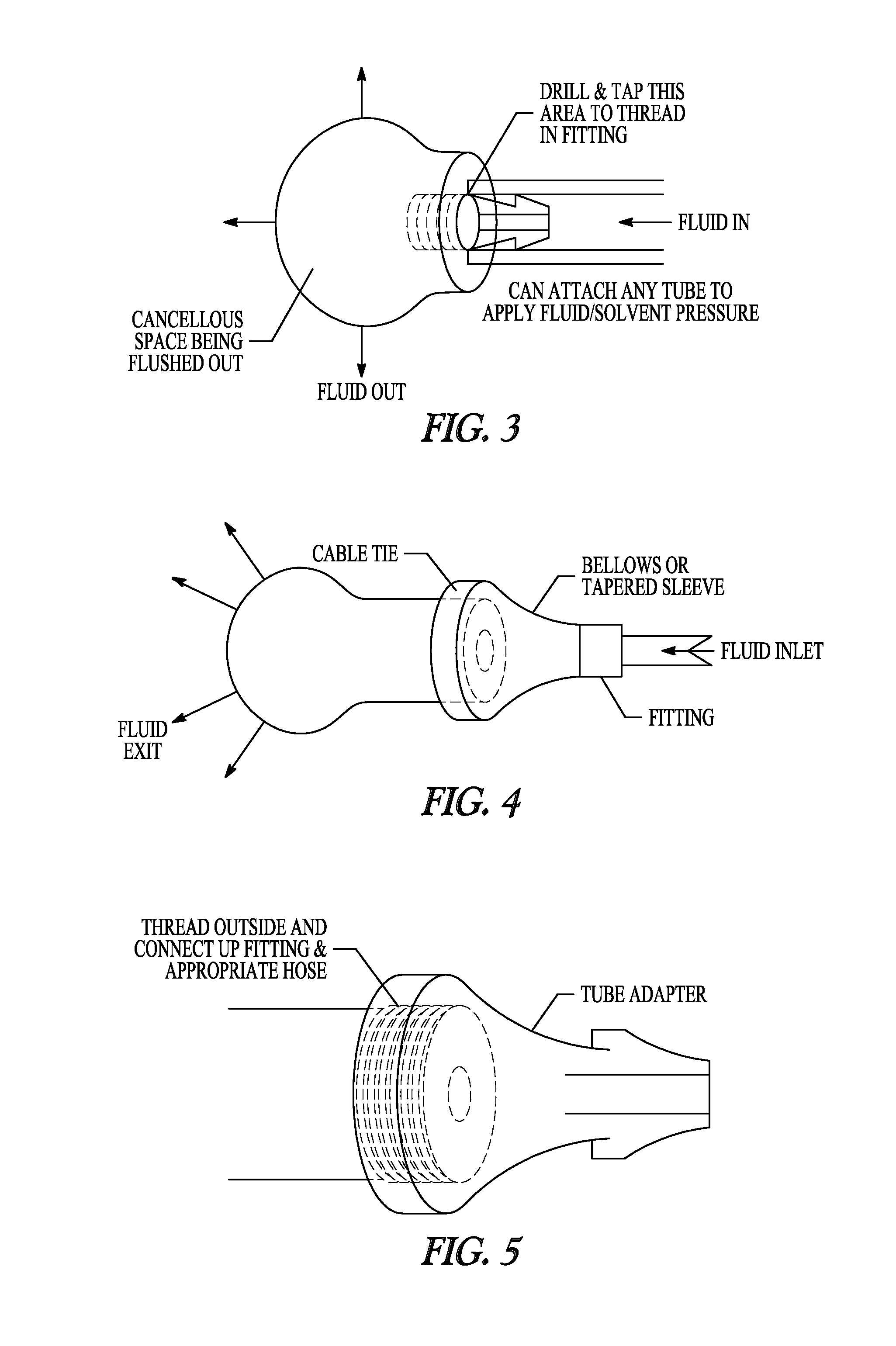
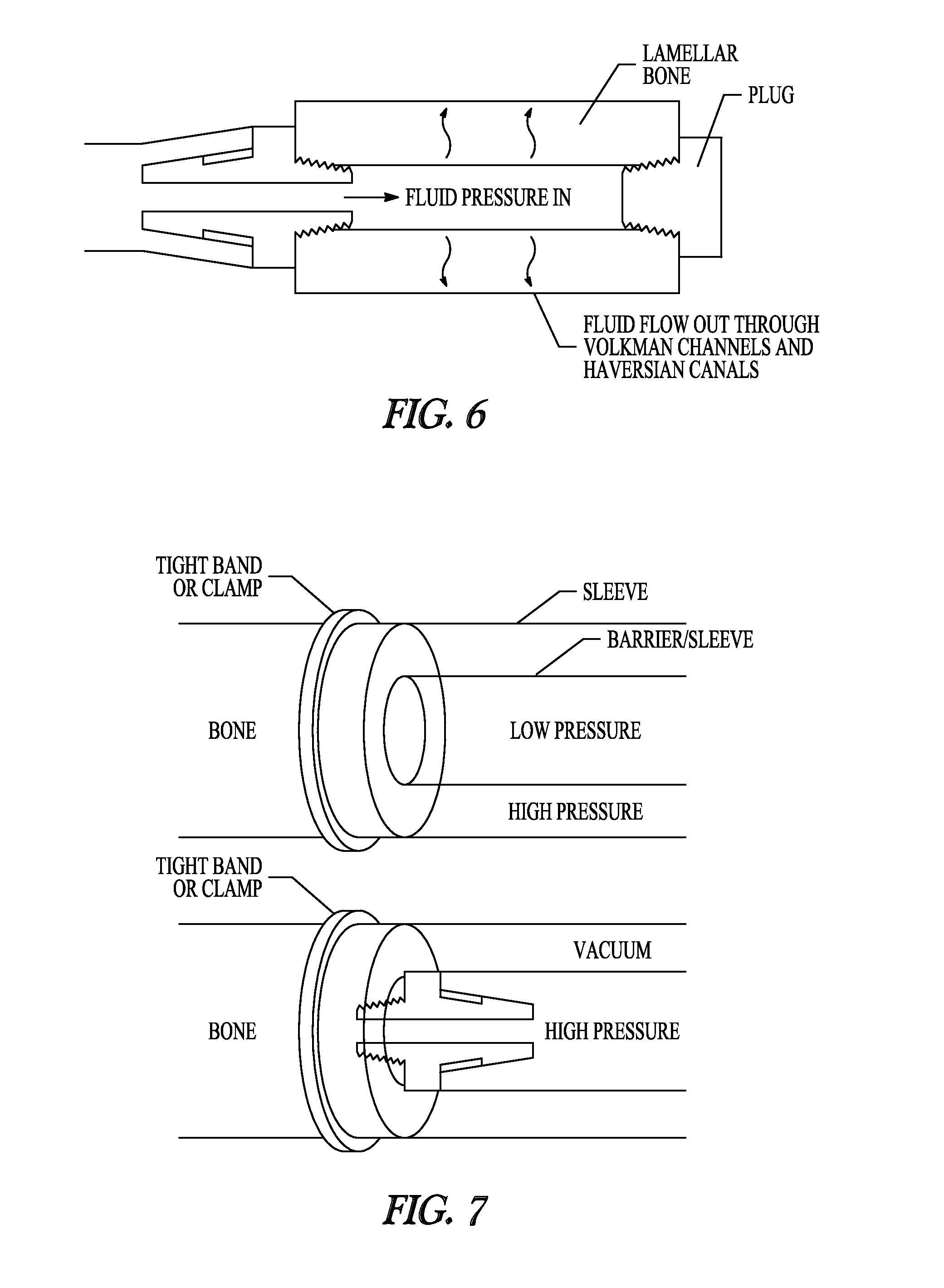
[0090]A 0.242 inch hole was drilled in the diaphysis of a porcine femur to facilitate the tapping and placement of a ⅛″ national pipe thread (NPT) tapered fitting. A ⅛″ ID tube was then connected to the fitting, and the bone was placed into 10 liters of 0.5% SDS solution and perfused at a constant pressure of 600 mmHg for 60 hours. Solutions were changed every 6-12 hours and allowed to recirculate between fluid changes. Pressure was maintained via a controlled system utilizing a pressure sensor and an automated voltage feedback loop to control the pump speed. During the 60 hours, the flow rate varied from 1.0-60 mL / min. Decellularization was evident by the active egress (exiting) of cellular material through the native bone vasculature.

[0091]It is to be understood that, while the methods and compositions of matter have been described herein in conjunction with a number of different aspects, the foregoing description of the various aspects is intended to illustrate and not limit the ...


PUM

PropertyMeasurementUnit
pressureaaaaaaaaaa
pressureaaaaaaaaaa
pressureaaaaaaaaaa

Abstract

The invention provides methods for decellularizing bone, e.g., human bone.

Description

CROSS-REFERENCE TO RELATED APPLICATIONS[0001]The present application claims the benefit of the filing date of U.S. application Ser. No. 61 / 659,046, filed on Jun. 13, 2012, the disclosure of which is incorporated by reference herein.BACKGROUND[0002]Biologic scaffolds composed of extracellular matrix (ECM) which represents the secreted products of resident cells of each tissue and organ, are increasingly used in regenerative medicine strategies for tissue and organ replacement. Preservation of the native ultrastructure and composition of ECM during the process of tissue decellularization is highly desirable (Ott et al., Nat. Med., 14:213 (2008); Uygun et al., Nat. Med., 16:814 (2010); Petersen et al., Science, 329:538 (2010); Nakayama et al., Tissue Eng. Part A, 16:2207 (2010); Allen et al., Tissue Eng. Part A, 16:3363 (2010); Simionescu et al., J. Heart Valve Dis., 12:226 (2003); Badylak et al., Acta Biomater., 5:1 (2009)). Numerous methods have been employed to decellularize tissues...

Claims


Application Information

Patent Timeline
Patent Type & Authority Patents(United States)
IPC IPC(8): A61F2/28A61F2/46C12N5/071C12N5/077
CPCC12N5/0654A61F2/4644A61F2002/4646
Inventor ROSS, JEFFREYPORTER, BLAISE
Owner MIROMATRIX MEDICAL
Learn More