
Micelle structure of nano preparation for diagnosis or treatment of cancer disease and preparation method thereof

a nano-preparation and micelle technology, applied in the direction of pharmaceutical delivery mechanism, organic active ingredients, drug compositions, etc., can solve the problems of reducing the ability to deliver drugs, reducing the effectiveness of drug delivery systems, and prolonging the substantial average survival time by only 12

Active Publication Date: 2016-07-19
KOREA UNIV RES & BUSINESS FOUND 50
View PDF2 Cites 0 Cited by
  • Summary
  • Abstract
  • Description
  • Claims
  • Application Information

AI Technical Summary

Benefits of technology

The nanopreparation described in this patent has a small size of 12 nm or less. It is made up of polymeric lipid DSPE-mPEG which forms micelles to encapsulate a photosensitizer called hyperricin. This nanopreparation can easily overcome the challenges of the blood-tumor barrier and interstitial fluid pressure. It also has high efficiency in photodynamic diagnosis and treatment of cancer, particularly brain tumors, due to its high relative coexistence coefficient to mitochondrion among intracellular organelles of cancer cells.

Problems solved by technology

However, the therapeutic method has a problem in that the substantial average survival time can be prolonged by only 12.1 to 14.6 months.
Various therapeutic methods for treating brain tumors have a problem in that a drug delivery system is less effective due to low drug permeability caused by a blood-brain barrier (BBB) and a blood tumor barrier (BTB) present in a brain; and a decrease in ability to deliver drug caused by an increase in pressure in tissue according to volumes and properties of brain tumors itself.

Method used


Image

  • Micelle structure of nano preparation for diagnosis or treatment of cancer disease and preparation method thereof
  • Micelle structure of nano preparation for diagnosis or treatment of cancer disease and preparation method thereof
  • Micelle structure of nano preparation for diagnosis or treatment of cancer disease and preparation method thereof

Examples


example 1

Preparation of Nanopreparation Having Micelle Structure According to Present Invention 1

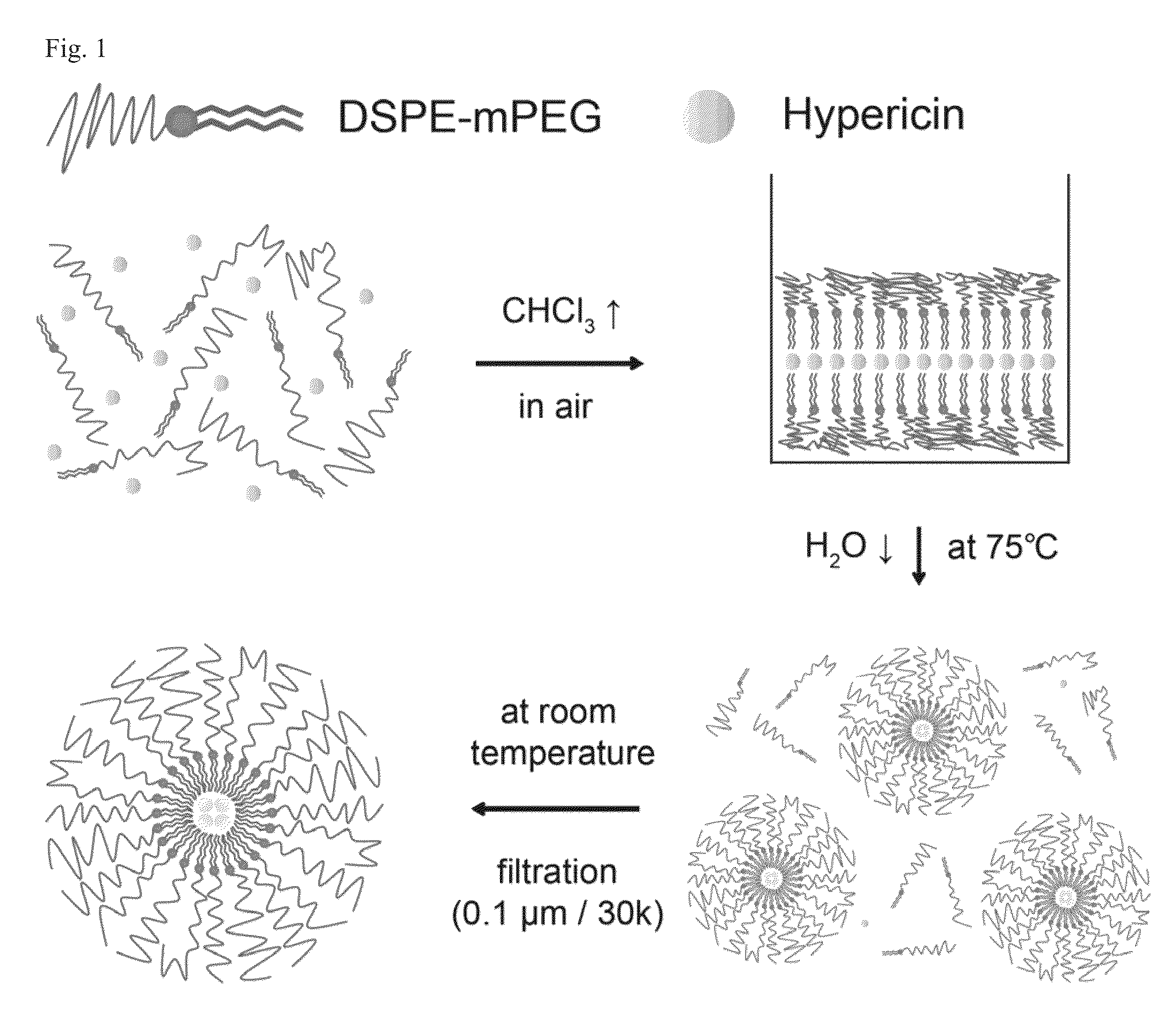
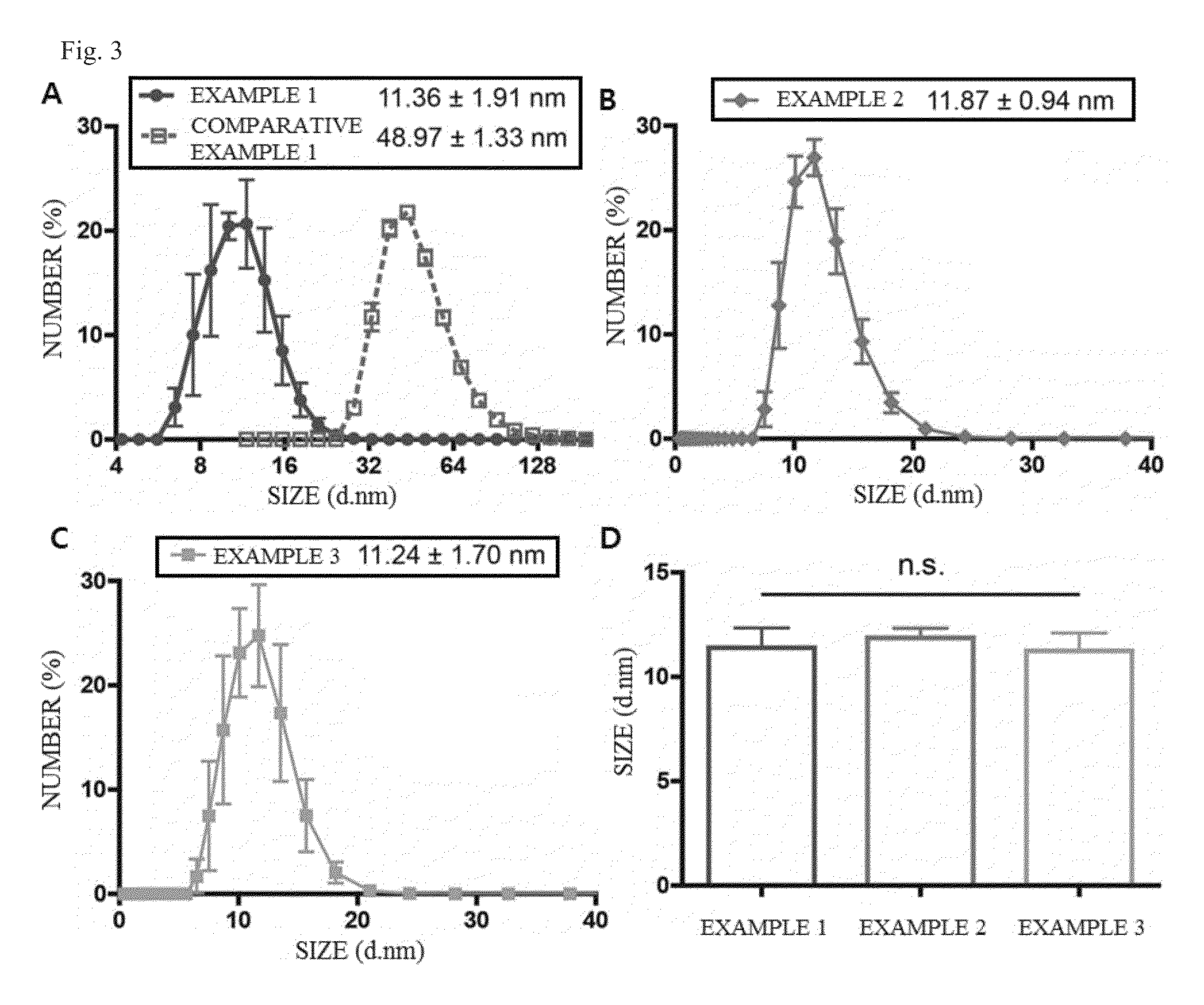
[0095]Hypericin (200 μL, 0.2 mg / mL) and DSPE-mPEG (1,2-distearoyl-sn-glycero-3-phosphoethanolamine-N-[methoxypoly(ethylene glycol), molecular weight 2000], 222 μL, 10 mg / mL) dissolved in chloroform were mixed at room temperature to have a molar ratio of 1:10. Subsequently, the resulting mixture was completely dried in a room temperature-light blocking state to form a polymeric lipid film, and the formed film was introduced into sonication equipment, and then hydrated in phosphate buffered saline (PBS, 1 mL) at 75° C. for 5 minutes. In this case, the polymeric lipid film was hydrated and sonicated simultaneously. After the sonicated solution was cooled to room temperature and filtered by a filter having the size of 0.1 μm, the filtrate was further filtered by centrifuge with a 30 k centrifugal filter (30000 MWCO centrifuge, Millipore, Billerica, Mass., USA) to prepare a nanopreparation having a mi...

example 2

Preparation of Nanopreparation Having Micelle Structure According to Present Invention 2

[0097]The same procedure as Example 1 was performed to prepare a nanopreparation having a micelle structure, except that HEPES buffered saline (HBS, 1 mL) was used instead of phosphate buffered saline (PBS, 1 mL) used in the Example 1.

[0098]The HEPES buffered saline (HBS) includes water; 10 to 20 mM 4-(2-hydroxyethyl)piperazine-1-ethanesulfonic acid; and 135 to 155 mM sodium chloride.

example 3

Preparation of Nanopreparation Having Micelle Structure According to Present Invention 3

[0099]The same procedure as Example 1 was performed to prepare a nanopreparation having a micelle structure, except that HEPES buffered glucose solution (HBG, 1 mL) was used instead of phosphate buffered saline (PBS, 1 mL) used in Example 1.

[0100]The HEPES buffered glucose solution (HBG) includes water; 10 to 20 mM 4-(2-hydroxyethyl)piperazine-1-ethanesulfonic acid; and 5% of glucose.


PUM

PropertyMeasurementUnit
temperatureaaaaaaaaaa
diameteraaaaaaaaaa
survival timeaaaaaaaaaa

Abstract

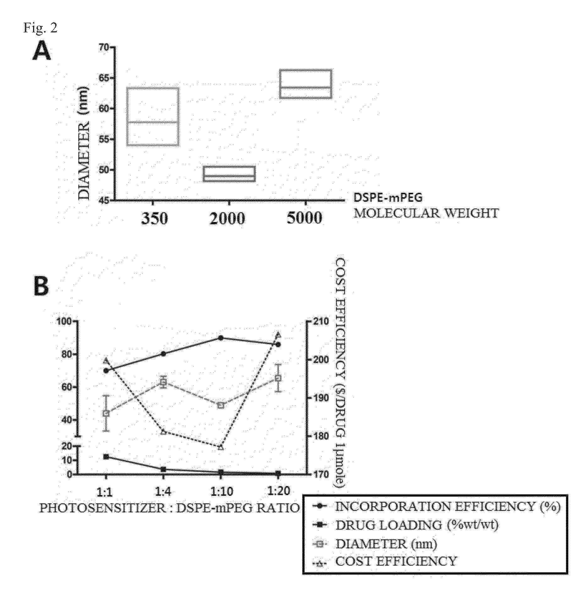
Disclosed herein are a nanopreparation having a micelle structure for diagnosis or treatment of cancer diseases, and a method of preparing the same, and more particularly, a nanopreparation having a micelle structure available for diagonosis or treatment of cancer diseases and a method for preparing the same, wherein the nanopreparation is prepared by encapsulating a photosensitizer by forming micelle with polymeric lipid DSPE-mPEG. The nanopreparation having the micelle structure according to the present invention has a size of 12 nm or less by encapsulating hypericin, which is a photosensitizer, by forming micelle with polymeric lipid DSPE-mPEG having a molecular weight of 1500 to 2500. Resultingly, the nanopreparation easily overcomes a blood-tumor barrier (BTB) and an interstitial fluid pressure and has light induced cytotoxicity efficiency that is about more than 2.5 times higher than that of the case where hypericin is used alone.

Description

CROSS-REFERENCES TO RELATED APPLICATION[0001]This patent application claims the benefit of priority from Korean Patent Application No. 10-2013-0143045, filed on, 22 Nov. 2013, the contents of which are incorporated herein by reference.BACKGROUND OF THE INVENTION[0002]1. Field of the Invention[0003]The present disclosure relates to a nanopreparation having a micelle structure for diagnosis or treatment of cancer diseases, and a method of preparing the same, and more particularly, to a nanopreparation having a micelle structure available for diagnosis or treatment of cancer diseases, and a method of preparing the same, wherein the nanopreparation is prepared by encapsulating anti-cancer drug, such as a photosensitizer, by forming micelle with polymeric lipid DSPE-mPEG.[0004]2. Description of the Related Art[0005]Photodynamic therapy is one of currently promising method for treating cancers. Photodynamic therapy is a therapeutic method using a following principle: when a photosensitize...

Claims


Application Information

Patent Timeline
Patent Type & Authority Patents(United States)
IPC IPC(8): A61K47/24A61K31/122A61K9/107
CPCA61K47/24A61K9/1075A61K31/122A61P35/00A61K9/08A61K9/127A61K49/08
Inventor CHOI, CHULHEECHONG, KYUHAPARK, JIHO
Owner KOREA UNIV RES & BUSINESS FOUND 50
Learn More