7-hydroxycoumarin-based cell-tracking reagents
- Summary
- Abstract
- Description
- Claims
- Application Information
AI Technical Summary
Benefits of technology
Problems solved by technology
Method used
Image
Examples
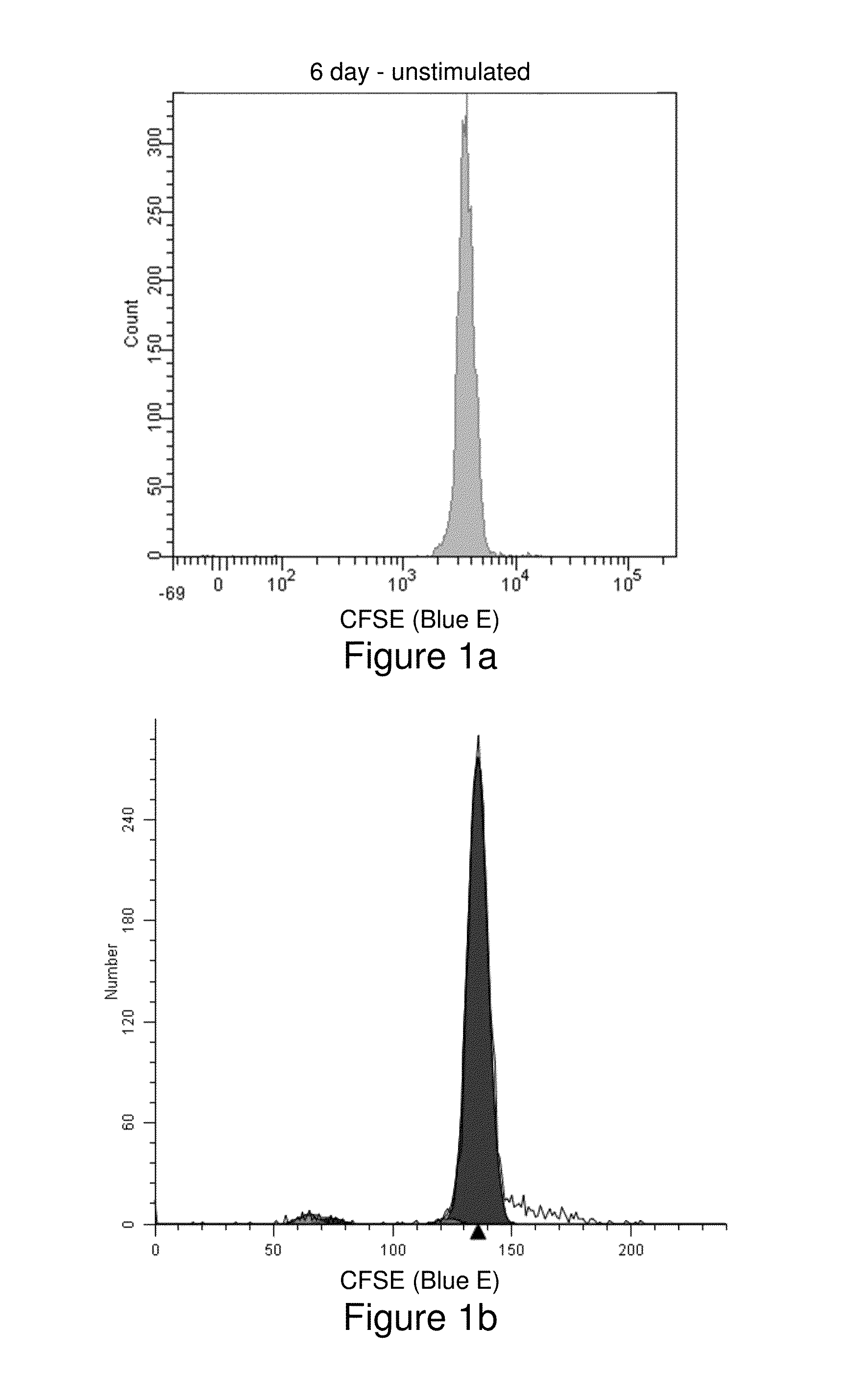
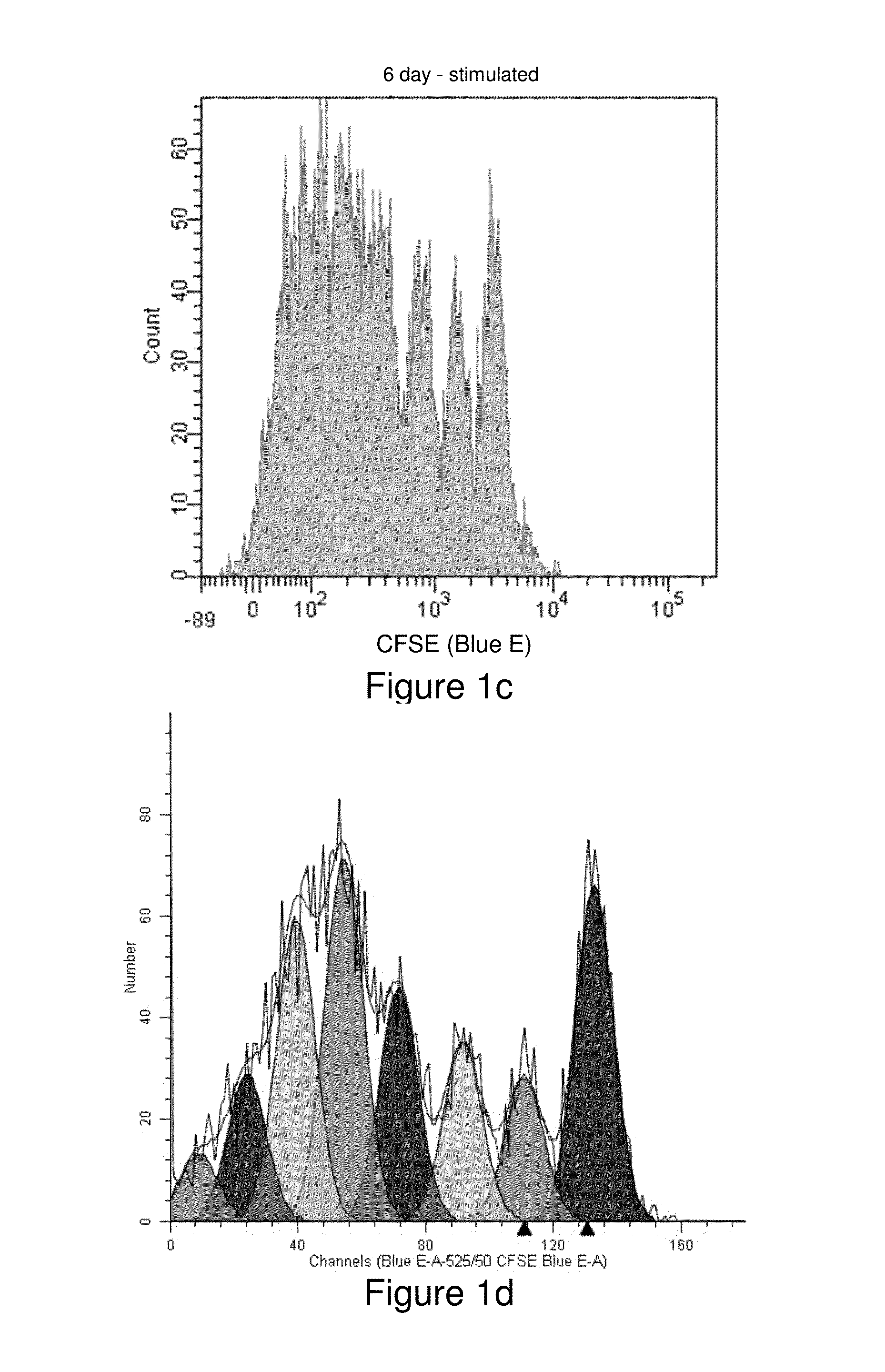
example 1
Experimental Protocol
[0137]Preparation of Culture Media
[0138]To 1 L of OpTimizer T-Cell Expansion Medium were added the following: 26 mL of T cell expansion supplement, 10 mL of 200 mM L-glutamine solution (final concentration=2 mM), and 1 ml of 50 mg / mL Gentamycin solution (final concentration=50 μg / mL). Complete media is stable for ˜4 weeks when stored at 2-8° C. in the dark.
[0139]Preparation of HIl-2 Stock Solution
[0140]Acetic acid, 100 mM, was prepared by diluting 5 μL glacial (17M) acetic acid into 800 μL of dH2O. Il-2, 40 μg, was dissolved in 400 μL of 100 mM acetic acid to make a 0.1 mg / mL solution; 20 μL aliquots of this solution were placed into microfuge tubes and stored at −20° C. One μL of this solution contains 100 ng of it-2.
[0141]CD3 Concentration
[0142]A 0.5 mL bottle of Caltag MHCD 0300 contains 100 μg of CD3; a 1 μL aliquot of this stock solution contains 200ng of CD3.
[0143]Ficoll Separation of Mononuclear Cells from Whole Blood
[0144]Human peripheral blood mononucle...
example 2
Experimental Protocol
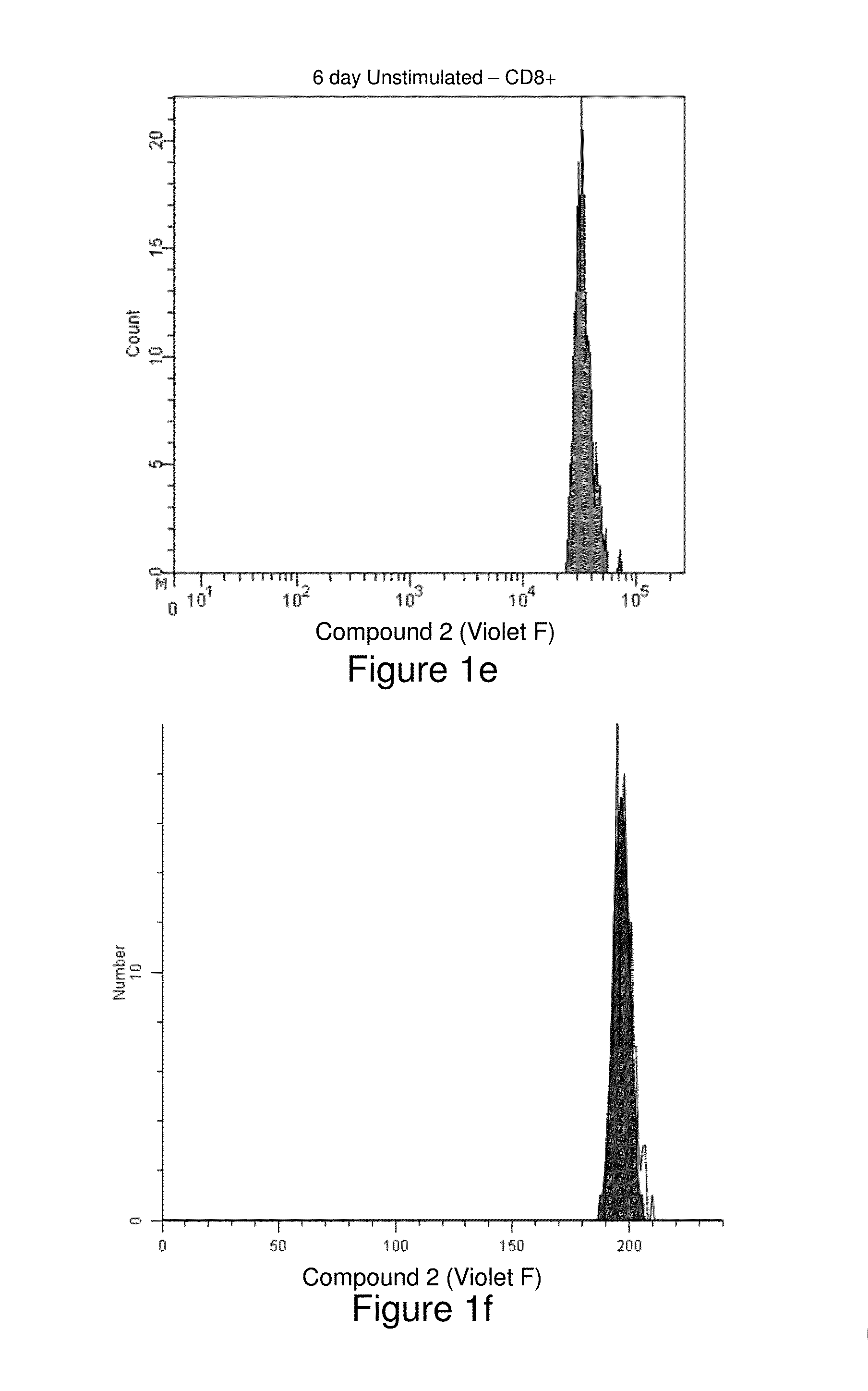
[0156]Human peripheral blood mononuclear cells were isolated from whole blood using a Ficoll density gradient, then washed and resuspended in phosphate buffered saline (PBS) at a concentration of 106 / mL (see, Example 1). Two samples, one more recently synthesized than the other, of the cell-tracking compound Compound Violet were each dissolved in anhydrous dimethylsulfoxide to a final concentration of 5 mM. Two 4-mL aliquots of cells were stained with 4 μL of either Compound Violet sample for final staining concentrations of 1 μM. Cells were mixed by vortexing and incubated with agitation at room temperature for 20 minutes. Then 2 mL of heat-inactivated Fetal Bovine Serum were added, followed by 5 more minutes of incubation. Cells were then washed twice with PBS and resuspended in 4 mL of OpTmizer T-Cell Expansion Buffer (GIBCO) containing 2 mM L-glutamine, and 100,000 units penicillin and 100 mg streptomycin per liter. Aliquots (1 mL) of cells stained with each...
example 3
Experimental Protocol
[0159]U2OS human osteosarcoma cells in complete media (McCoys media plus 10% FBS) were plated down at approximately 5000 cells / cm2 and allowed to adhere to four MacTec dishes. Two dishes of cells were transfected with Cellular Lights™ Talin-GFP at a volume to volume of 10% for 24 hours prior to analysis. A 5 μM solution of Compound Violet was created by diluting 2 μL of 5 mM dye in anhydrous dimethylsulfoxide into 2 mL of phosphate buffered saline (PBS) containing calcium and magnesium. Growth medium was removed from two plates of cells and replaced with dye solution. Cells were stained at room temperature for 20 minutes and washed twice with PBS. Imaging analysis was performed on a Delta Vision microscope with filters for DAPI and FITC.
Results (Shown in FIGS. 3a-3c):
[0160]Compound Violet and GFP each appeared brightly fluorescent in their respective emission filters, and there did not appear to be fluorescence overlap into the opposite emission filters.
PUM
 Abstract
Description
Claims
Application Information
 - R&D
- Intellectual Property
- Life Sciences
- Materials
- Tech Scout
- Unparalleled Data Quality
- Higher Quality Content
- 60% Fewer Hallucinations
Browse by: Latest US Patents, China's latest patents, Technical Efficacy Thesaurus, Application Domain, Technology Topic, Popular Technical Reports.
© 2025 PatSnap. All rights reserved.Legal|Privacy policy|Modern Slavery Act Transparency Statement|Sitemap|About US| Contact US: help@patsnap.com


