Method for fabricating simulated tissue structures by means of multi material 3D printing

a 3d printing and tissue technology, applied in the field of anatomical models, can solve the problems of not being readily available, unable to develop the degree of detail, and unable to read textbooks or watch instructional videos

Active Publication Date: 2016-09-06
BIONIKO CONSULTING
View PDF22 Cites 17 Cited by
  • Summary
  • Abstract
  • Description
  • Claims
  • Application Information

AI Technical Summary

Benefits of technology

[0024]The anatomical tissue types can include loose connective tissue/muscle, dense connective tissue, supportive connective tissue, and cellular tissue. The synthetic tissue type corresponding to loose connective tissue/muscle has a material property type with a hardness between Shore A 5 and Shore A 30; the synthetic tissue type corresponding to dense connective tissue has a hardness between Shore A 30 and Shore A 95; the synthetic tissue type corresponding to the supportive connective tissues has a hardness of Shore D 20 or greater, and the synthetic tissue

Problems solved by technology

This dexterity cannot be developed by reading textbooks or watching instructional videos.
Animal models or cadavers have been the default method for hands-on surgical training in the past, but they are costly, not readily available and may pose biohazard issues.
Systems of computerized virtual reality or electromechanical devices may be useful and effective but their cost restricts their reach and thus their impact in

Method used


Image

  • Method for fabricating simulated tissue structures by means of multi material 3D printing
  • Method for fabricating simulated tissue structures by means of multi material 3D printing
  • Method for fabricating simulated tissue structures by means of multi material 3D printing

Examples


Embodiment Construction

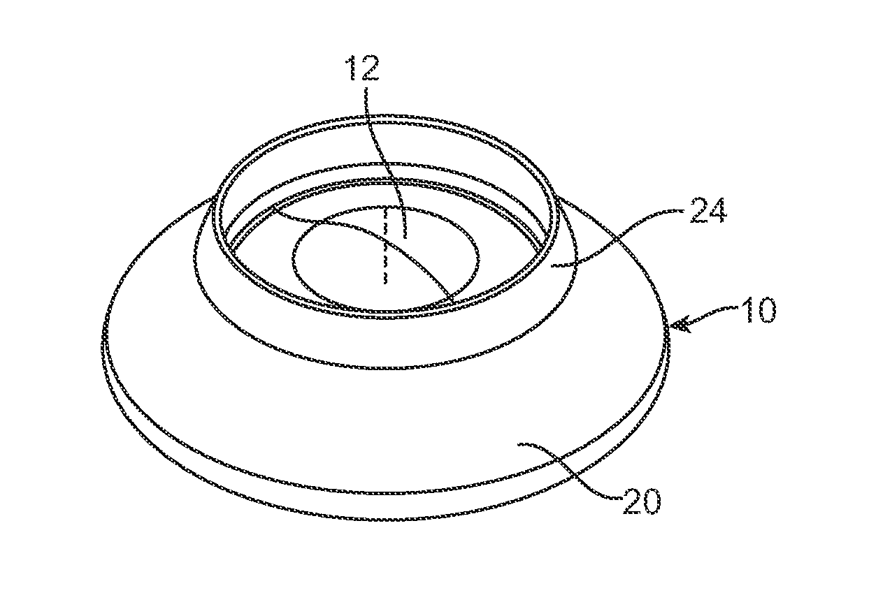
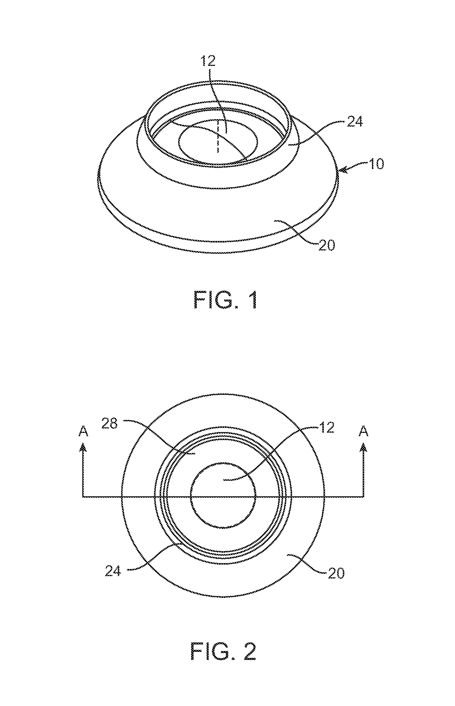
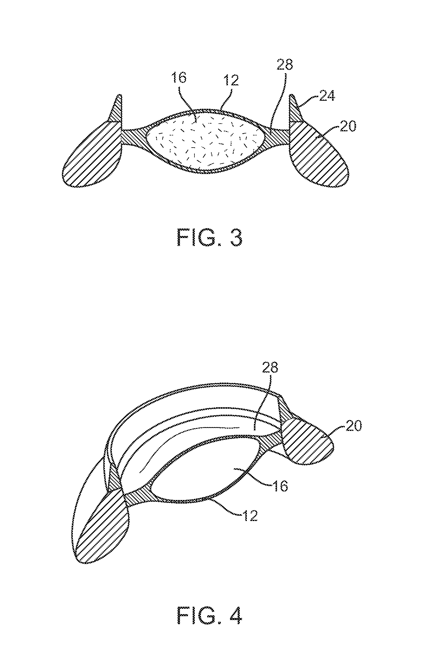
[0070]This invention relates to a method to create devices which mimic the mechanical properties of tissue structures and their pathologies, for surgical training, demonstration, teaching aids or surgical instrument calibration, validation and research and development. The invention is directed particularly to the development of a surgical model of the anterior segment of the eye, to serve as a model for cataract surgery. Other applications using the same principle may include but are not limited to: joint models for arthroplasty; cardio-vascular models for cardiac surgery; spinal models for spinal surgery; models for craneo-facial surgery such as cleft palate; crash test dummy components; and ballistic impact models.

Tissue

[0071]The human body, as well as any other organism, is a natural “assembly” of multiple sub-structures having different mechanical properties; hard bones, soft muscles, tough membranes, elastic skin and many others. These sub-structures are called tissues. There ...


PUM


Abstract

A synthetic eye model includes an enclosed lens capsule, a removable cortex material within the lens capsule, and an outer support for the lens capsule. A synthetic eye model assembly includes an eye segment comprising a lens capsule and an outer support and a base for detachably engaging the eye segment. The synthetic eye model and assembly can be made by 3D printing. A method of practicing eye surgery and a method of making a three-dimensional synthetic tissue model of an anatomical tissue structure are also disclosed.

Description

CROSS REFERENCE TO RELATED APPLICATIONS[0001]This application claims the priority of U.S. Provisional Application Ser. No. 61 / 644,248 filed May 8, 2012, and also claims the priority of Provisional Application Ser. No. 61 / 644,257 filed May 8, 2012, the disclosures of which are hereby incorporated fully by reference.FIELD OF THE INVENTION[0002]This invention relates generally to anatomical models, and more particularly to anatomical models that are useful to practice surgical techniques.BACKGROUND OF THE INVENTION[0003]According to a study by the American Board of Surgery and the Association of Program Directors in Surgery, a graduating general surgeon should be competent to perform 121 “Level-A” procedures after a 5 year residency program. The same study shows the level of experience in these procedures for the 2005 graduating class of surgeons. The study found that for sixty-three (63) procedures, the mode (most commonly reported) experience was 0. In addition, there was significant...

Claims


Application Information

Patent Timeline
IPC IPC(8): G09B23/30
CPCG09B23/30G09B23/34
Inventor BERNAL, ANDRES
Owner BIONIKO CONSULTING
Why Eureka
Try Eureka