Assays for detecting neutralizing autoantibodies to biologic therapy

a technology of autoantibodies and autoantibodies, which is applied in the field of autoantibodies detection assays for biologic therapy, can solve the problems of autoimmune disorders that are a significant and widespread medical problem, joint destruction and functional disability, and can not be detected in time, so as to improve the accuracy of optimizing therapy, monitor the efficacy of therapeutic treatment, and reduce toxicity

Active Publication Date: 2016-10-11
PROMETHEUS BIOSCI INC
View PDF76 Cites 5 Cited by
  • Summary
  • Abstract
  • Description
  • Claims
  • Application Information

AI Technical Summary

Benefits of technology

[0007]The present invention provides assays for detecting and measuring the presence or level of neutralizing and non-neutralizing autoantibodies to biologics such as anti-TNFα drug therapeutics in a sample. The present invention is useful for monitoring the formation of neutralizing and / or non-neutralizing anti-drug antibodies over time while a subject is on biologic therapy (e.g., anti-TNFα drug therapy). The present invention is also useful for predicting and / or determining the cross-reactivity of neutralizing anti-drug antibodies in a subject's sample with alternative biologic therapies (e.g., alternative anti-TNFα therapies). As such, the present invention provides information for guiding treatment decisions for those subjects receiving therapy with a biologic agent and improves the accuracy of optimizing therapy, reducing toxicity, and / or monitoring the efficacy of therapeutic treatment to biologic therapy.

Problems solved by technology

Autoimmune disorders are a significant and widespread medical problem.
RA causes chronic inflammation of the joints and typically is a progressive illness that has the potential to cause joint destruction and functional disability.
Also during disease flare ups, joints frequently become red, swollen, painful and tender, due to inflammation of the synovium.
Many of these drugs, however, can have detrimental side-effects.
While these mouse anti-hTNF-α antibodies often displayed high affinity for hTNF-α and were able to neutralize hTNF-α activity, their use in vivo has been limited by problems associated with the administration of mouse antibodies to humans, such as a short serum half-life, an inability to trigger certain human effector functions, and elicitation of an unwanted immune response against the mouse antibody in a human (the “human anti-mouse antibody” (HAMA) reaction).
While such biologic therapies have demonstrated success in the treatment of rheumatoid arthritis and other autoimmune disorders such as CD, not all subjects treated respond, or respond well, to such therapy.

Method used


Image

  • Assays for detecting neutralizing autoantibodies to biologic therapy
  • Assays for detecting neutralizing autoantibodies to biologic therapy
  • Assays for detecting neutralizing autoantibodies to biologic therapy

Examples


example 1

Development of a Novel Assay to Monitor Neutralizing Anti-Drug Antibody Formation in IBD Patients

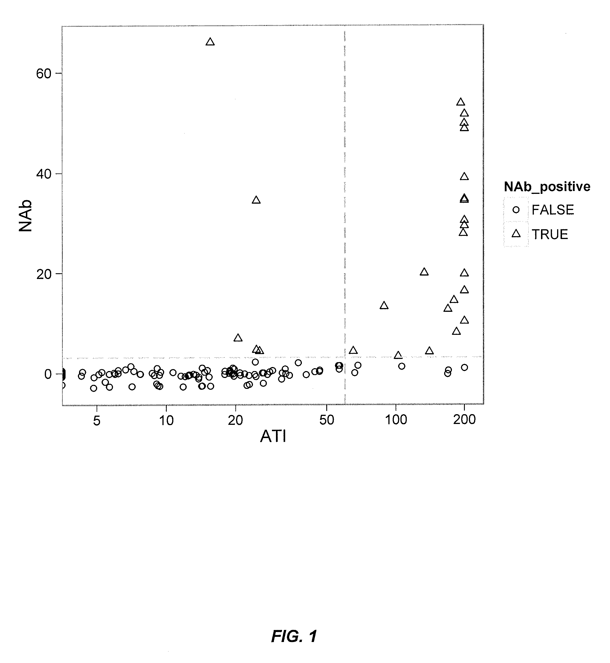
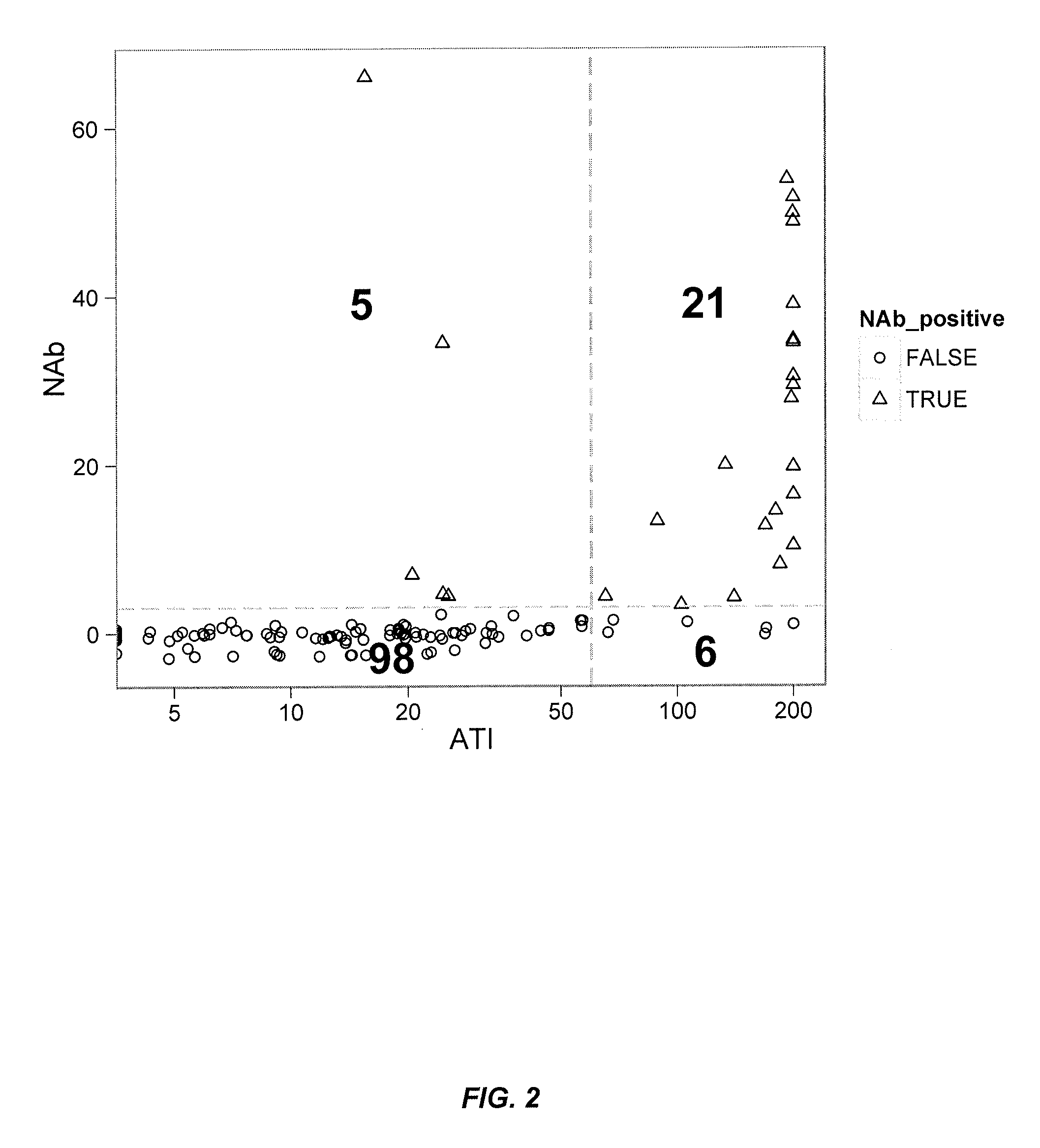
[0336]This example illustrates a novel homogeneous assay for detecting or measuring the presence or level of neutralizing and / or non-neutralizing anti-drug autoantibodies (ADA) in a patient sample (e.g., serum) using size exclusion chromatography in the presence of labeled (e.g., fluorescently labeled) anti-TNFα drug and labeled TNFα. In particular embodiments, this assay is advantageous because it obviates the need for wash steps which remove low affinity ADA, uses distinct labels (e.g., fluorophores) that allow for detection on the visible and / or IR spectra which decreases background and serum interference issues, increases the ability to detect neutralizing and / or non-neutralizing ADA in patients with a low titer due to the high sensitivity of fluorescent label detection, and occurs as a liquid phase reaction, thereby reducing the chance of any changes in the epitope by attachment to ...

example 2

Patient Case Studies for Monitoring the Formation of Neutralizing Anti-Drug Antibodies Over Time

[0345]This example illustrates additional embodiments of a novel homogeneous assay for detecting or measuring the presence or level of neutralizing and / or non-neutralizing anti-drug autoantibodies (ADA) in a patient sample (e.g., serum) using size exclusion chromatography in the presence of labeled (e.g., fluorescently labeled) anti-TNFα drug and labeled TNFα. In addition, this example demonstrates time course case studies of IBD patients on anti-TNFα drug therapy for monitoring the formation of neutralizing and / or non-neutralizing anti-drug antibodies and / or a shift from non-neutralizing to neutralizing anti-drug antibodies while the patient is on therapy.

1. Drug and Anti-Drug Antibody Assays

[0346]FIG. 4 illustrates detection of ATI (i.e., antibody to IFX; “HACA”) by the fluid phase mobility shift assay described herein. For example, 444 ng of Alexa488 labeled IFX (18.8 μg / ml in 100% ser...

example 3

Detection of Neutralizing Antibody (NAb) Activity via an HPLC Mobility Shift Competitive Ligand-Binding Assay

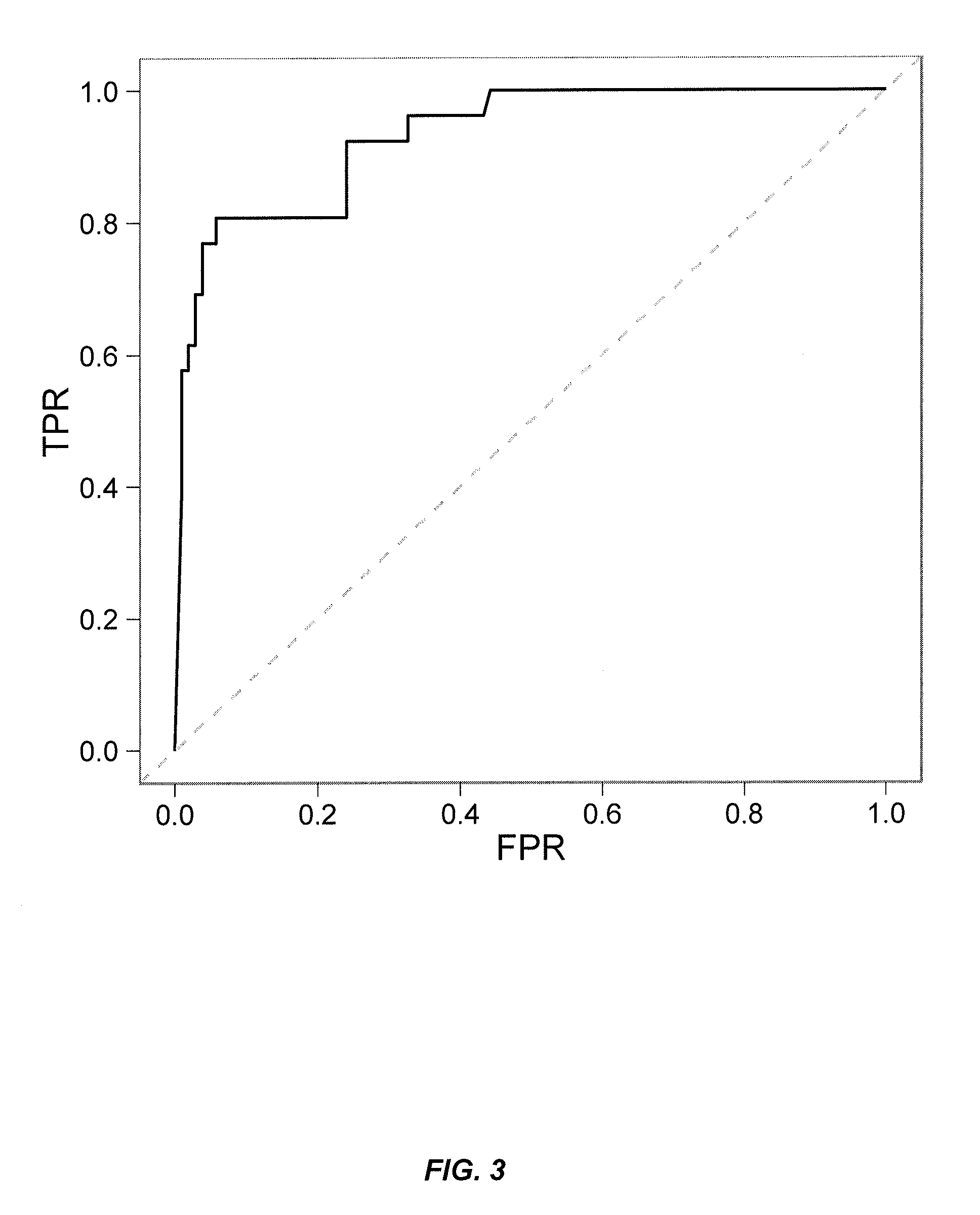
[0353]This example illustrates yet additional embodiments of a novel homogeneous assay for detecting or measuring the presence or level of neutralizing and / or non-neutralizing anti-drug autoantibodies (ADA) in a patient sample (e.g., serum) using an HPLC size exclusion chromatography assay. In addition, this example demonstrates methods for predicting and / or determining the cross-reactivity of NAb with alternative biological drugs such as other anti-TNF drugs.

[0354]In some embodiments, a multi-tiered approach to immunogenicity testing comprises first screening both drug and anti-drug antibodies by a rapid, sensitive screening assay. This approach is recommended by both the FDA and the EMEA and is a useful management tool for large clinical trials and multiple time points per patient. After confirming the presence of ADA such as ATI, patient samples are then further examined f...


PUM

PropertyMeasurementUnit
timeaaaaaaaaaa
timeaaaaaaaaaa
timeaaaaaaaaaa

Abstract

The present invention provides assays for detecting and measuring the presence or level of neutralizing and non-neutralizing autoantibodies to biologics such as anti-TNFα drug therapeutics in a sample. The present invention is useful for monitoring the formation of neutralizing and / or non-neutralizing anti-drug antibodies over time while a subject is on biologic therapy. The present invention is also useful for predicting and / or determining the cross-reactivity of neutralizing anti-drug antibodies in a subject's sample with alternative biologic therapies. As such, the present invention provides information for guiding treatment decisions for those subjects receiving therapy with a biologic agent and improves the accuracy of optimizing therapy, reducing toxicity, and / or monitoring the efficacy of therapeutic treatment to biologic therapy.

Description

CROSS-REFERENCES TO RELATED APPLICATIONS[0001]This application is a continuation of PCT / US2012 / 045794, filed Jul. 6, 2012, which application claims priority to U.S. Provisional Application No. 61 / 505,031, filed Jul. 6, 2011, U.S. Provisional Application No. 61 / 528,072, filed Aug. 26, 2011, and U.S. Provisional Application No. 61 / 535,884, filed Sep. 16, 2011, the disclosures of which are hereby incorporated by reference in their entirety for all purposes.BACKGROUND OF THE INVENTION[0002]Autoimmune disorders are a significant and widespread medical problem. For example, rheumatoid arthritis (RA) is an autoimmune disease affecting more than two million people in the United States. RA causes chronic inflammation of the joints and typically is a progressive illness that has the potential to cause joint destruction and functional disability. The cause of rheumatoid arthritis is unknown, although genetic predisposition, infectious agents and environmental factors have all been implicated i...

Claims


Application Information

Patent Timeline
Patent Type & Authority Patents(United States)
IPC IPC(8): G01N33/53G01N33/564G01N33/68
CPCG01N33/5308G01N33/564G01N33/6854G01N2333/525A61P19/02A61P37/02B01D15/34G01N33/538
Inventor HAUENSTEIN, SCOTTOHRMUND, LINDASINGH, SHARATWANG, SHUI LONG
Owner PROMETHEUS BIOSCI INC
Why Eureka
Try Eureka