

Compositions and methods for blood-brain barrier delivery of IgG-decoy receptor fusion proteins
a technology of fusion proteins and igg-decoy, which is applied in the direction of fusion cells, drug compositions, peptides, etc., can solve the problems that the fusion protein cannot be prescribed for cns conditions and the variability is not evenly distributed throughout the variable domain
- Summary
- Abstract
- Description
- Claims
- Application Information
AI Technical Summary
Benefits of technology
Problems solved by technology
Method used
Image

Examples
example 1
Cloning and Expression of the Human TNFR ECD cDNA
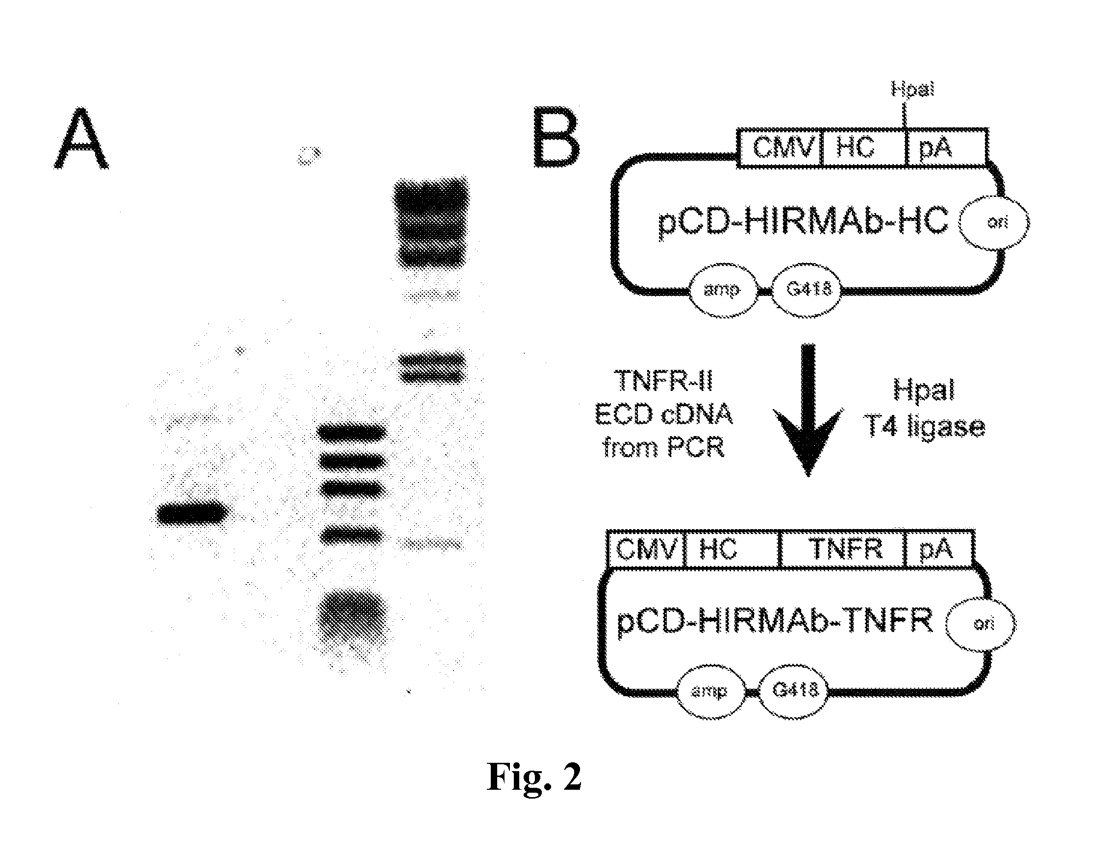
[0190]The human TNFR-II extracellular domain (ECD), corresponding to amino acids 23-257 of NP_001057, was cloned by the polymerase chain reaction (PCR) using the oligodeoxynucleotides (ODNs) described in Table 2 and cDNA derived from reverse transcription of polyA+RNA isolated from human U87 glial cells. The TNFR cDNA was cloned by PCR using 25 ng polyA+RNA-derived cDNA, 0.2 μM forward and reverse ODN primers (Table I, SEQ ID NO 1, SEQ ID NO 2, respectively), 0.2 mM deoxynucleosidetriphosphates, and 2.5 U PfuUltra DNA polymerase in a 50 μl Pfu buffer. The amplification was performed in a Mastercycler temperature cycler with an initial denaturing step of 95° C. for 2 min followed by 30 cycles of denaturing at 95° C. for 30 sec, annealing at 55° C. for 30 sec and amplification at 72° C. for 1 min; followed by a final incubation at 72° C. for 10 min. PCR products were resolved in 0.8% agarose gel electrophoresis, and the expected major s...
example 2
Genetic Engineering of Expression Plasmids Encoding Heavy Chain-TNFR Fusion Protein Wherein the TNFR is Fused to the Carboxyl Terminus of the HIRMAb Heavy Chain
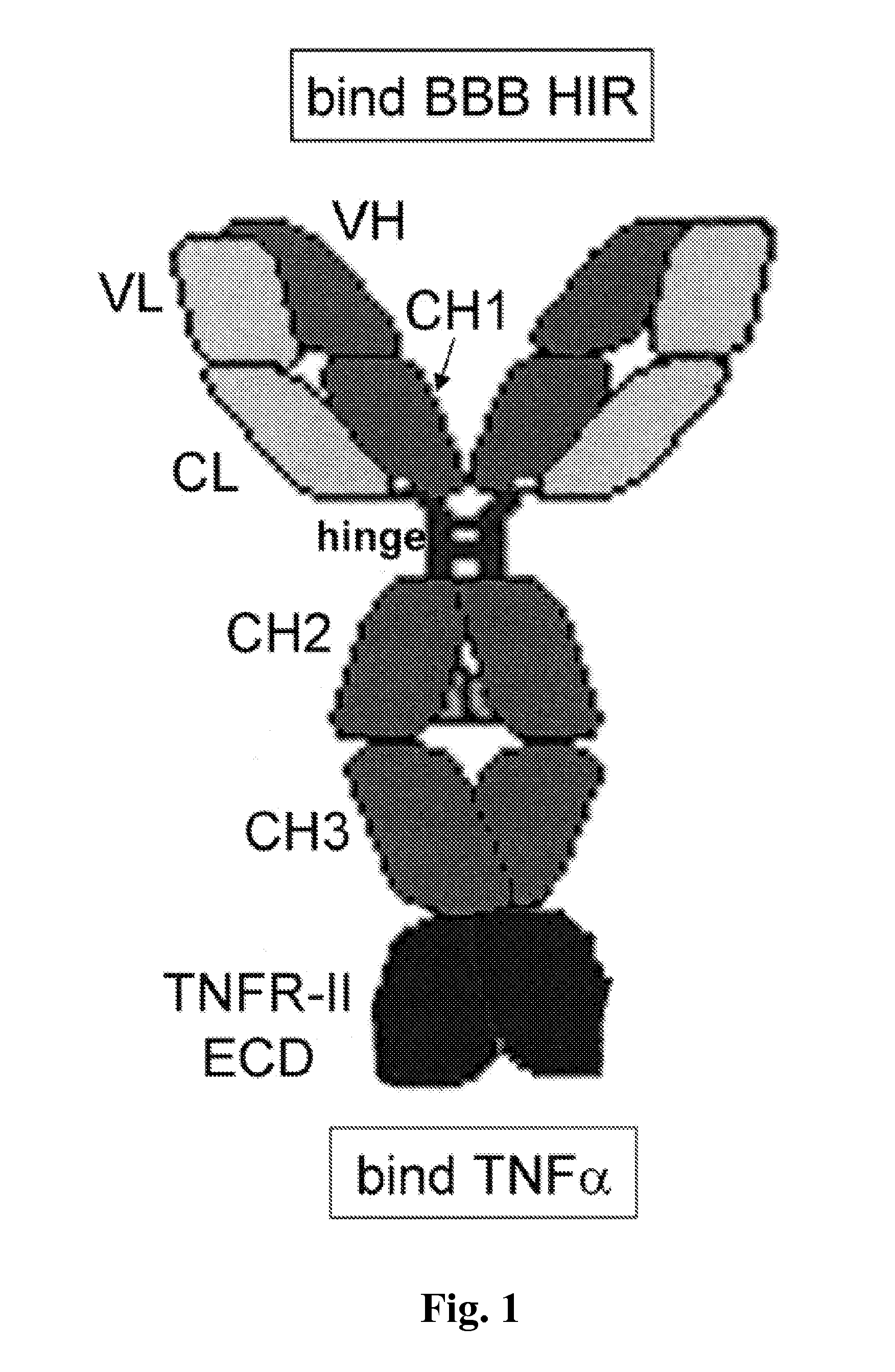
[0192]The expression plasmid expressing the fusion protein of the heavy chain (HC) and the TNFR ECD is designated pCD-HIRMAb-TNFR. This plasmid was engineered by insertion of the mature human TNFR cDNA, corresponding to amino acids Leu23-Asp257 of the human TNFR-II ECD (NP_001057), at the HpaI site of the pCD-HIRMAb-HC expression plasmid (FIG. 2B) to produce pCD-HIRMAb-TNFR (FIG. 2B). The pCD-HIRMAb-HC plasmid encodes the HC of the chimeric HIRMAb, and dual transfection of COS cells with this plasmid and a light chain (LC) expression plasmid, pHIRMAb-LC, allows for transient expression of the chimeric HIRMAb. The TNFR forward (FWD) PCR primer (Table I) introduces “CA” nucleotides to maintain the open reading frame and to introduce a Ser-Ser-Ser linker between the carboxyl terminus of the CH3 region of the HIRMAb HC and the am...
example 3
Secretion of HIRMAb-TNFR Fusion Protein by Transfected COS Cells
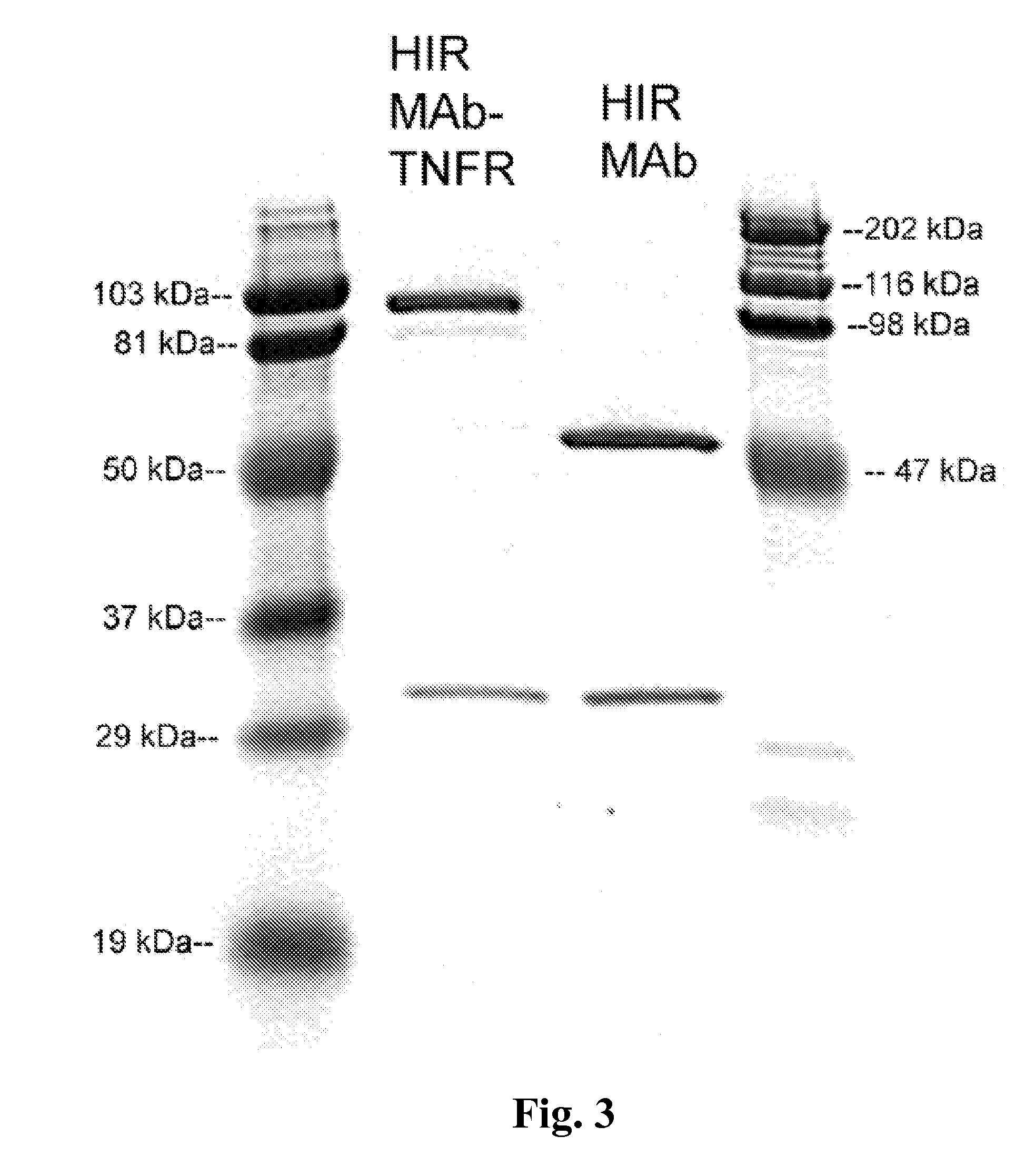
[0196]COS cells were dual transfected with pCD-HIRMAb-LC and pCD-HIRMAb-TNFR using Lipofectamine 2000, with a ratio of 1:2.5, μg DNA:uL Lipofectamine, where pCD-HIRMAb-LC is an expression plasmid encoding the light chain of the chimeric HIRMAb, which is the same light chain incorporated into the HIRMAb-TNFR fusion protein. Following transfection, the cells were cultured in serum free medium. COS cells were initially plated in 6-well cluster dishes for screening for expression with a human IgG specific ELISA. Subsequently, the transfection was scaled up for plating of transfected COS cells in 10×T500 flasks. The conditioned serum free medium was collected at 3 and 7 days. The fusion protein was purified by protein A affinity chromatography.
[0197]Human IgG ELISA was performed in Immulon 2 high binding plates with COS cell conditioned medium. A goat anti-human IgG primary antibody was plated in 0.1 M NaHCO3 (100 μA, 2 μg / m...
PUM
| Property | Measurement | Unit |
|---|---|---|
| dissociation constant | aaaaa | aaaaa |
| dissociation constant | aaaaa | aaaaa |
| dissociation constant | aaaaa | aaaaa |
Abstract
Description
Claims
Application Information