Image processing device and method for operating endoscope system

an image processing device and endoscope technology, applied in the field of image processing devices and methods for operating endoscope systems, can solve the problems of difficult to determine the staging of atrophy, the border between the normal portion and the portion with gastritis, and the inability to enhance the changes in mucosa color, so as to enhance the color of the mucosa or the like in the image due to the atrophy of stomach caused by atrophic gastritis

Active Publication Date: 2017-01-10
FUJIFILM CORP
View PDF10 Cites 2 Cited by
  • Summary
  • Abstract
  • Description
  • Claims
  • Application Information

AI Technical Summary

Benefits of technology

[0014]An object of the present invention is to provide an image processing device that enhances changes in color of mucosa or the like in an image due to atrophy of stomach caused by atrophic gastritis and a method for operating an endoscope system.
[0020]According to an aspect of the present invention, the changes in color of the mucosa or the like in the image due to the atrophy of stomach caused by the atrophic gastritis are enhanced.

Problems solved by technology

However, in a case where the atrophy is in an intermediate stage (for example, in a case where the atrophy is included in the group B or the group C in the ABC screening), there is little difference between the atrophic portion and the normal portion in the endoscopic image, so that it may be difficult to determine the staging of atrophy and the border between the normal portion and the portion with the gastritis.
However, the method described in the Japanese Patent No. 3228627 enhances only the redness of a reddish area in an image and cannot enhance the changes in color of mucosa with the progress of the atrophy of the stomach and cannot enhance and display the blood vessels that are located beneath the mucosa and seen through the mucosa as the atrophy progresses.

Method used


Image

  • Image processing device and method for operating endoscope system
  • Image processing device and method for operating endoscope system
  • Image processing device and method for operating endoscope system

Examples


first embodiment

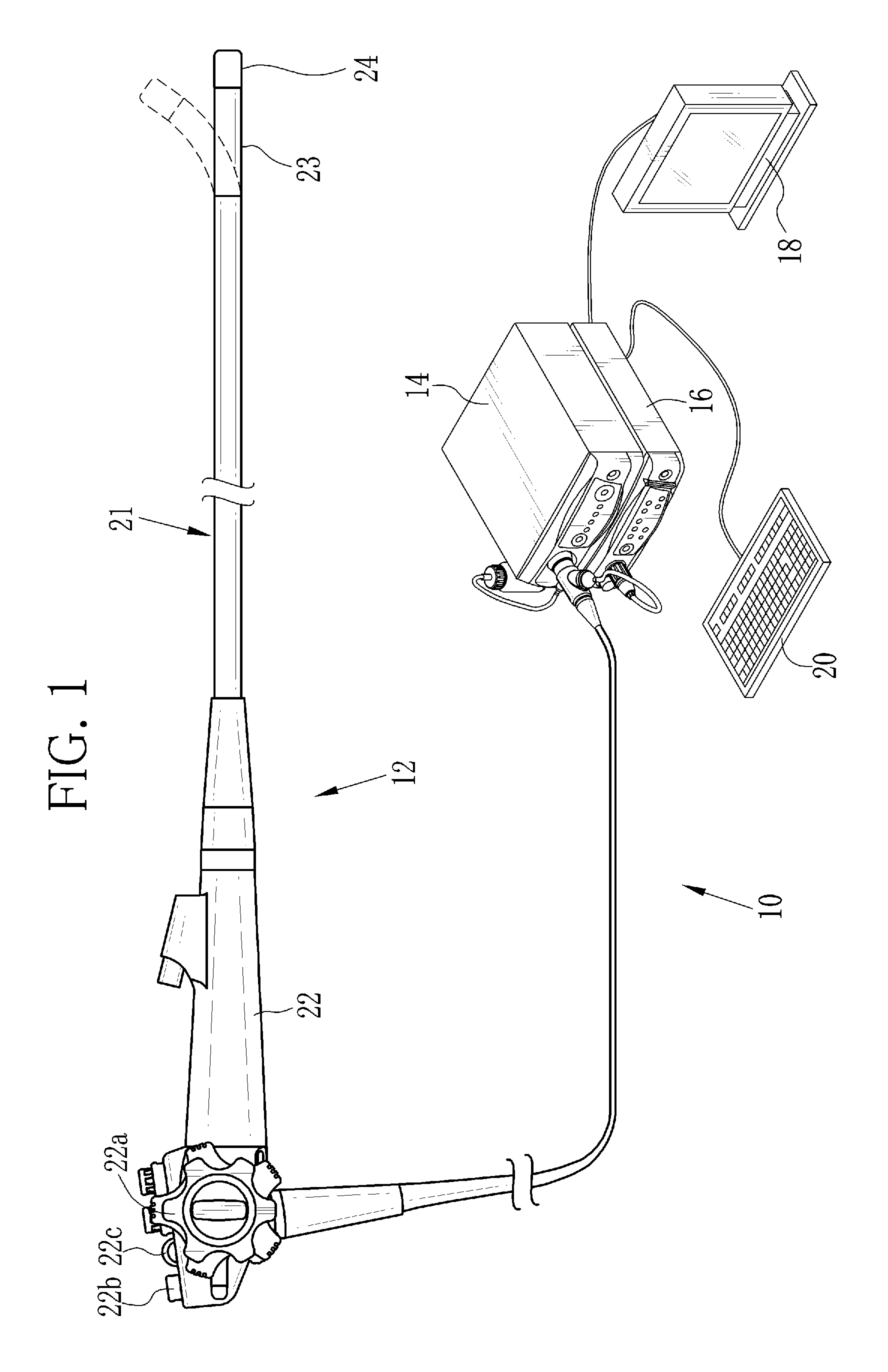
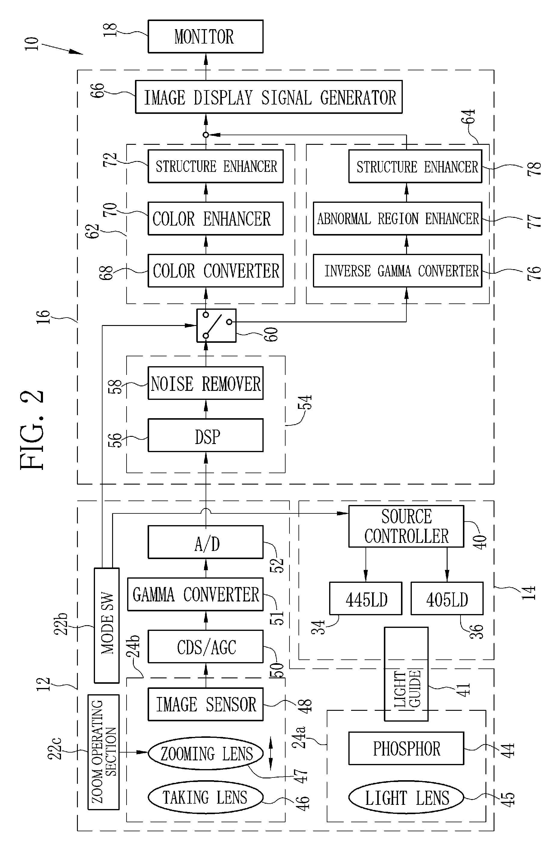
[0053]In FIG. 1, an endoscope system 10 according to a first embodiment comprises an endoscope 12, a light source device 14, a processor device 16, a monitor 18, and a console 20. The endoscope 12 is connected optically to the light source device 14 and electrically to the processor device 16. The endoscope 12 comprises an insertion section 21 to be inserted into a body cavity, a control handle unit 22 provided at the proximal end of the insertion section 21, a flexible portion 23, and a distal portion 24. The flexible portion 23 and the distal portion 24 are provided on the distal side of the insertion section 21. The flexible portion 23 is bent by operating an angle knob 22a of the control handle unit 22. The distal portion 24 is directed to a desired direction by bending the flexible portion 23.

[0054]The control handle unit 22 is provided with the angle knob 22a, a mode switch (SW) 22b, and a zoom operating section 22c. The mode SW 22b is operated to switch between two observatio...

second embodiment

[0116]In the first embodiment, the RGB image signals are generated simultaneously by the color image sensor. Ina second embodiment, the RGB image signals are generated sequentially by a monochrome image sensor. As illustrated in FIG. 21, the light source device 14 of an endoscope system 200 of the second embodiment comprises a broadband light source 202, a rotary filter 204, and a filter switcher 205, instead of the blue laser 34, the blue violet laser 36, and the source controller 40. The illumination optical system 24a of the endoscope 12 eliminates the phosphor 44. The imaging optical system 24b is provided with a monochrome image sensor 206, which eliminates the color filters, in place of the color image sensor 48. Other than those, the endoscope system 200 is similar to the endoscope system 10 of the first embodiment.

[0117]The broadband light source 202 comprises a xenon lamp, a white LED, or the like, and emits white light in the wavelength range from blue to red. The rotary f...

third embodiment

[0122]The endoscope system 10 of the first embodiment uses the B image signal, being the narrowband signal containing narrowband wavelength information of the blue laser beams and the blue violet laser beams, to produce the special light image. The endoscope system 200 of the second embodiment uses the Bn image signal, being the narrowband signal containing the narrowband wavelength information of the blue narrowband light, to produce the special light image. In a third embodiment, a blue narrowband image signal is generated by spectral calculation based on a broadband image such as a white light image. Based on the blue narrowband image signal, the special light image is produced.

[0123]In the special observation mode of the synchronization-type endoscope system 10 in the third embodiment, white light, being the broadband light, is emitted instead of the special light. As illustrated in FIG. 23, a spectral calculator 300, which is disposed between the receiver 54 and the inverse gam...


PUM


Abstract

A first signal ratio (−log(B / G)) between a B image signal and a G image signal is calculated. A second signal ratio (−log(G / R)) between the G image signal and an R image signal is calculated. A difference between first and second signal ratios in a first area and first and second signal ratios in a specific area is increased to enhance a color difference between normal mucosa and an abnormal region (an atrophic mucosal region and a deep blood vessel region). The color difference between the normal mucosa and the abnormal region in a case where at least one of the RGB image signals is a narrowband image signal is greater than the color difference in a case where all of the RGB image signals are broadband image signals.

Description

CROSS-REFERENCE TO RELATED APPLICATIONS[0001]This application is a Continuation of PCT International Application No. PCT / JP2014 / 057714 filed on Mar. 20, 2014, which claims priority under 35 U.S.C. §119(a) to Japanese Patent Application No. 2013-066283, filed Mar. 27, 2013 and Japanese Patent Application No. 2013-201275, filed Sep. 27, 2013. Each of the above application(s) is hereby expressly incorporated by reference, in its entirety, into the present application.BACKGROUND OF THE INVENTION[0002]1. Field of the invention[0003]The present invention relates to an image processing device for processing images used for diagnosing atrophic gastritis and a method for operating an endoscope system.[0004]2. Description Related to the Prior Art[0005]Diagnoses using endoscope systems have been widely performed in medical fields. The endoscope system comprises a light source device, an electronic endoscope, and a processor device. Due to high resolution achieved by a high-definition imaging s...

Claims


Application Information

Patent Timeline
Patent Type & Authority Patents(United States)
IPC IPC(8): G06T7/00A61B5/103A61B1/00A61B1/06G06T7/40A61B1/273
CPCA61B1/00009A61B1/00045A61B1/0638A61B1/0653A61B5/1032G06T7/0012G06T7/90A61B1/063A61B1/2736G06T2207/10024G06T2207/10068G06T2207/30092A61B1/000094
Inventor CHUN, MINKYUNG
Owner FUJIFILM CORP
Why Eureka
Try Eureka