Device for guiding a medical imaging probe and method for guiding such a probe

a technology for medical imaging and probes, applied in the direction of sensors, catheters, diagnostics, etc., can solve the problems of difficult to accurately guide devices, etc., to speed up the process of image acquisition, improve the accuracy of positioning of the probe relative to the prostate, and improve the effect of accuracy

Active Publication Date: 2017-01-10
KOELIS
View PDF11 Cites 0 Cited by
  • Summary
  • Abstract
  • Description
  • Claims
  • Application Information

AI Technical Summary

Benefits of technology

[0025]Another advantage of the invention is therefore that a smaller amount of information from the acquisition means is necessary compared with guide devices of the prior art, based only on processing of the images in order to estimate the position of the probe relative to the anatomical volume in question. This makes it possible to speed up the process of image acquisition by the image acquisition means, and consequently to know the position of the probe relative to the anatomical volume sufficiently rapidly after the start of the acquisition of a new image, so that the positioning of the probe relative to the prostate is precise.
[0026]Thus, the combination of a sensor generating signals representing a partial position of the probe, image processing means and estimation means makes it possible both to obtain rapid monitoring of the position of the probe relative to the anatomical volume, by virtue of the sensor, and at the same time to obtain reliable monitoring of said position even in the event of significant shifts of the targeted anatomical volume, by virtue of the image estimation means.
[0027]Estimating the plausible positions of the probe relative to the anatomical volume on the basis of information about the position of the probe in space, provided by the sensor, makes it possible to limit the processing of the images and avoid situations in which the processing of the images does not make it possible to obtain the real position of the probe relative to the anatomical volume, as with certain devices of the prior art.
[0028]According to a preferred embodiment, the sensor is capable of generating only signals representing the rotation of the probe in space.
[0029]Thus, the estimation means determine the plausible positions of the probe relative to the anatomical volume only on the basis of information about the rotation of the probe in space, i.e. without information about the translation of the probe in space. When a patient is lying down during the intervention, it is quite likely that he will shift by a few centimeters on his bed, but it is far less likely that the anatomical volume will turn by more than a few degrees. By virtue of this embodiment, only information relating to the rotation of the probe is used, i.e. the information which has less likelihood of being greatly modified during the intervention. The estimation means then make it possible to estimate the plausible positions of the probe with respect to the anatomical volume, and therefore to estimate the translations of the probe in space, which have a high probability of corresponding to the real position of the probe relative to the anatomical volume.
[0030]Here, an anatomical volume is intended to mean both an organ such as the kidney, the breast, the uterus, the thyroid, the liver or the prostate, and a cavity, for example the Douglas' cul-de-sac.

Problems solved by technology

So-called “open” operations may prove stressful for a patient.
However, the use of a two-dimensional image to carry out three-dimensional positioning, the relatively indiscriminate nature of an ultrasound scan as well as the rather symmetrical shape of the prostate and its mobility often lead to a significant positioning error of the needle relative to the prostate, so that the samples cannot in fact be taken regularly or in a targeted fashion, depending on the approach.
However, such a guide device is particularly bulky.
Moreover, such a guide device only takes into account the movements of the instrument.
However, the patient may also shift during the intervention, which leads to movement of the prostate.
In addition, the bladder fills and enlarges during the intervention.
Hematomas, or liquid accumulations, caused or not caused by the intervention, may also occur and cause movement of the prostate.
Thus, even if the position of the instrument in the reference frame is correctly determined, the prostate may shift so that the clinician cannot reach the initially intended region with the needle.
In the same way, however, such a guide device is bulky and takes into account only the movements of the instrument, and in no way the possible movements of the prostate: the position of the prostate relative to the instrument is in fact rapidly displaced, and therefore lost.
In other cases, it is possible to add additional sensors on an anatomical volume targeted by an intervention, in order to resolve this problem, but this invasive solution is not applicable to an intervention on the prostate, which is a soft internal organ.
However, ultrasound probes acquiring images in three dimensions present the drawback of requiring several seconds for the acquisition of an image.
Furthermore, since the three-dimensional images contain a large amount of information, the processing of said images consequently also takes a few seconds.
It is therefore not possible to know the position of the probe relative to the prostate rapidly enough after the start of the acquisition of a new image, so that the positioning of the instrument relative to the prostate still remains too imprecise.
In addition, this approach lengthens the duration of the intervention.
In addition, the receiver is often bulky.
Furthermore, external perturbations such as magnetic interference may hamper the localization of the receivers.
However, such devices prove unreliable, particularly in the event of large movements of the prostate between the capture of a reference image and of a subsequent monitoring image.
However, if the organ moves significantly because the patient shifts during the intervention, the position provided by the sensor may lie outside what is referred to as the “capture range” of the intended solution (i.e. the actual position of the probe relative to the prostate) and it then becomes impossible for a local optimization calculation to reach this solution.
In this case, these devices then perform as poorly as the guide devices comprising only a single sensor, since they can provide extremely erroneous and possibly dangerous positions.
Furthermore, it proves necessary to calibrate the guide device regularly in order to reset it to a position which is known relative to the prostate, which is time-consuming and onerous for the operator.

Method used


Image

  • Device for guiding a medical imaging probe and method for guiding such a probe
  • Device for guiding a medical imaging probe and method for guiding such a probe
  • Device for guiding a medical imaging probe and method for guiding such a probe

Examples


Embodiment Construction

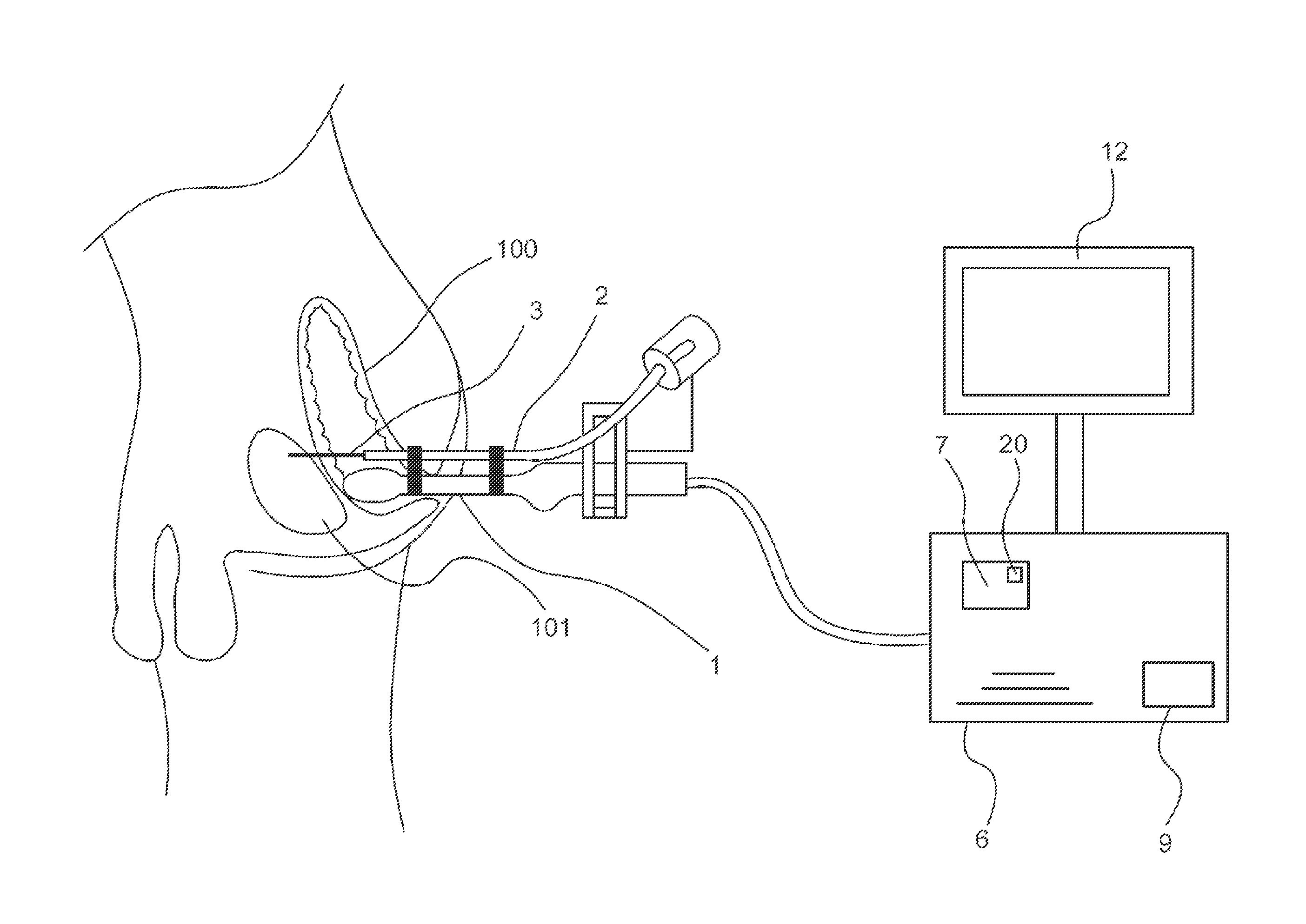
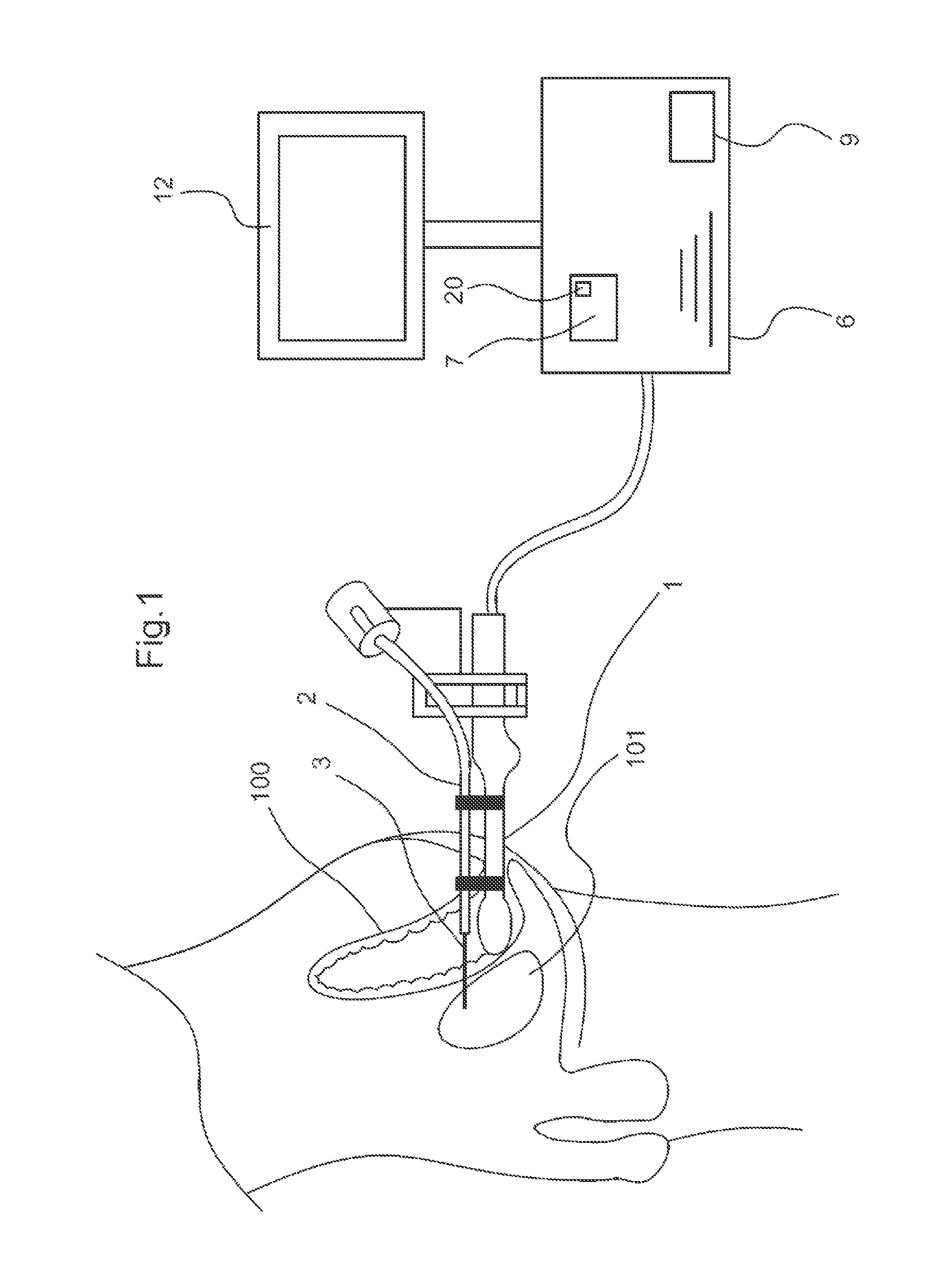
[0039]The invention is illustrated in its application to a prostate biopsy. This application is, of course, not limiting. Furthermore, the invention is illustrated in its application to a needle holder. This application is also not limiting.

[0040]Referring to FIG. 1, the guide device according to the invention comprises a medical imaging probe 1, which is illustrated as being inserted into the rectum 100 of a patient. Here, the guide device is intended to guide the probe 1 in proximity to the prostate 101 of the patient. The probe 1 comprises means for acquiring images of the prostate 101.

[0041]According to one particular embodiment, the probe 1 is rigidly connected to a medical instrument, here comprising a needle holder 2 for carrying out a biopsy of the prostate 101. The needle holder 2 holds a needle 3. Since the probe 1 is fixed to the needle holder 2, the position of the probe 1 relative to the needle holder 2 is known.

[0042]The guide device comprises a control unit 6, to whic...


PUM


Abstract

A device for guiding a medical imaging probe in order to move the probe close to an anatomical space, the probe having a mechanism for acquiring images of the anatomical space, and the device having at least one sensor mounted to the probe. The device includes a processing mechanism for deducing a rotation of the probe in the space from the signals generated by the sensor and for estimating a plurality of plausible positions of the probe relative to the anatomical space by deducing the rotation of the probe in the space. The processing mechanism is arranged such as to determine the position of the probe relative to the anatomical space from the plausible positions. Also disclosed is a method for guiding the probe.

Description

CROSS-REFERENCE TO RELATED APPLICATIONS[0001]This is a National Stage of International Application No. PCT / EP2013 / 061382 filed Jun. 3, 2013, claiming priority based on French Patent Application No. 12 55130 filed Jun. 1, 2012, the contents of all of which are incorporated herein by reference in their entirety.[0002]The invention relates to a device for guiding a medical imaging probe and to a method for guiding such a probe.BACKGROUND OF THE INVENTION[0003]So-called “open” operations may prove stressful for a patient. Thus, practitioners are resorting more and more to so-called “minimally invasive” operations, during which medical instruments are inserted through the skin (percutaneously), or into a natural passage of the patient (vagina, rectum, auditory canal, etc.), or into an artificial passage connected to the body of the patient (cannula, artificial vein, trocar, etc.). To this end, practitioners are assisted by images of the organ or organs in question, the images being taken...

Claims


Application Information

Patent Timeline
Patent Type & Authority Patents(United States)
IPC IPC(8): A61B5/05A61B17/34A61B8/00A61B8/12A61B8/08A61B17/00
CPCA61B8/4254A61B8/0841A61B8/12A61B8/4461A61B8/4488A61B8/5215A61B17/3403A61B34/20A61B2017/00274A61B2017/3405A61B2017/3413A61B2034/2048A61B2090/3784
Inventor BAUMANN, MICHAELLEROY, ANTOINE
Owner KOELIS
Why Eureka
Try Eureka