Preparation for repairing cartilage defects or cartilage/bone defects in human or animal joints

a technology for cartilage defects and human or animal joints, applied in the field of medical technology, can solve the problems of articular cartilage damage, very little vascularization, and damage to the bone tissue lying below the articular cartilage, and achieve the effect of reducing resorbability and being resistant to resorption

Inactive Publication Date: 2012-10-02
ZIMMER ORTHOBIOLOGICS
View PDF137 Cites 13 Cited by
  • Summary
  • Abstract
  • Description
  • Claims
  • Application Information

AI Technical Summary

Benefits of technology

[0023]It is an object of the invention to provide a device for repairing cartilage defects or cartilage / bone defects in human or animal joints, which device prevents the above described risk of an implanted cartilage layer to sink into the region of the native bone tissue. The device according to the invention allows production and implantation in a manner equally simple as for known devices comprising a bone part and a cartilage layer calescent with the bone part.
[0027]On the other hand the bone part of the device may be equipped with columns having a reduced resorbability. These columns extend axially through the bone part up to the subchondral bone plate. The resorbability of the columns relative to the resorbability of the bone part regions between the columns may be reduced by way of a suitable chemical treatment or by way of producing axial bores in the bone part of the device and filling these with an artificial material more resistant to resorption.

Problems solved by technology

Damage to articular cartilage by way of injuries or involution caused by aging or disease is particularly common in humans.
Very often such damage also takes its toll on the bone tissue lying below the articular cartilage.
However, it contains very little vascularization in the fully grown condition and, therefore, has a very limited regeneration capability.
Due to the described, three-phase transformation process comprising a middle phase in which the cartilage layer of the implant is not carried by the bone part capable to do so but by a mechanically inferior cyst, there exists a high risk that the cartilage layer is pressed into this cyst where it can neither fulfill its mechanical nor its biological function and from where it cannot be displaced during the following phases of the healing process.
This risk significantly reduces the chances of healing success.
Healing with a badly positioned cartilage layer causes negative after-effects.
This means that by cutting off the natural blood supply on harvesting the implant even when harvesting it from viable tissue and even when it is implanted directly after harvesting, the bone tissue loses its viability.

Method used


Image

  • Preparation for repairing cartilage defects or cartilage/bone defects in human or animal joints
  • Preparation for repairing cartilage defects or cartilage/bone defects in human or animal joints
  • Preparation for repairing cartilage defects or cartilage/bone defects in human or animal joints

Examples


Embodiment Construction

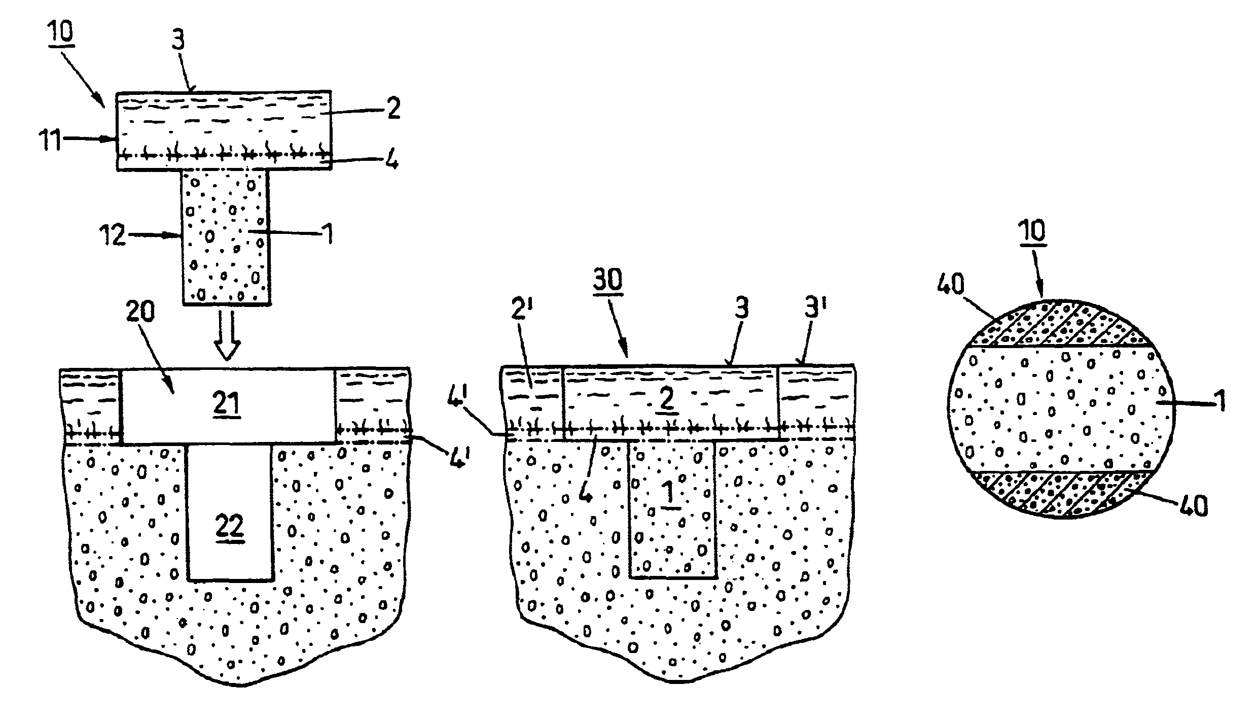
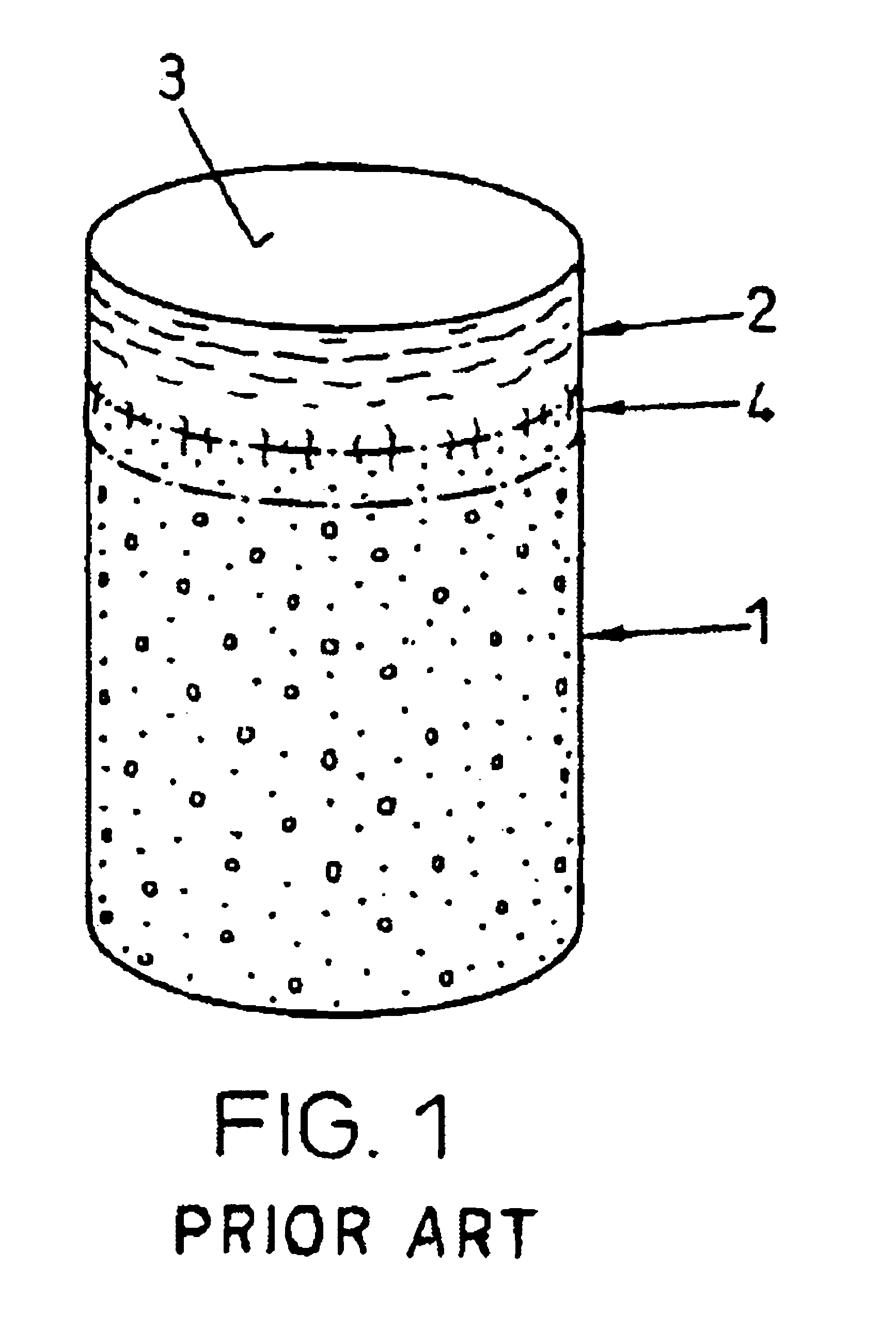
[0034]FIG. 1 shows a device as used according to the state of the art for repairing cartilage defects or cartilage / bone defects in human or animal joints. The device is cylindrical, advantageously with a circular cross section, and comprises a bone part 1 and cartilage layer 2 grown on one end face onto the bone part 1. The cartilage layer 2 forms a cartilage surface 3. Between the bone part 1 and the cartilage layer 2 there extends a subchondral bone plate 4. The transitions from the bone part 1 to the subchondral bone plate 4 and from the subchondral bone plate 4 to the cartilage layer 2 are not visible as lines, as is shown in FIG. 1 in a simplified manner, but they are natural, rather continuous transitions.

[0035]As already mentioned a device as shown in FIG. 1, is harvested from advantageously vital tissue using a hollow drill and is implanted if possible immediately after harvesting (auto-transplants and homo-transplants), or it is removed from joints of slaughtered animals (e...


PUM

PropertyMeasurementUnit
lengthsaaaaaaaaaa
diameteraaaaaaaaaa
diameteraaaaaaaaaa

Abstract

Repairs of cartilage defects or of cartilage / bone defects in human or animal joints with the help of devices including a bone part (1), a cartilage layer (2) and a subchondral bone plate (4) or an imitation of such a plate in the transition region between the cartilage layer (2) and the bone part (1). After implantation, the bone part (4) is resorbed and is replaced by reparative tissue only after being essentially totally resorbed. In a critical phase of the healing process, a mechanically inferior cyst is located in the place of the implanted bone part (1). In order to prevent the cartilage layer (2) from sinking into the cyst space during this critical phase of the healing process the device has a top part (11). and a bottom part (12), wherein the top part (11) consists essentially of the cartilage layer (2) and the subchondral bone plate (4) and the bottom part (12) corresponds essentially to the bone part (1) and wherein the top part (11) parallel to the subchondral bone plate (4) has a larger diameter than the bottom part (12). After implantation in a suitable opening or bore (20), the cartilage layer (2) and the subchondral bone plate (4) are supported not only on the bone part (1) but also on native bone tissue having a loading capacity not changing during the healing process. Therefore, the implanted cartilage layer cannot sink during the healing process.

Description

BACKGROUND OF THE INVENTION[0001]1. Field of the Invention[0002]The present invention lies in the field of medical technology and generally relates to a device for repairing cartilage defects and / or cartilage / bone defects in human or animal joints. More specifically, the device serves to repair defects in the cartilage layer, which in joints covers the bone surface, or of defects that concern this articular cartilage layer and also bone tissue lying thereunder.[0003]2. Description of Related Art[0004]Damage to articular cartilage by way of injuries or involution caused by aging or disease is particularly common in humans. Very often such damage also takes its toll on the bone tissue lying below the articular cartilage. The degree of damage to articular cartilage defects and / or cartilage / bone defects is determined with the help of the Outerbridge scale, with the following categories: superficial fraying (approx. 10% of all cases), cartilage fissure (approx. 28%), fissure down to the ...

Claims


Application Information

Patent Timeline
Patent Type & Authority Patents(United States)
IPC IPC(8): A61F2/02A61B17/00A61L27/00A61B17/16A61B17/32A61F2/00A61F2/28A61F2/30A61F2/46
CPCA61B17/1637A61B17/32053A61B2017/00969A61F2/28A61F2/30756A61F2/4601A61F2002/2839A61F2002/30032A61F2002/30062A61F2002/30113A61F2002/30143A61F2002/30153A61F2002/30154A61F2002/30156A61F2002/30172A61F2002/30224A61F2002/30233A61F2002/30299A61F2002/30327A61F2002/30733A61F2002/30751A61F2002/30759A61F2002/30808A61F2002/30929A61F2002/30971A61F2002/4631A61F2002/4649A61F2002/4685A61F2210/0004A61F2230/0006A61F2230/0017A61F2230/0019A61F2230/0021A61F2230/0023A61F2230/0052A61F2230/0069A61F2230/0093A61F2240/001A61F2250/003A61F2250/0039A61F2310/00293A61F2310/00964
Inventor NADLER, DANIELBITTMANN, PEDROAKENS, MARGARETE K.VON RECHENBERG, BRIGITTEAUER, JORG
Owner ZIMMER ORTHOBIOLOGICS
Why Eureka
Try Eureka