
Subcutaneous embedding type ventricle external drainage bursa

A ventricle and scalp technology, applied in the field of shunt devices, can solve problems such as shunt system obstruction, achieve the effect of solving increased intracranial pressure and avoiding complications of conventional shunt operations

Inactive Publication Date: 2007-12-05
傅相平 +1
View PDF0 Cites 3 Cited by
  • Summary
  • Abstract
  • Description
  • Claims
  • Application Information

AI Technical Summary

Problems solved by technology

Especially for hydrocephalus caused by cerebral hemorrhage, brain tumor and intracranial infection, because the cerebrospinal fluid contains a lot of protein, shunt system obstruction often occurs after shunt operation, even if the shunt tube is replaced repeatedly, it is difficult to completely relieve the increase of intracranial pressure in patients

Method used


Image

  • Subcutaneous embedding type ventricle external drainage bursa

Examples


Embodiment Construction

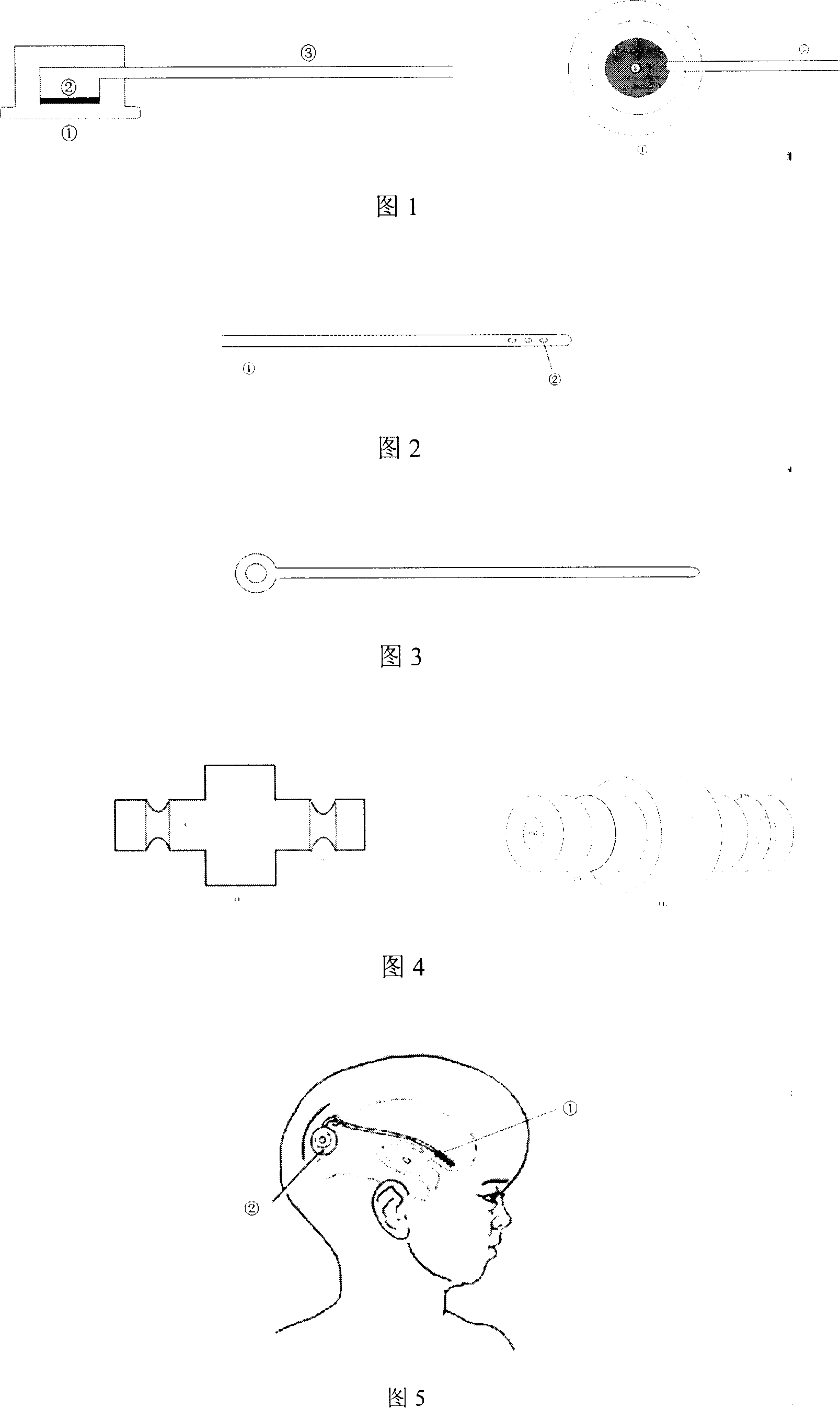
[0012] Instructions:

[0013] 1. According to different conditions, choose different ventricle puncture points.

[0014] 2. Cut the scalp, and use the mastoid retractor to separate the incision after hemostasis to expose the skull.

[0015] 3. Use a cranial drill to drill a small hole in the skull, use bipolar electric coagulation to stop bleeding and cauterize the dura mater. Use a sharp knife to open the dura mater and cauterize it with bipolar electric coagulation to form a small hole of 3×3mm.

[0016] 4. Use the guide needle to puncture the ventricle with the intracranial end of the ventricular shunt tube, control the puncture distance according to the scale on the shunt tube, until the cerebrospinal fluid flows out of the shunt tube, withdraw the guide pin, and send the shunt tube 2cm inward.

[0017] 5. Remove the mastoid retractor, embed the subcutaneous shunt sac in a suitable position under the aponeurosis next to the incision, connect the tail end of the shunt tub...


PUM

PropertyMeasurementUnit
Diameteraaaaaaaaaa
Diameteraaaaaaaaaa
Diameteraaaaaaaaaa

Abstract

The shunt device for shunting cerebrospinal fluid from lateral ventricle to vitro includes one ventricle shunting pipe, one embedded subcutaneous shunting silica gel bag, one puncture guide needle and one shunting duct interface. It is applied in shunting cerebrospinal fluid from lateral ventricle to reduce intracranial pressure, especially for brain tumor patient with hydrocephalus and increased protein content in cerebral hydrops. The shunting bag has fluent drainage of cerebrospinal fluid, and may be applied in extracting fluid, injecting medicine, flushing. It has less transfer of bacteria and tumor cell along the shunting duct, and can make up the deficiency of ventriculo-peritoneal shunt.

Description

1. Technical field [0001] The invention is a shunt device for shunting the cerebrospinal fluid of the lateral ventricle out of the body by subcutaneously embedding a shunt bag. inject medicine. 2. Background technology [0002] At present, shunts are mainly used in the treatment of hydrocephalus, including ventriculo-peritoneal shunt, ventricle-cerebellar cisterna magna shunt, ventricle-atrial shunt, among which ventriculo-peritoneal shunt is the most. Although this treatment method is simple and effective, according to statistics, about 42% of patients after shunt surgery have complications, including shunt function failure, intracranial and abdominal infection, intestinal obstruction, ascites formation, and brain tumor metastasis. Especially for hydrocephalus caused by cerebral hemorrhage, brain tumor and intracranial infection, because the cerebrospinal fluid contains a lot of protein, shunt system obstruction often occurs after shunt operation, even if the shunt tube is...

Claims


Application Information

Patent Timeline
IPC IPC(8): A61M1/00A61M39/00A61B17/00
Inventor 傅相平赵明
Owner 傅相平
Learn More