Therapeutic agent for disease with apoptotic degeneration in eye tissue cell containing PEDF and FGF2

A technique for tissue cells and diseases, applied in the field of therapeutic drugs containing PEDF and FGF2 for diseases accompanied by apoptosis and degeneration of eye tissue cells

Active Publication Date: 2008-04-09
DNAVEC CORP +1
View PDF3 Cites 10 Cited by
  • Summary
  • Abstract
  • Description
  • Claims
  • Application Information

AI Technical Summary

Problems solved by technology

[0004] At present, there is no effective treatment for retinitis pigmentosa, only symptomatic therap

Method used


Image

  • Therapeutic agent for disease with apoptotic degeneration in eye tissue cell containing PEDF and FGF2
  • Therapeutic agent for disease with apoptotic degeneration in eye tissue cell containing PEDF and FGF2
  • Therapeutic agent for disease with apoptotic degeneration in eye tissue cell containing PEDF and FGF2

Examples


Embodiment 1V

[0098] Example 1 Construction of VSV-G pseudotyped SIV vector

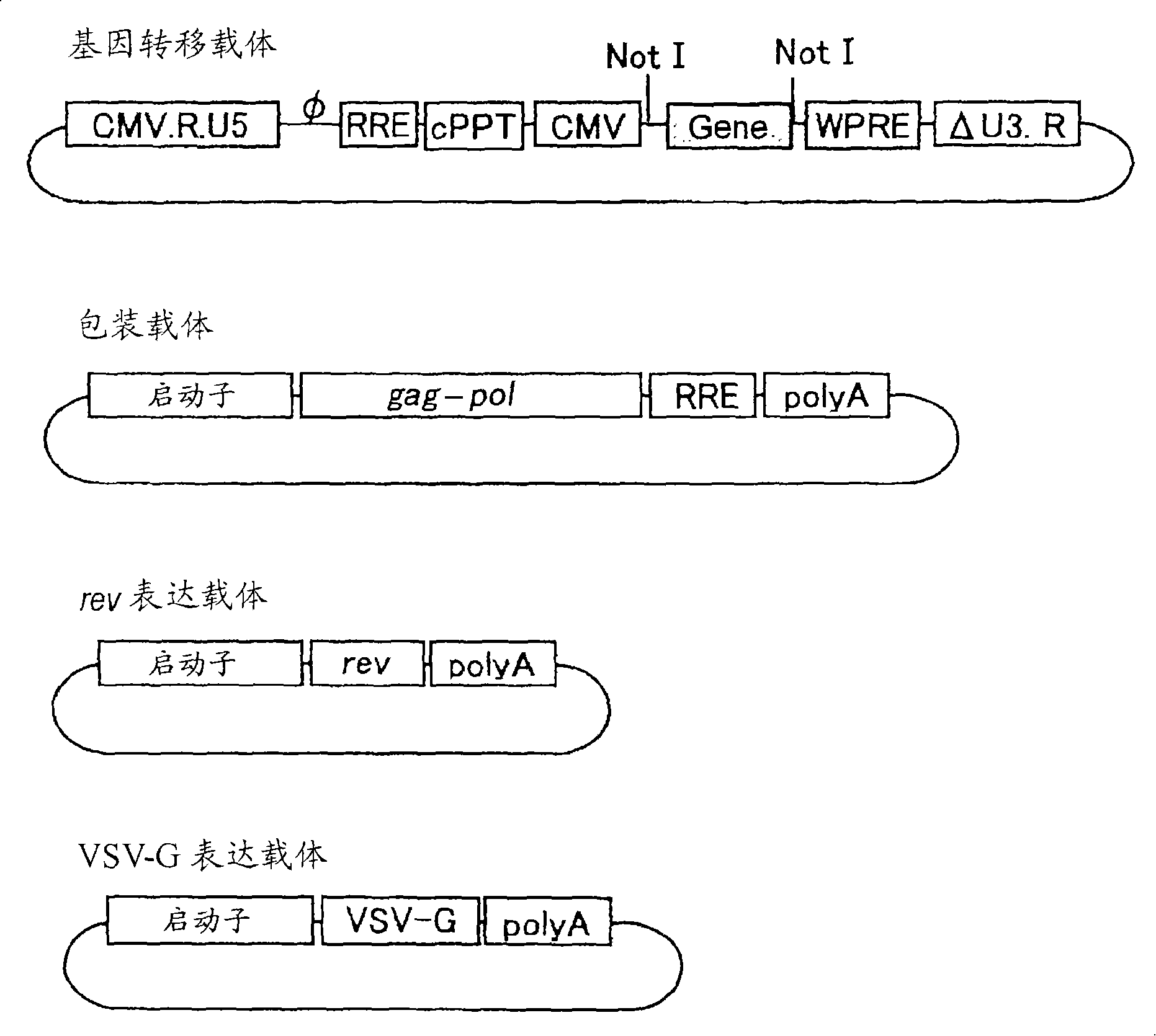
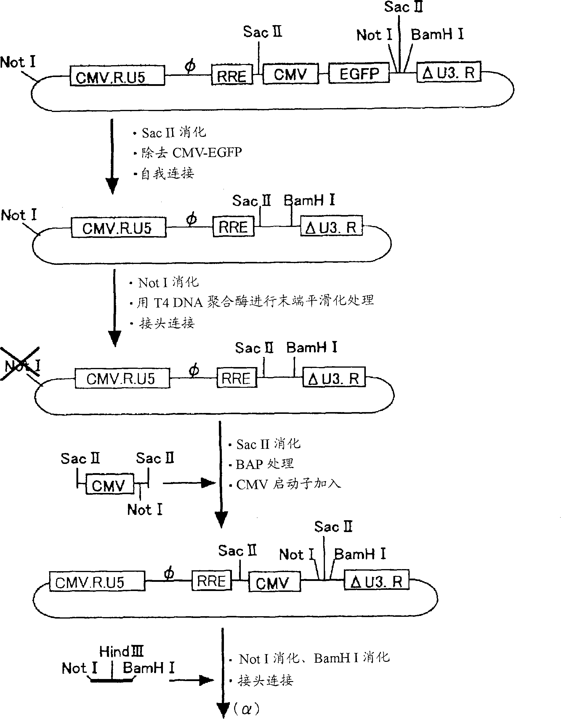
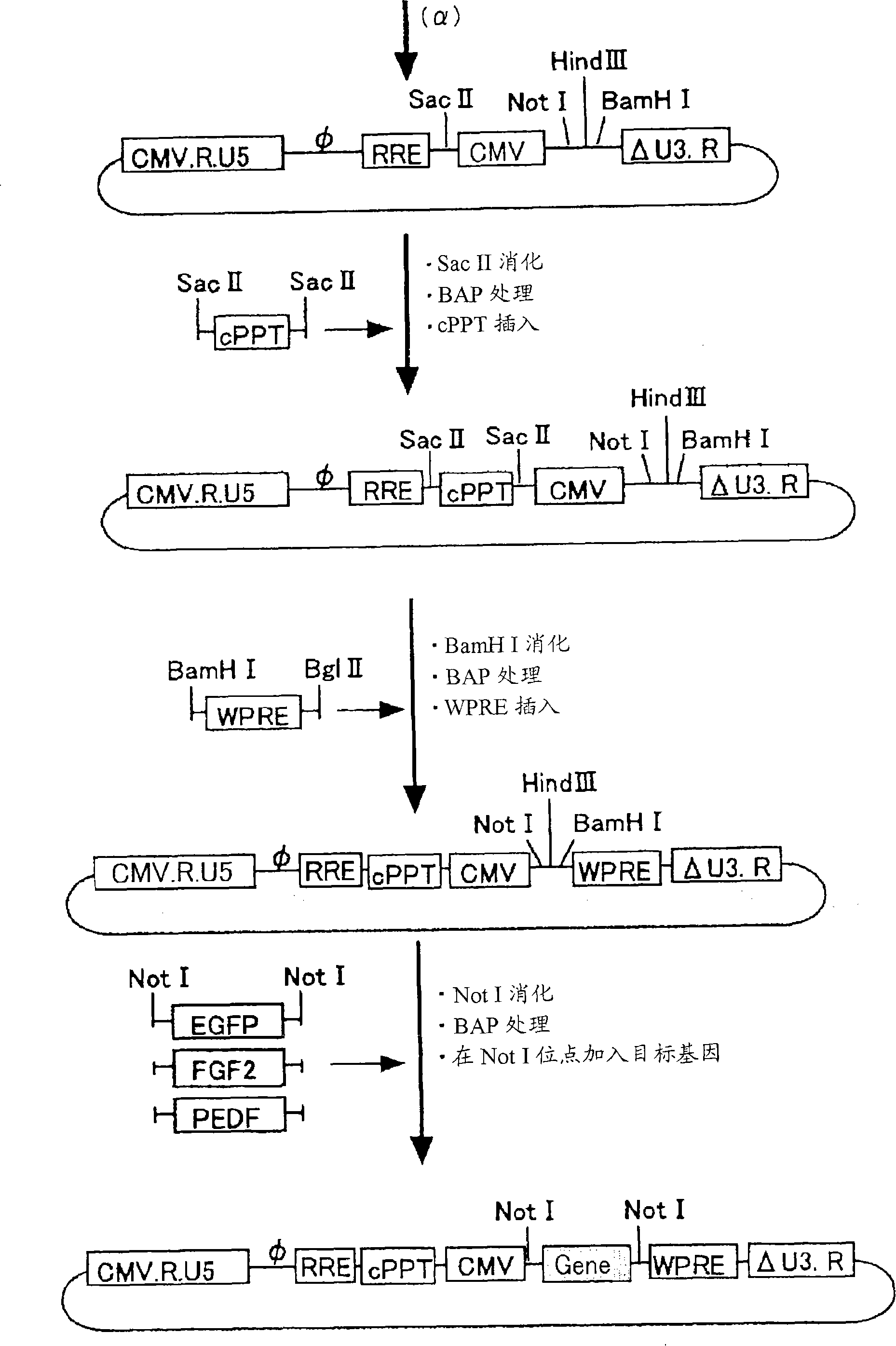
[0099] The vector was constructed using such as figure 1 The 4 plasmids shown in (gene transfer vector, packaging vector, rev expression vector, VSV-G expression vector). Among them, the gene transfer vector, the packaging vector, and the rev expression vector were prepared by modifying the original vector plasmid (PCT / JP00 / 03955). For the VSV-G expression vector, the original vector without modification was used.

[0100] For plasmid preparation, various commercially available kits were used. Restriction endonucleases were produced by New England Biolabs, and plasmid DNA was extracted, purified, and recovered using QIAGEN kits (QIAquick PCR purification kit, QIAquick Nucleotide Removal kit, QIAquick Gel extraction kit, Plasmid Maxi kit). For PCR, TaKaRa's EX Taq enzyme was used, and the primers used were synthesized by an external contractor (SIGMA GENOSYS JAPAN). Dephosphorylation of DNA ends used Takara's a...

Embodiment 2

[0118] Example 2 Functional evaluation of the SIV vector carrying cPPT, WPRE

[0119] In order to investigate the introduction effect of cPPT and WPRE, in addition to carrying cPPT and WPRE at the same time, a vector carrying cPPT alone and WPRE alone was also produced, and compared with the original control. All gene transfer vectors used carried EGFP. The original type (sequence number: 33) was used as the packaging carrier.

[0120] 2-1. Preparation of SIV vector

[0121] The cell line 293T cells from human fetal kidney cells were divided into about 1×10 per 15cm plastic culture dish 7 Inoculate (70-80% density on the next day) and culture in 20 ml of D-MEM medium (Gibco BRL) containing 10% fetal bovine serum for 24 hours. After 24 hours of culture, the medium was replaced with 10 ml of OPTI-MEM medium (Gibco BRL) and used as transfected cells for later use.

[0122] Dissolve 6 μg of gene transfer vector, 3 μg of packaging vector, and 1 μg of VSV-G expression vector in ...

Embodiment 3

[0134] Example 3 Large-scale preparation and concentration of SIV vectors carrying therapeutic genes

[0135] like figure 1 As shown, the SIV vector was prepared as follows on the basis of four kinds of plasmids: a modified gene transfer vector, a packaging vector, a rev expression vector, and a VSV-G expression vector. The vectors carrying the therapeutic genes of PEDF and FGF2 are produced in units of 20 15cm dishes.

[0136] According to each 15cm plastic Petri dish about 1×10 7 The 293T cells were inoculated (at a density of 70-80% on the next day), and cultured in 20 ml of D-MEM medium containing 10% fetal bovine serum for 24 hours. After 24 hours of cultivation, the medium was replaced with 10 ml of OPTI-MEM medium for transfection. Dissolve 10 μg of gene transfer vector, 5 μg of packaging vector, 2 μg of rev expression vector, and 2 μg of VSV-G expression vector in 1.5 ml of OPTI-MEM medium for each culture dish, then add 40 μl of PLUS Reagent reagent (Invitrogen) fo...


PUM


Abstract

It is intended to provide a novel therapeutic method for a disease with apoptotic degeneration in eye tissue cells such as retinal pigment degeneration. Attention was paid on the concomitant administration of two neurotrophic factors: pigment epithelium-derived factor (PEDF) and fibroblast growth factor 2 (FGF2). The sites of actions are considered to be different between PEDF and FGF2. An SIV-PEDF vector and an SIV-FGF2 vector were constructed and administered to the subretinal space of an RCS rat, which is a disease model of retinal pigment degeneration, and their effects were evaluated. At 4 weeks, 8 weeks and 12 weeks after the administration of the vectors, a significantly higher visual cell protection effect was obtained in a group with concomitant administration than in a group with sole administration. Further, the functional evaluation of retina was carried out by electroretinogram and an effect in the group with concomitant administration was significantly higher than in the group with sole administration. From the above results, it was found for the first time that the concomitant administration of PEDF and FGF2 has higher effects in the treatment of retinal pigment degeneration than conventional methods.

Description

technical field [0001] The invention relates to a therapeutic drug for treating diseases accompanied by apoptosis and degeneration of ocular tissue cells by using a lentiviral vector carrying a neurotrophic factor. technical background [0002] For diseases in the field of ophthalmology, inappropriate treatment can lead to severe blindness. Since there is no fundamental treatment, there are many diseases that have to rely on symptomatic therapy. Recently, it has been found that the occurrence and exacerbation of some severe diseases in the field of ophthalmology is related to apoptosis. [0003] Retinitis pigmentosa is a multiple retinal visual cell layer and pigment epithelium lesions, which can lead to apoptosis and difficult to cure hereditary disease. Visual cells are roughly divided into two types: rods and cones. Rods are mainly distributed in parts slightly deviated from the center of the retina, which are related to object discrimination under scotopic conditions ...

Claims


Application Information

Patent Timeline
IPC IPC(8): A61K48/00A61K35/76A61K38/00A61K38/22A61P9/10A61P27/02A61P27/06A61P43/00A61K38/27C12N15/09
CPCA61K38/57A61K38/1825A61P9/10A61P27/02A61P27/06A61P43/00A61K2300/00
Inventor 宫崎胜德米满吉和池田康博居石克夫田畑寿晃饭田章博上田泰次长谷川护
Owner DNAVEC CORP
Why Eureka
Try Eureka