
Method and device for fundus oculi affection early diagnosis using time discrimination autofluorescence lifetime imaging

An autofluorescence and fluorescence lifetime technology, applied in diagnosis, ophthalmoscope, eye testing equipment, etc., can solve the problems such as subtle changes that cannot be clearly observed, changes in the shape of fundus tissue are not obvious, and the magnification of fundus images is limited. Achieve the effect of strong single photon counting ability, fast time response and high sensitivity detection

Inactive Publication Date: 2008-05-21
SHENZHEN UNIV
View PDF0 Cites 23 Cited by
  • Summary
  • Abstract
  • Description
  • Claims
  • Application Information

AI Technical Summary

Problems solved by technology

However, due to the limited magnification of the fundus image, the subtle changes in the early stage of the disease cannot be observed clearly, and in the early stage of the disease, the changes in the shape of the fundus tissue are often not obvious or difficult to measure

Method used


Image

  • Method and device for fundus oculi affection early diagnosis using time discrimination autofluorescence lifetime imaging
  • Method and device for fundus oculi affection early diagnosis using time discrimination autofluorescence lifetime imaging
  • Method and device for fundus oculi affection early diagnosis using time discrimination autofluorescence lifetime imaging

Examples


Embodiment Construction

[0025] The implementation of the technical solution will be further described in detail below in conjunction with the accompanying drawings.

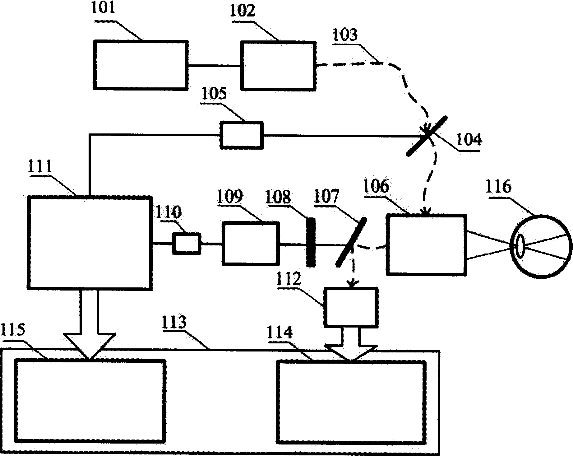
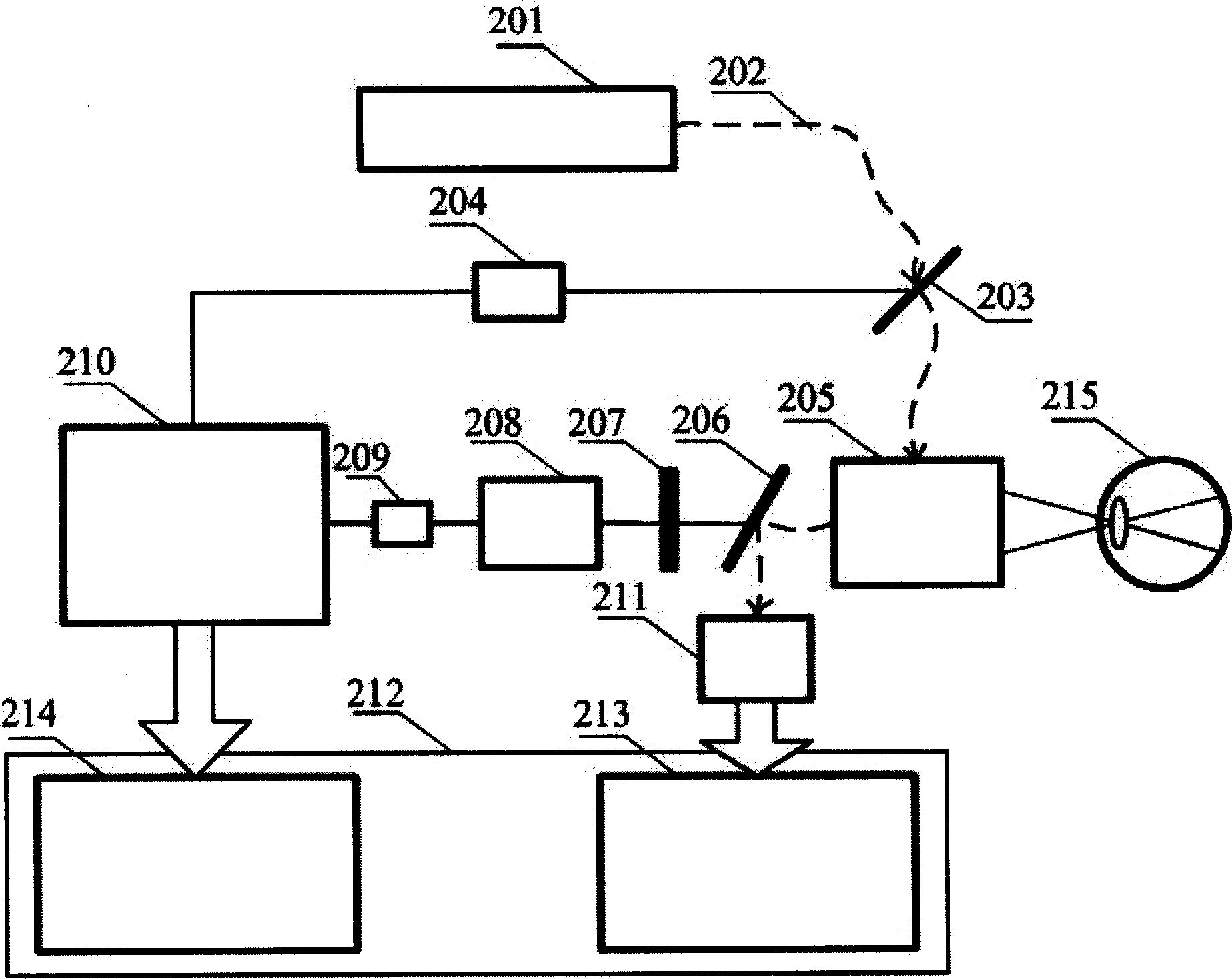
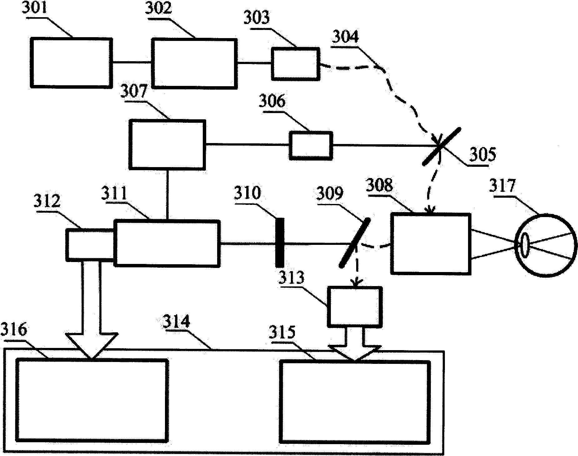
[0026] figure 1 Among them, the near-infrared ultrashort pulse output by a high repetition rate ultrashort pulse laser such as a titanium sapphire mode-locked femtosecond laser 101 is divided into two beams after passing through a frequency doubler 102 and a beam splitter 104, one of which is The energy is relatively low, and after being detected by the photodiode 105 , it is input into the time-correlated single photon counter 111 . This beam of synchronous signal triggers the time-to-amplitude converter inside 111 in the inverted pulse reference mode, and generates a high level as the start of timing. The other beam of light is the main beam, which is input to the scanning head of the laser scanning confocal ophthalmoscope 106 through the optical fiber 103, and is focused on a single point in the fundus to complete the scanning of ea...


PUM


Abstract

The invention relates to a novel measurement method and the device thereof, which combines the time-resolved self-fluorescence lifetime imaging technology and a laser scanning confocal ophthalmoscopeand realizes the early diagnosis of the ocular fundus diseases by obtaining and analyzing the self-fluorescence intensity and the service life information of the ocular fundus. The light which is emitted by a pulse laser is sent to the scanning head of the laser scanning confocal ophthalmoscope to excite the ocular fundus inner-source flgorgen, and the fluorescence which is emitted from the ocularfundus is collected by a time-correlated single photon counter or a high-repetition-rate synchronous scanning camera, so as to realize the service life measurement and the imaging of the self-fluorescence. As the service life of the self-fluorescence of the ocular fundus is very sensitive to the micro-environment and the cell metabolism state of flgorgen molecule and is not affected by the difference of the intensity of the flgorgen of the ocular fundus, the measurement of the fluorescence service life provides a brand new route for the detection of ocular fundus diseases and the obtainment of the function information of ocular fundus. The invention can become the solution and the basis of the early diagnosis of various ocular fundus diseases, which plays an important role in pathogeny research, clinical diagnosis, staging and typing, prognosis judgement, real-time observation and the analysis of the development of ocular fundus diseases, therefore the invention can be widely appliedin the field of medical research and clinical medical diagnosis.

Description

technical field [0001] The invention relates to a method and a device for realizing early diagnosis of fundus diseases by measuring and analyzing the time-resolved autofluorescent lifetime change of the fundus. Modern ophthalmology has confirmed that autofluorescence lifetime is very sensitive to the type of fundus fluorophore and the metabolic state of retinal pigment epithelial cells. The measurement and imaging can be used as an important basis for judging whether there are fundus diseases. The present invention aims to realize the acquisition of fundus time-resolved autofluorescence lifetime and its spatial distribution information, which will provide important functional information for ophthalmic medical detection and analysis, and has important application value in the field of medical research and clinical medical diagnosis. Background technique [0002] There are a large number of endogenous fluorophores in the human fundus, which can be excited by light of a certa...

Claims


Application Information

Patent Timeline
IPC IPC(8): A61B3/10A61B3/00
CPCA61B3/1233A61B3/1025
Inventor 屈军乐孙怡雯许改霞赵羚伶牛憨笨
Owner SHENZHEN UNIV
Learn More