Mass spectrum reagent kit and method for detecting and prognosis judging CEA negative gastric cancer

A detection kit and the technology of the kit, which are applied in the field of protein detection, can solve the problems of lack of early monitoring methods in the prognosis evaluation of CEA-negative gastric cancer patients, etc.

Inactive Publication Date: 2008-09-24
许洋
View PDF2 Cites 3 Cited by
  • Summary
  • Abstract
  • Description
  • Claims
  • Application Information

AI Technical Summary

Problems solved by technology

[0004] The traditional tumor marker CEA is of great value in evaluating the prognosis of gastric cancer, but it is also found in clinical work that a considerable proportion of gastric cancer patients cannot be detected due to the negative expression of CEA. At the same time, there is a lack of effective early monitoring methods for the prognosis assessment of CEA-negative gastric cancer patients
When we performed protein mass spectrometry analysis, we found that there was no standardized kit for mass spectrometry of clinical CEA-negative gastric cancer

Method used


Image

  • Mass spectrum reagent kit and method for detecting and prognosis judging CEA negative gastric cancer
  • Mass spectrum reagent kit and method for detecting and prognosis judging CEA negative gastric cancer
  • Mass spectrum reagent kit and method for detecting and prognosis judging CEA negative gastric cancer

Examples


Embodiment 1

[0078] Example 1 Distinguishing between normal and CEA-negative gastric cancer patients and preparation of a mass spectrometry kit

[0079] (1) Experimental method

[0080] The preoperative sera of 280 patients with CEA-negative gastric cancer were collected, with an average age of 47 years. 280 healthy subjects, with an average age of 45 years old, were from a population with normal liver and kidney function tests. Collect 1mL of venous blood from the subject on an empty stomach, immediately after collection, let it stand in a refrigerator at 4°C for 2 hours, centrifuge at 4,000r / min at 4°C for 10 minutes to separate the serum, and centrifuge the serum again at 12,000r / min at 4°C for 5 minutes to remove all residual cell debris and insoluble matter, the serum was divided into 100 μL / tubes on ice, a total of 5 tubes, and stored in a -80°C refrigerator. Avoid repeated freeze-thaw cycles.

[0081] Protein chip and magnetic beads operation steps

[0082] Serum sample processi...

Embodiment 2

[0095] The double-blind test of embodiment 2 kit

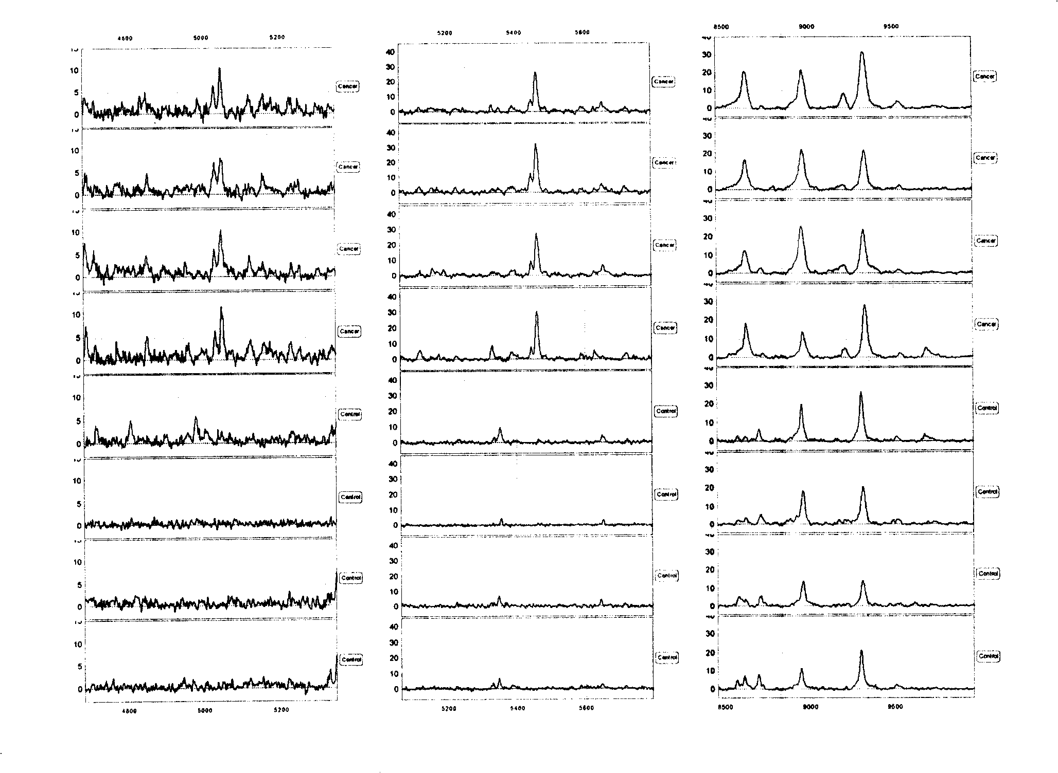
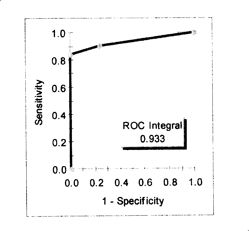
[0096] Screen out several characteristic protein peaks from embodiment 1, the test model that 5047,5461,8628 ± 15Da 3 difference peaks form ( figure 1 The top four cases are gastric cancer patients, and the bottom four cases are healthy people) Blind screening test and ROC curve were used to analyze the mass spectrometry results of 71 CEA negative gastric cancer samples with CEA negative gastric cancer patients and healthy people, and 64 of them were correctly distinguished , 7 were misclassified, and the sensitivity was 90%; 69 of 71 control samples were correctly distinguished, 2 were misclassified, and the specificity was 97% (see Table 2, figure 2 ):

[0097] Table 2: Discrimination of test models in biological samples

[0098]

[0099] Note: Sensitivity 90% (64 / 71); Specificity 97% (69 / 71)

[0100] The experimental results using C8 and C18 hydrophobic matrix magnetic beads or chips are consistent with the experime...

Embodiment 3

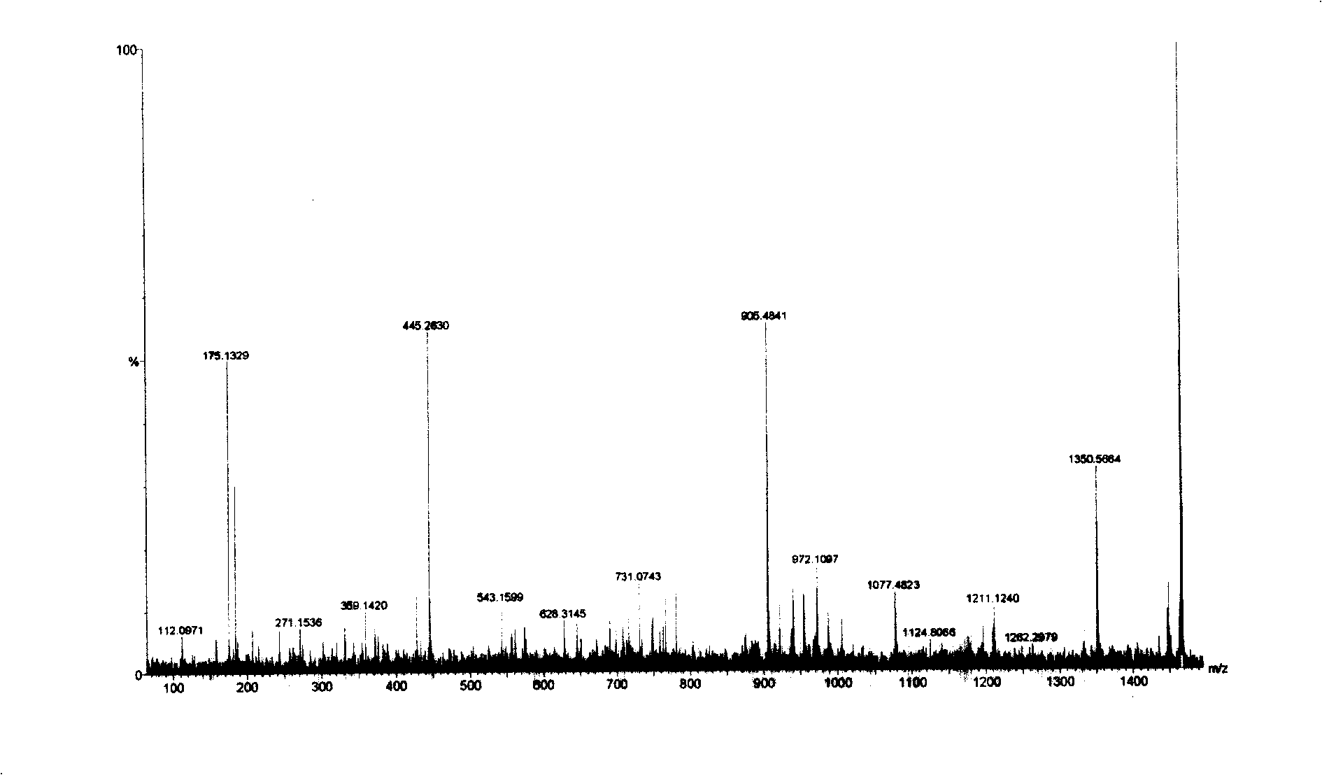
[0101] Example 3 Sequencing and identification of 1465.6Da protein

[0102] The 1465.6Da biomarkers were sequenced using multiple-stage mass spectrometry (MS / MS), post-source fragmentation (PSD) and protein ladder sequencing. By breaking molecules into pieces, protein ladders can be generated. This gradient is then analyzed by mass spectrometry. The 1465±1Da protein was identified as a variant fibrinopeptide A (Fibrinopeptide A with alanine truncation at N-terminal) ( image 3 ). Its chemical structure is (15 amino acids arranged from N-terminal to C-terminal):

[0103] N-terminal Asp-Ser-Gly-Glu-Gly-Asp-Phe-Leu-Ala-Glu-Gly-Gly-Gly-Gly-Val-Arg C-terminal.

[0104] Check the chemical structure of the normal fibrinopeptide A molecule in the known genome or cDNA library database (16 amino acids arranged from the N-terminal to the C-terminal, the molecular weight is 1536Da):

[0105] N-terminal Ala-Asp-Ser-Gly-Glu-Gly-Asp-Phe-Leu-Ala-Glu-Gly-Gly-Gly-Gly-Val-Arg C-terminal.


PUM

PropertyMeasurementUnit
molecular weightaaaaaaaaaa
Sensitivityaaaaaaaaaa

Abstract

The invention relates to a method of a magnetic bead support matrix used for capturing gastric cancer proteome in an in vitro biological sample, by a magnetic separator separating the magnetic bead and the sample which does not need to be centrifuged. Then, the mass spectrometric method is used for carrying out an analysis of the protein fingerprint technique. The method of the invention can be used in a kit of protein fingerprint or mass spectrum peptide atlas for the in vitro sample detection and the prognosis judgment of normal people and people with negative CEA gastric cancer. The method is accurate, convenient and rapid.

Description

technical field [0001] The invention relates to a new reagent kit for a protein analysis method in a CEA negative gastric cancer biological sample. One captures biomarkers via protein-binding substrates and detects CEA-negative gastric cancer biomarkers using quantitatively controlled mass spectrometry. The invention mentioned here relates to the field of protein detection and is a new non-invasive in vitro mass spectrometry detection method. The present invention can be applied to the detection method or kit of the CEA-negative gastric cancer biomarker combination in the body fluid that has been separated from the human body. Background technique [0002] Both normal function and pathological characteristics of cells depend to some extent on the function of proteins expressed by cells. Therefore, the identification of differences in proteins expressed in the human body can be used for diagnosis and screening of disease samples in vitro, and ultimately for drug development...

Claims


Application Information

Patent Timeline
Patent Type & Authority Applications(China)
IPC IPC(8): G01N30/02G01N30/86
Inventor 许洋
Owner 许洋
Why Eureka
Try Eureka