Mass spectrum kit and method for evaluating prognosis from screening lung cancer

A technology of kits and detection kits, applied in the field of protein detection, can solve problems such as difficult to accurately measure the growth rate of small nodules, difficult to detect early lung cancer, correct staging of lung cancer, and unevaluated lung cancer mortality.

Inactive Publication Date: 2007-12-26
GENERAL HOSPITAL OF TIANJIN MEDICAL UNIV +1
View PDF0 Cites 12 Cited by
  • Summary
  • Abstract
  • Description
  • Claims
  • Application Information

AI Technical Summary

Problems solved by technology

In the 1950s, four non-randomized and non-controlled screening studies of "Philadelphia Pulmonary Neoplasm Research Project", "Veterans Administration Trial", "Tokyo Metropolitan Government Study" and "South London Lung Cancer Study" showed that chest radiography for lung cancer Screening, survival improved, but lung cancer mortality not assessed
[0017] Although LDCT is one of the most promising tools for lung cancer screening, there are still some prob

Method used


Image

  • Mass spectrum kit and method for evaluating prognosis from screening lung cancer
  • Mass spectrum kit and method for evaluating prognosis from screening lung cancer

Examples


Embodiment 1

[0099] Example 1 The distinction between normal and lung cancer patients and the preparation of mass spectrometry kits

[0100] (1) Experimental method

[0101] The preoperative serum samples of 262 patients with lung cancer were collected, with an average age of 65 years. A total of 262 healthy subjects, with an average age of 63 years, were obtained from a population with normal liver and kidney function tests. Collect 1mL of venous blood from the subject on an empty stomach, immediately after collection, let it stand in the refrigerator at 4°C for 2 hours, centrifuge at 4000r / min at 4°C for 10 minutes to separate the serum, and centrifuge the serum again at 12000r / min at 4°C for 5 minutes to remove all residual cell debris and insoluble matter, the serum was divided into 100 μL / tubes on ice, a total of 5 tubes, and stored in a -80°C refrigerator. Avoid repeated freeze-thaw cycles.

[0102] Protein chip and magnetic beads operation steps

[0103]Serum sample processing: ...

Embodiment 2

[0115] Double-blind test of embodiment 2 kit (early detection and staging of lung cancer)

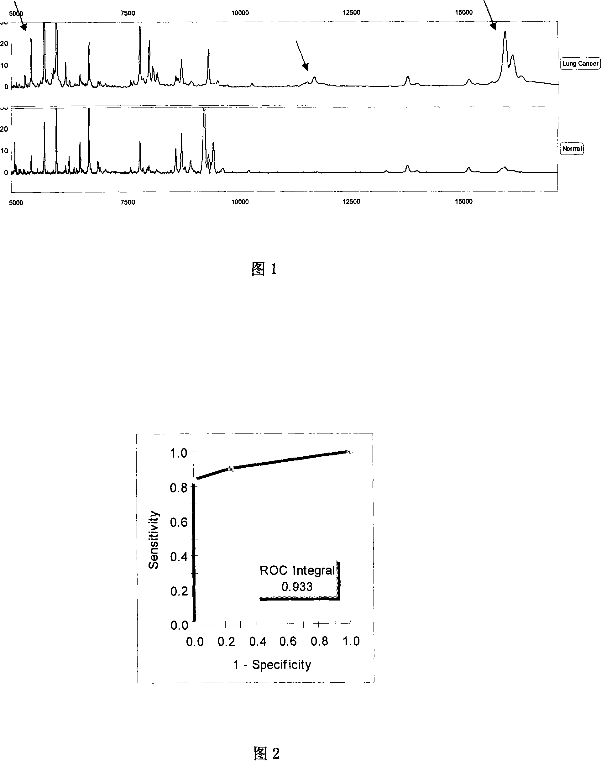
[0116] Screen out several characteristic protein peaks from embodiment 1, the test model (Fig. 1) that 2538, 5335, 3286, 15938 ± 15Da 4 difference peaks forms is to lung cancer patient and healthy crowd blind screening test and ROC curve, use this Classification method was used to analyze the mass spectrometry results of 71 lung cancer samples (11 cases of stage I lung cancer; 23 cases of stage II lung cancer; 26 cases of stage III lung cancer; 11 cases of stage IV lung cancer), of which 68 samples were correctly distinguished and 3 samples were wrongly distinguished. The sensitivity was 95.8%; 69 of 71 control samples were correctly distinguished, 2 were wrongly distinguished, and the specificity was 97.2% (see Table 2, Figure 2):

[0117] Table 2: Discrimination of test models in biological samples

[0118] group

lung cancer

healthy person

total

lung canc...

Embodiment 3

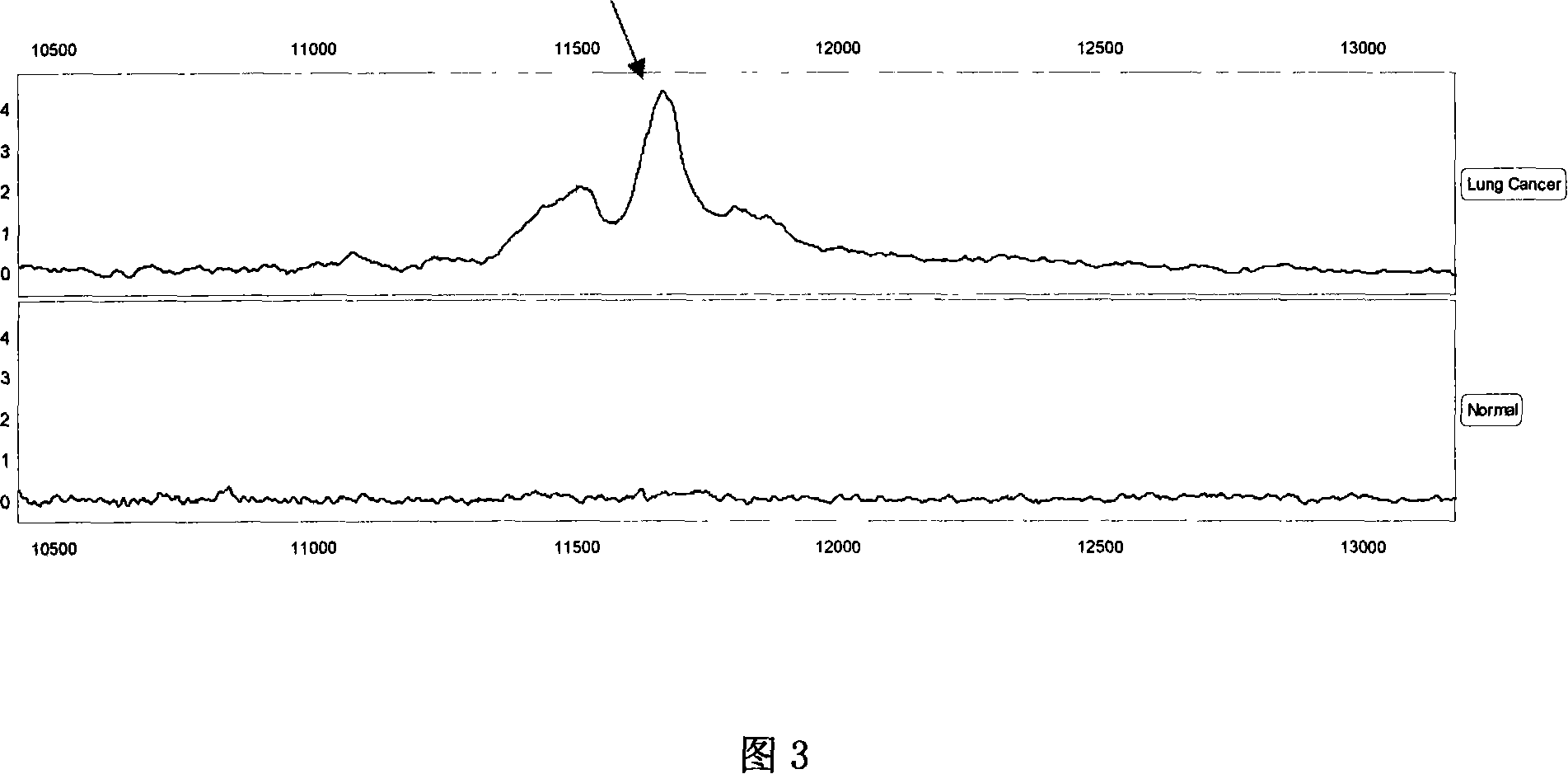
[0122] Example 3 Sequencing and identification of 11683.2 Da protein

[0123] The 11683.2 biomarkers were sequenced using multiple-stage mass spectrometry (MS / MS), post-source fragmentation (PSD) and protein ladder sequencing. By breaking molecules into pieces, protein ladders can be generated. This gradient is then analyzed by mass spectrometry. The 11683.2 Da protein was identified as variant serum amyloid A. Its chemical structure is (amino acids arranged from N-terminal to C-terminal):

[0124] N-terminal

[0125] RSFFSFLGEAFDGARDMWRAYSDMREANYIGSDKYFHARGNYDAAKRGPG

[0126] GVWAAEAISDARENIQRFFGHGAEDSLADQAANEWGRSGKDPNHFRPAGL

[0127] PEKY

[0128] C-terminal.


PUM


Abstract

A method for screening and prognosis-estimating lung cancer includes catching lung cancer protein set in external biological sample by utilizing means of using magnetic bead to support substrate, using magnetic separator to separate magnetic bead and sample then using mass spectrography to carry out analysis. The mass spectrum kit used for screening and prognosis-estimating lung cancer is also disclosed.

Description

technical field [0001] The invention relates to a new kit for analyzing protein in biological samples of lung cancer. A method to capture biomarkers via a protein-binding matrix and detect lung cancer biomarkers using quantitatively controlled mass spectrometry. The invention mentioned here relates to the field of protein detection and is a new non-invasive in vitro mass spectrometry detection method. The present invention can be applied to the detection method or kit of the combination of lung cancer biomarkers in the body fluid that has been separated from the human body. Background technique [0002] Both normal function and pathological characteristics of cells depend to some extent on the function of proteins expressed by cells. Therefore, the identification of differences in proteins expressed in the human body can be used for diagnosis and screening of disease samples in vitro, and ultimately for drug development and disease treatment. However, differential analysi...

Claims


Application Information

Patent Timeline
IPC IPC(8): G01N30/02G01N30/86
Inventor 许洋周清华
Owner GENERAL HOSPITAL OF TIANJIN MEDICAL UNIV
Why Eureka
Try Eureka