
Drug eluting vascular closure devices and methods

A blood vessel and blood vessel wall technology, applied in the field of drug-eluting blood vessel closure devices for hemostasis at blood vessel puncture points, can solve problems such as serious complications, substances entering the bloodstream, dependence, etc., and achieve the effects of complete hemostasis and reduction of bleeding

Active Publication Date: 2008-12-17
CARDIVA MEDICAL
View PDF39 Cites 1 Cited by
  • Summary
  • Abstract
  • Description
  • Claims
  • Application Information

AI Technical Summary

Problems solved by technology

This device has several disadvantages such as causing serious complications
For example, occlusive elements may not be sufficient to prevent entry of these highly thrombogenic materials into blood vessels
Also, injection of the mixture is often poorly controlled and highly technique-dependent, which in turn may allow these substances to enter the bloodstream

Method used


Image

  • Drug eluting vascular closure devices and methods
  • Drug eluting vascular closure devices and methods
  • Drug eluting vascular closure devices and methods

Examples


Embodiment Construction

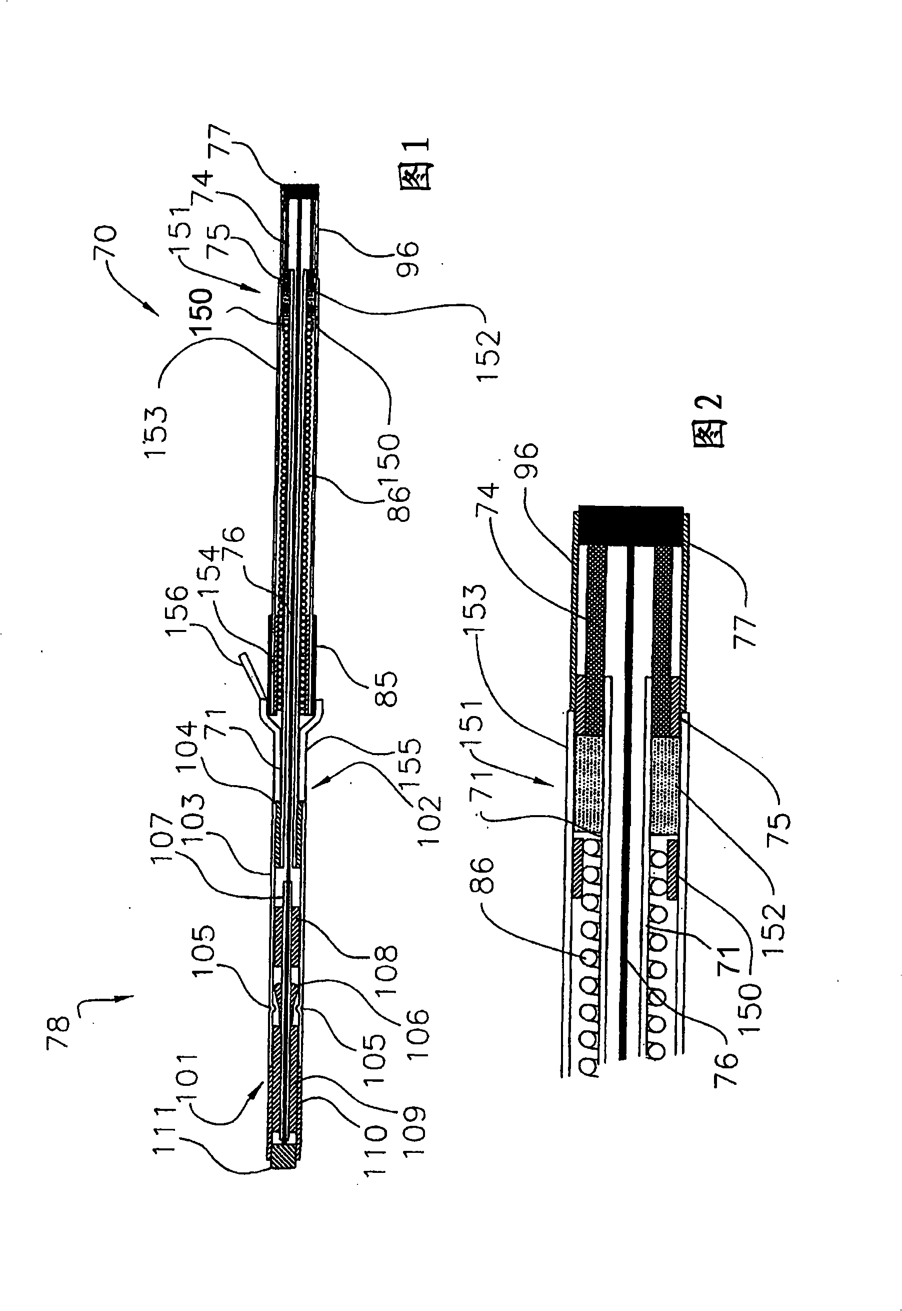
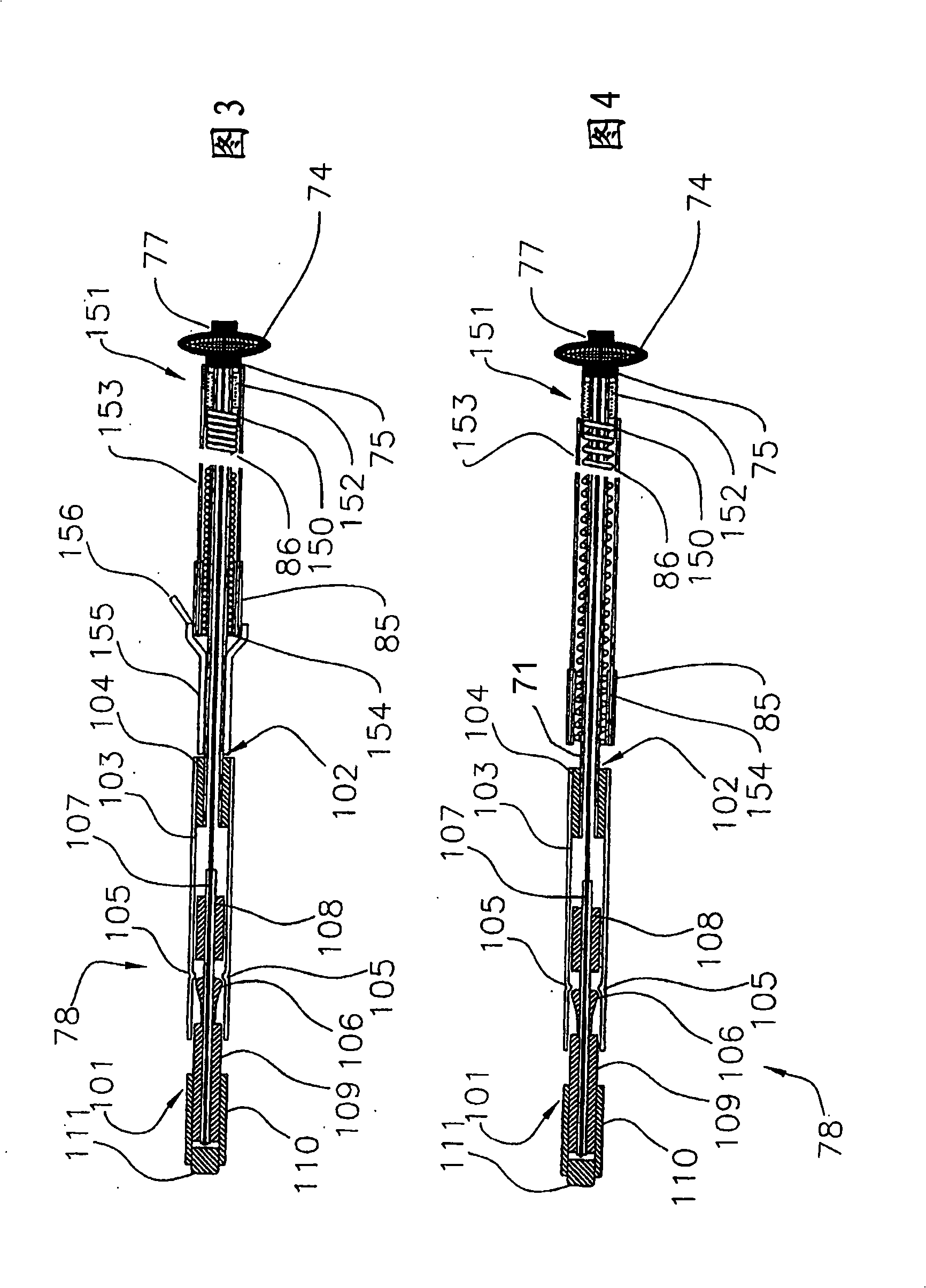
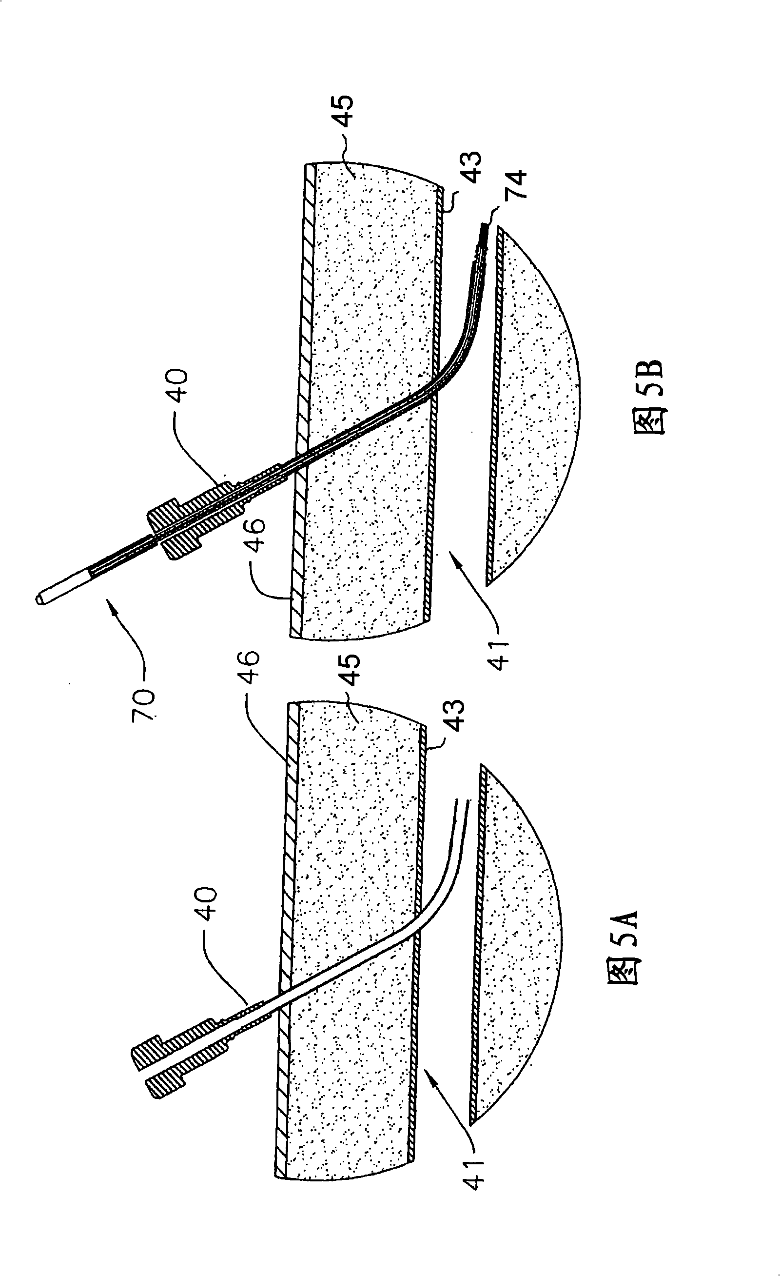
[0037] Referring now to FIG. 1 , there is shown a first embodiment of a drug-eluting, self-tensioning vaso-occlusive device 70 for hemostasis at a vascular puncture site, wherein at least one biochemical agent 152 is in a biochemical region or chamber 151 in combination with device as a whole. The device 70 typically consists of stainless steel coiled tubing or, for example, nylon, polyurethane, polyimide, PEEK 、PEBAX A first flexible elongated tubular element 71 formed of a polymer material such as the like. Tubular member 71 may have a length ranging from about 5 cm to about 50 cm, typically from about 10 cm to about 30 cm, and a diameter ranging from about 0.25 mm to about 5 mm, typically from about 0.5 mm to about 2 mm. An expandable occlusive element 74 is placed on the distal end of the tubular element 71 . Biochemical sealing element 153 is slidably positioned over tubular element 71 and adjacent expandable element 74 . A biochemical region 151 containing a bioche...


PUM


Abstract

Drug eluting vascular closure devices and methods for closing a blood vessel puncture site disposed at a distal end of a tissue tract are described. The devices and methods rely on a combination of the body's own natural mechanism to achieve hemostasis with bio-chemical agents to accelerate the hemostatic process. One method includes the steps of introducing a closure device through the tissue tract and deploying an expansible member at a distal end of the device within the blood vessel to occlude the puncture site. A bio-chemical sealing member disposed proximal the expansible member is then displaced so as to expose a bio-chemical region or release region of the device. At least one bio-chemical agent is thereafter released from the device and into the tissue tract to accelerate the occlusion process in the tract.

Description

technical field [0001] The present invention generally relates to devices and methods for percutaneous sealing of puncture points in body lumens or tissue tracts. More particularly, the present invention relates to drug-eluting vessel closure devices and methods for hemostasis at vessel puncture sites. Background technique [0002] Typically performed for diagnostic or interventional procedures such as coronary and peripheral angiography, angioplasty, atherectomy, vasodilation, coronary retrograde perfusion and infusion, cerebral angiography, percussion therapy, cerebral aneurysm Transcutaneous access to human blood vessels. Patients undergoing these procedures are often treated with anticoagulants, such as heparin, thrombolytics, etc., which make sealing and hemostasis of the puncture site in the vessel wall upon completion of such interventional procedures more difficult to achieve. [0003] Various devices have been introduced to provide hemostasis, however none have be...

Claims


Application Information

Patent Timeline
Patent Type & Authority Applications(China)
IPC IPC(8): A61F13/00
CPCA61B17/00491A61B17/0057A61B2017/00893A61B2017/3484A61B2090/032A61M29/00A61M25/00A61F13/00
Inventor Z·亚辛扎德
Owner CARDIVA MEDICAL
Learn More