Eureka AIR delivers breakthrough ideas for toughest innovation challenges, trusted by R&D personnel around the world.

Vascular endothelial cells, and preparation and use thereof

A vascular endothelial and cell technology, applied in the field of vascular endothelial cells and their preparation, can solve the problems of easy damage to endothelial cells and difficulty in controlling strength, and achieve the effect of vigorous proliferation in vitro, high purity, and simple operation

Inactive Publication Date: 2009-01-28
INST OF BASIC MEDICAL SCI ACAD OF MILITARY MEDICAL SCI OF PLA
View PDF0 Cites 10 Cited by
  • Summary
  • Abstract
  • Description
  • Claims
  • Application Information

AI Technical Summary

Problems solved by technology

The mechanical scraping method is to cut open the umbilical vein, scrape the endothelial cells with the back of a scalpel, etc. or use a separation tube with a nylon mesh, but this method is difficult to control the strength, easily damages the endothelial cells, and is often mixed with other cells such as adult cells. Fibroblasts, etc.

Method used


Image

  • Vascular endothelial cells, and preparation and use thereof
  • Vascular endothelial cells, and preparation and use thereof
  • Vascular endothelial cells, and preparation and use thereof

Examples


Embodiment 1

[0030] Embodiment 1, separation and cultivation of endothelial cells

[0031] In this experiment, endothelial cells were isolated from fresh umbilical cord veins of healthy full-term neonates. When operating, first cut off the fresh umbilical cord of the newborn.

[0032] The raw materials used in this experiment are as follows: type II collagenase (Gibco), IMDM medium (Gibco, Cat.No.12200-036)), fetal bovine serum (Hyclone), horse serum (Gibco), bovine serum albumin BSA (Fluka), vascular endothelial growth factor VEGF (Prepro Tech), basic fibroblast growth factor bFGF (Prepro Tech), heparin sodium (Shanghai Sinopharm Chemical Reagent Co., Ltd.).

[0033] Type II collagenase solution is prepared by dissolving type II collagenase dry powder in IMDM medium.

[0034] 1. Isolation and cultivation under condition I

[0035] The condition I is as follows: the initial concentration of the type II collagenase solution used is 342U / ml, digested for 10 minutes; the cell culture condi...

Embodiment 2

[0052] Embodiment 2, identification of endothelial cells

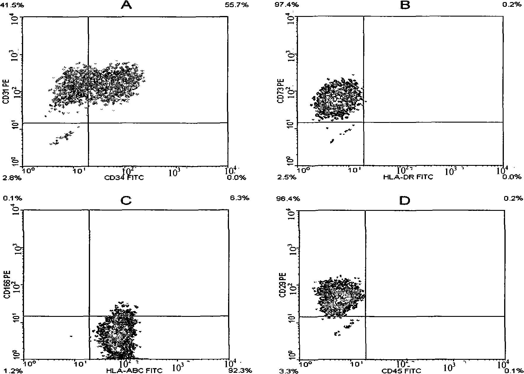
[0053] Take the cells obtained from the primary generation to the 10th passage in Example 1, the cells obtained from the primary generation to the 8th passage in Example 1 two, and the cells of each generation obtained from the primary generation to the 9th passage in Example 1 3, respectively. The following identification experiments.

[0054] 1. Observation of cell morphology

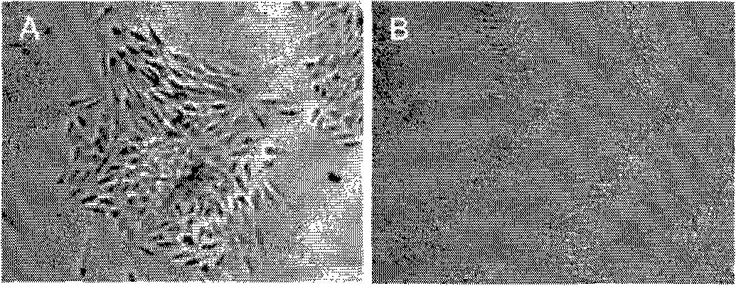
[0055] During the growth process of each endothelial cell, several clones were formed in the 24-well plate, and the cells were in the shape of a spindle. When the cells covered the 24-well plate, the cells became thicker and shorter, and were in the shape of pebbles, such as obtained in Example 1-1. The morphology of the primary cells is as figure 1 as shown ( figure 1 A is fusiform, figure 1 B is pebble-like). It shows that, from the point of view of morphology, the cells obtained in the present invention conform to the morphological cha...


PUM


Abstract

The invention discloses a vascular endothelial cell and a preparation method and application thereof; the method for separating the vascular endothelial cell disclosed by the invention comprises the following steps: II type collagenase is used for digesting umbilical vein of umbilical cord in vitro, cells are collected, then culture is carried out and adherent cells are obtained, namely the vascular endothelial cell. The separation method of the invention has simple operation and convenience and practicability; the obtained vascular endothelial cell has high purity, large amount, strong exogenic propagation and a plurality of times of passage, and not only has the functions of phagotrophy and tube formation, but also has the function of inhibiting progenitor cells to develop into dendritic cells; therefore, the invention establishes a stable technical system for the separation and culture of the vascular endothelial cell and lays the foundation for the study and the application of the endothelial cell.

Description

technical field [0001] The invention relates to a vascular endothelial cell and its preparation method and application. Background technique [0002] Vascular endothelial cells not only participate in the regulation of vascular permeability and blood coagulation, but also play an important role in many processes such as immune regulation, transplant rejection, and tumor metastasis. In these research applications, the isolation and in vitro culture of endothelial cells are critical. [0003] Currently, endothelial cells are mostly isolated from human umbilical cord veins. However, the obtained human umbilical vein endothelial cells have poor growth ability in vitro, slow proliferation, primary culture is not easy to succeed, and subculture can only be passed on for 3-4 generations. [0004] Endothelial cells are mostly obtained by mechanical scraping and enzymatic digestion. The mechanical scraping method is to cut open the umbilical vein, scrape the endothelial cells with...

Claims


Application Information

Patent Timeline
IPC IPC(8): C12N5/08C12N5/071
Inventor 毛宁江小霞刘元林张毅朱恒苏永锋李秀森
Owner INST OF BASIC MEDICAL SCI ACAD OF MILITARY MEDICAL SCI OF PLA
Why Eureka
Eureka Blog
Learn More