Treatment of peripheral vascular disease using postpartum-derived cells
A peripheral vascular disease, cell-based technology for cell-based or regenerative therapy that addresses issues such as insufficient cord blood volume or cell numbers, cell population heterogeneity, and lack of characterization
- Summary
- Abstract
- Description
- Claims
- Application Information
AI Technical Summary
Problems solved by technology
Method used
Image
Examples
Embodiment 1
[0133] Umbilical cord cell culture and isolation Umbilical cord-derived cells (UDCs) were prepared according to US Patent Publication No. 2005 / 0058631 or 2005 / 0054098. Cells were cultured to passage 10 or 11 (approximately 20-25 population doublings) and then stored frozen.
[0134] Ischemia model treatment group:
[0135] Sample preparation for injection Cells were thawed immediately before injection (groups 3-5) or cultured for 24-30 hours (group 6). Cells were counted and viability was determined by trypan blue staining and counting with a hemocytometer. The entire dose of cells or plasmids (100 μg) was resuspended in 100 μl PBS and loaded into a 300 μl tuberculin syringe with a 27 gauge needle for injection into mice.
[0136] Surgery On day 0, acute hindlimb ischemia was induced in athymic nude mice by surgical unilateral ligation and resection of the left iliofemoral artery. Mice were divided into 6 groups of at least n=8 for treatment with UDCs or controls. For grou...
Embodiment 2
Endothelial network formation assay
[0144] Angiogenesis, or the formation of new blood vessels, is required for the growth of new tissue. Induction of angiogenesis is an important therapeutic goal in many pathological conditions. To identify the potential angiogenic activity of postpartum-derived cells in an in vitro assay, an established method was followed: Endothelial cells were seeded on plates coated with a biological cell culture substrate, MATRIGEL (BD Discovery Labware, Bedford, MA), a basement membrane extract (Nicosia and Ottinetti (1990) In Vitro Cell Dev. Biol. 26(2):119-28). Treatment of endothelial cells on MATRIGEL (BD Discovery Labware, Bedford, MA) with angiogenic factors stimulates the cells to form a network resembling capillaries. This is a general in vitro assay for testing stimulators and inhibitors of angiogenesis (Ito et al. (1996) Int. J. Cancer 67(1):148-52). The experiments utilized this co-culture system in which postpartum-derived cells were ...
Embodiment 3
Effects of hUTCs on proliferation and migration of endothelial cells in vitro
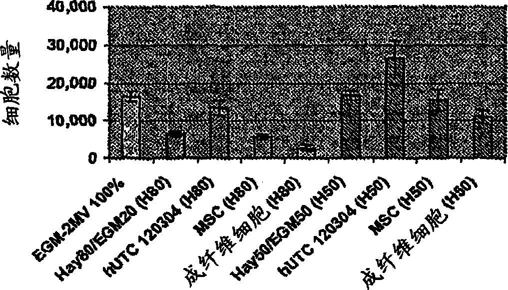
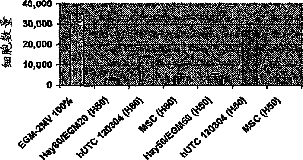
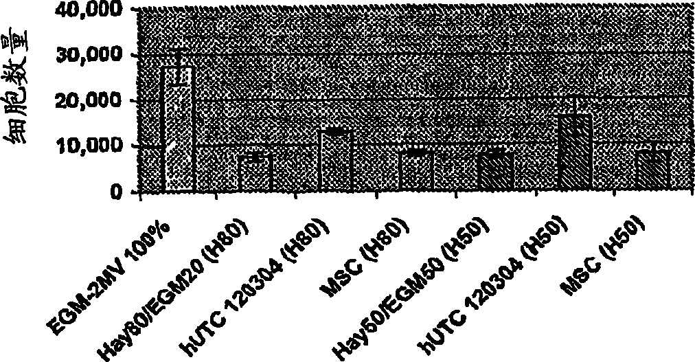
[0158] Studies were performed to determine the effect of human umbilical cord tissue-derived cells (hUTCs) on the proliferation and migration of endothelial cells in vitro. These effects were investigated by co-culturing hUTCs with endothelial cells and by incubating cultures of human umbilical vein endothelial cells (HUVECs) with hUTC lysates. The results presented here show that hUTCs induce increased proliferation and migration of endothelial cells. Furthermore, the data also suggest that these effects are mediated in part by fibroblast growth factor (FGF) and hepatocyte growth factor (HGF).
Materials and methods
[0159] Refrigerated human umbilical cord tissue-derived cells (hUTCs) of lot number 120304 were thawed between passages 8-9, inoculated into gelatin-coated bottles, and grown in Hayflick growth medium (DMEM-low glucose [Gibco, catalog number 11885-084] , 15% v / v ...
PUM
| Property | Measurement | Unit |
|---|---|---|
| diameter | aaaaa | aaaaa |
Abstract
Description
Claims
Application Information
 - R&D
- Intellectual Property
- Life Sciences
- Materials
- Tech Scout
- Unparalleled Data Quality
- Higher Quality Content
- 60% Fewer Hallucinations
Browse by: Latest US Patents, China's latest patents, Technical Efficacy Thesaurus, Application Domain, Technology Topic, Popular Technical Reports.
© 2025 PatSnap. All rights reserved.Legal|Privacy policy|Modern Slavery Act Transparency Statement|Sitemap|About US| Contact US: help@patsnap.com


