


Quick Research
Generate reliable direction feasibility study reports for your R&D in just a few steps.

Technical Q&A
Discover and master advanced knowledge NOW. Basics, ideas, possibilities, all at once.

Find Solutions
As an expert in R&D theories, this can generate solutions to your technical problems instantly.

Evaluate Feasibility
Analyze your overall solution with one click, know your potential R&D risks in advance.

Monitor Landscape
Get weekly tech updates, stay abreast of the latest tech innovations and key insights.
Method for quantitatively testing MG7-Ag in serum
A quantitative detection method and serum technology, applied in the biological field, to achieve the effect of improving sensitivity, ensuring universality and repeatability, and eliminating inter-chamber differences
- Summary
- Abstract
- Description
- Claims
- Application Information
AI Technical Summary
Problems solved by technology
Method used
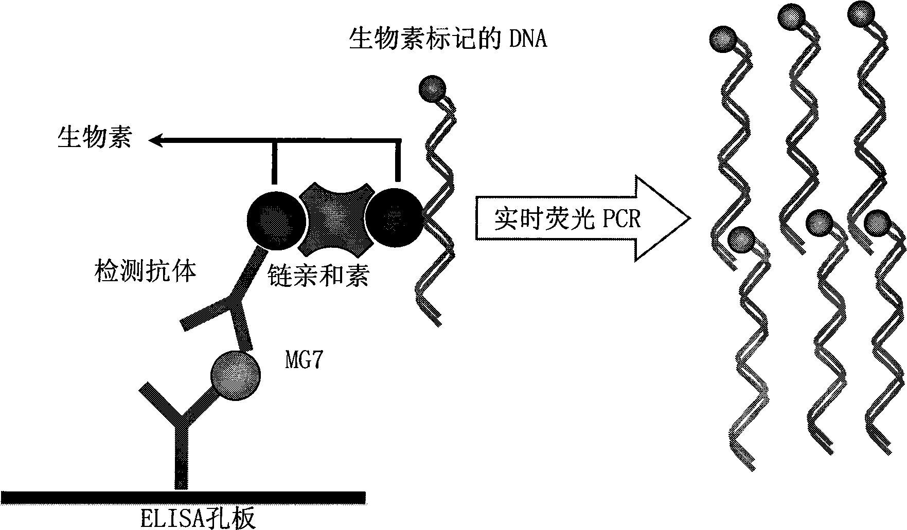
Image

Examples
preparation example Construction
[0051] 1) Preparation of tissue homogenate: Wash fresh gastric cancer tissue with normal saline in a water bath or ice bath at 4°C to remove blood stains and dirt, remove capsule or connective tissue, and cut the washed tissue into 0.3-0.5 cm 3 Small pieces, add an appropriate amount of saline, put into the barrel of a mashing machine to make a tissue homogenate. After the tissue homogenate was centrifuged at 3000r / min for 10min, the cells and tissue debris were removed, and the supernatant was kept for use.
[0052] Or cell crushing: collect gastric cancer cell line SGC7901 or MKN45, count the cells, place the cells in liquid nitrogen for 10 minutes, then take them out and thaw them at 37°C, repeat the freeze-thaw operation three times; remove cell debris, and centrifuge at 3000r / min for 10min , and the supernatant was kept for later use.
[0053] 2) Crude extraction of MG7-Ag: Salt out the tissue homogenate or cell lysate supernatant with saturated ammonium sulfate to make ...
Embodiment
[0056] Using MG7-Ag in gastric cancer cell SGC7901 as a standard, the quantitative detection of the MG7-Ag content in the serum to be tested specifically includes the following steps:
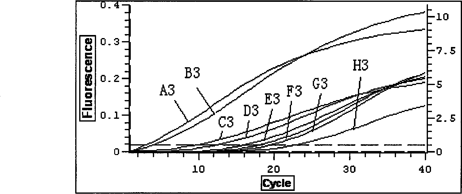
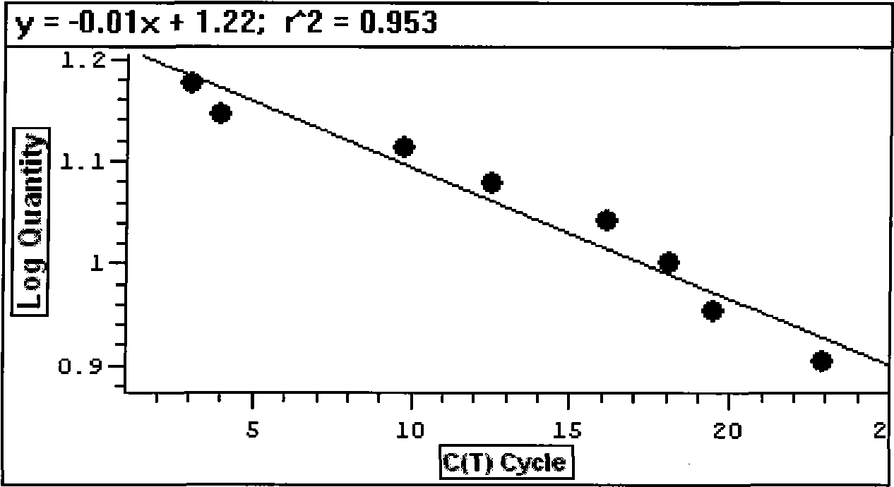
[0057] 1) Coating: Divide the wells of the ELISA plate into standard wells and detection sample wells; standard wells: gradient concentration diluted MG7-Ag standard, 100 μl per well; detection sample wells: add 90 μl of serum to be tested and 10 μl of 10 Double-dilute coating buffer, mix well; ELISA plate well overnight at 4°C, discard liquid, wash 3 times with PBS-T solution, 5 min each time.
[0058] Specifically: MG7-Ag in gastric cancer cell SGC7901 was used as a standard, and 10 7 、10 6 、10 5 、10 4 、10 3 、10 2 、10 1 The concentration gradient dilution of 0 cells was added to the standard wells (A3, B3, C3, D3, E3, F3, G3, H3); to prepare the serum to be tested: draw 2ml of blood, let it stand at room temperature for 30 minutes, 3000rpm for 20 minutes, and draw the upper Set aside a...
PUM
 Abstract
Description
Claims
Application Information
 - R&D Engineer
- R&D Manager
- IP Professional
- Industry Leading Data Capabilities
- Powerful AI technology
- Patent DNA Extraction
Browse by: Latest US Patents, China's latest patents, Technical Efficacy Thesaurus, Application Domain, Technology Topic, Popular Technical Reports.
© 2024 PatSnap. All rights reserved.Legal|Privacy policy|Modern Slavery Act Transparency Statement|Sitemap|About US| Contact US: help@patsnap.com