Method for detecting proliferation of improved GFP expression cell

A detection method and cell technology, applied in the determination/inspection of microorganisms, biochemical equipment and methods, fluorescence/phosphorescence, etc., to achieve the effects of simple operation, easy detection, subsequent multiple labeling, and good fluorescent signals

Inactive Publication Date: 2012-05-30
SHANDONG UNIV QILU HOSPITAL
View PDF0 Cites 0 Cited by
  • Summary
  • Abstract
  • Description
  • Claims
  • Application Information

AI Technical Summary

Problems solved by technology

[0006] for the above purpose, the invention provides a kind of improved GFP expression cell proliferation detection method, solves the problem that the GFP color weakens after the traditional fixation and staining method processing, Make GFP bright in color and easy to detect; and avoid the disadvantages of complicated steps such as formamide and hydrochloric acid acidification

Method used


Image

  • Method for detecting proliferation of improved GFP expression cell
  • Method for detecting proliferation of improved GFP expression cell
  • Method for detecting proliferation of improved GFP expression cell

Examples


Embodiment

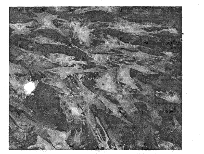
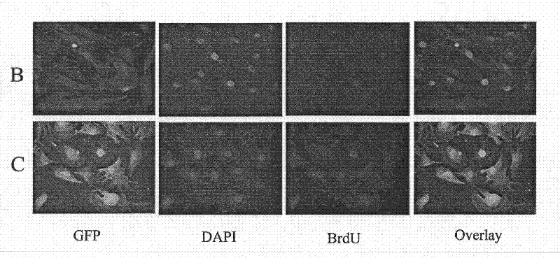
[0039] Embodiment: To detect the proliferation of GFP expressing cells, the steps are as follows:

[0040] 1) Add BrdU to the GFP-expressing cells to a final concentration of 30 μg / ml, and incubate at 37°C for 1 hour;

[0041] 2) Prepare 0.1% formaldehyde (volume concentration) with Hank's solution, and fix the cells for 12 hours under culture conditions;

[0042] 3) Wash with Hank's solution 3 times, 5 minutes each time;

[0043] 4) 0.2% Triton X-100 (polyethylene glycol octylphenyl ether) for 20 minutes;

[0044] 5) Rinse with Hank's solution 3 times, 5 minutes each time;

[0045] 6) 100U / ml DNase I (deoxyribonuclease I), treated at room temperature for 5 minutes;

[0046] 7) Wash with TBS 3 times, 5 minutes each time;

[0047] 8) Block with normal goat serum for 1 hour at room temperature;

[0048] 9) Add anti-BrdU primary antibody, and place in a humid box at 4°C overnight;

[0049] 10) Wash with TBS 3 times, 5 minutes each time;

[0050] 11) Add goat anti-mouse sec...


PUM


Abstract

The invention discloses a method for detecting the proliferation of an improved GFP expression cell, comprising the following steps: carrying out BrdU percolation dyeing and DAPI redyeing on a cell taking a green fluorescent protein (GFP) as a reporter protein; then sealing slices by a conventional fluorescence resistant quencher; observing by a fluorescent microscope; and taking photos. The method for detecting the GFP expression cell keeps a GFP color bright, has no obvious change of signal strength compared with a cultivation state and thoroughly solves the problem that the GFP color is faded after being processed by the traditional fixing and dyeing method, thereby enabling the GFP color to be bright and being easy to detection and follow multiple marks and preventing the shortages ofcomplex method steps, i.e. formamide denaturation, hydrochloric acid acidification, and the like.

Description

technical field [0001] The invention relates to an improved detection method for the proliferation of GFP expressing cells. [0002] Background technique [0003] Green fluorescent protein (GFP) is a light-emitting protein isolated from jellyfish, which absorbs blue light and emits green light, and does not require a light-emitting cofactor. Because GFP is stable in mammalian cells and easily detected by fluorescence microscopy, GFP has been a commonly used marker molecule and is currently the most useful research tool for molecular and cell biologists to track and quantify target proteins. However, after routine fixation (commonly used fixatives include: 70% ethanol, 4% paraformaldehyde, acetone, methanol, etc.), nucleic acid denaturation (formamide, hydrochloric acid), and multiple staining of cells expressing GFP, the GFP signal was significantly weakened. Not even detectable. Image results often require post-processing. [0004] BrdU is used to mark proliferating and ...

Claims


Application Information

Patent Timeline
Patent Type & Authority Patents(China)
IPC IPC(8): C12Q1/68C12Q1/00G01N21/64
Inventor 王旭平
Owner SHANDONG UNIV QILU HOSPITAL
Why Eureka
Try Eureka