System for detecting pulmonary malignant tumour and benign protuberance based on PET/CT image texture characteristics

A texture feature, CT image technology, applied in the field of medical digital image processing

Inactive Publication Date: 2010-03-17
FUDAN UNIV
View PDF0 Cites 60 Cited by
  • Summary
  • Abstract
  • Description
  • Claims
  • Application Information

AI Technical Summary

Problems solved by technology

Similarly, in PET / CT fusion images, experienced doctors can visually judge whether cancer has occurred, so cancerous tissues and benign tissues should have certain differences in texture; the texture features of CT / PET fusion images should be used as A basis for cancer judgment, but there is no mature technology application using texture features to distinguish lung cancer from benign nodules (such as inflammation, etc.)

Method used


Image

  • System for detecting pulmonary malignant tumour and benign protuberance based on PET/CT image texture characteristics
  • System for detecting pulmonary malignant tumour and benign protuberance based on PET/CT image texture characteristics
  • System for detecting pulmonary malignant tumour and benign protuberance based on PET/CT image texture characteristics

Examples


Embodiment Construction

[0075] Further describe the present invention below in conjunction with accompanying drawing and example.

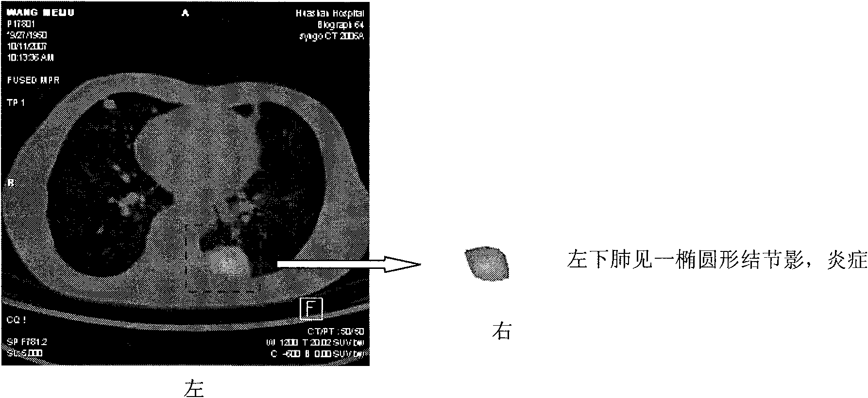
[0076] Patients A and B are scanned on the PET / CT workbench. In actual use, an interface can be built between the system and the PET / CT graphics workstation, so that the system can directly obtain PET / CT fusion images from the graphics workstation. figure 2 The left is the PET / CT fusion image of patient A, image 3 is the PET / CT fusion image of patient B.

[0077] The original fused image obtained from the PET / CT graphics workstation is the preprocessed image A obtained through the processing of the image preprocessing module 2 . Preprocessing includes image edge sharpening, contrast enhancement and other operations. Module 2 also provides a manual labeling interface and a graphical operation interface for manual labeling of lesion areas.

[0078]The preprocessed image A is further processed by the image segmentation and ROI labeling module 3 . This module extracts ...


PUM


Abstract

The invention relates to a system for detecting a pulmonary malignant tumour and a benign protuberance based on PET / CT image texture characteristics, belonging to the technical field of medical digital image processing. The invention has the major function of searching useful texture characteristics from a PET / CT picture to better distinguish a pulmonary tumour tissue and a benign tissue. The system comprises the following steps: firstly, dividing a region of interest ROI from the PET / CT image and then extracting five texture characteristics of roughness, contrast, busyness, complexity and intensity of the ROI; then, carrying out classifying discrimination on the characteristics by distance calculation and a characteristic classifier and distinguishing the malignant tumour and the benign protuberance effectively by the combined data of various characteristics.

Description

technical field [0001] The invention belongs to the technical field of medical digital image processing, in particular to a detection system for lung malignant tumors and benign nodules based on PET / CT image texture features. Background technique [0002] Lung cancer is one of the deadliest cancers worldwide. Positron emission tomography / computed tomography (PET / CT), as a non-innovative imaging diagnostic technique, has been widely used in the diagnosis and preoperative staging of lung cancer in recent years. big development. Compared with traditional computerized tomography (computed tomography, CT), magnetic resonance imaging (Magnetic Resonance Imaging, MRI) or simple PET (positron emission tomography, positron emission tomography) and other technologies, the advantages of PET / CT technology is very obvious. The contrast resolution of CT images in soft tissues is very low, and many malignant tumor tissues exist in soft tissues, and the difference between them and the su...

Claims


Application Information

Patent Timeline
Patent Type & Authority Applications(China)
IPC IPC(8): A61B6/03G06T7/40
Inventor 张文强管一晖潘岩王长梅袁永盛周垣施翔沈域
Owner FUDAN UNIV
Why Eureka
Try Eureka