Method and system for processing images based on photographing in vivo by wireless capsule endoscopy or video endoscope

An image processing and image processing device technology, which is applied in image data processing, 3D image processing, in-vivo radio detectors, etc., can solve problems such as complicated information, inability of diagnosis results to be applied clinically in time, and affecting the speed of diagnosis and identification of diseases, etc. Achieve the effect of improving image quality, economical effect and development, and enhancing image resolution

Active Publication Date: 2010-06-02
XIAN JIAOTONG LIVERPOOL UNIV
View PDF1 Cites 24 Cited by
  • Summary
  • Abstract
  • Description
  • Claims
  • Application Information

AI Technical Summary

Problems solved by technology

[0016] The purpose of the present invention is to provide an image processing method based on wireless capsule endoscope imaging in vivo, which solves the problem that the complexity of wireless capsul...

Method used


Image

  • Method and system for processing images based on photographing in vivo by wireless capsule endoscopy or video endoscope
  • Method and system for processing images based on photographing in vivo by wireless capsule endoscopy or video endoscope
  • Method and system for processing images based on photographing in vivo by wireless capsule endoscopy or video endoscope

Examples


Embodiment Construction

[0045] The above solution will be further described below in conjunction with specific embodiments. It should be understood that these examples are used to illustrate the present invention and not to limit the scope of the present invention. The implementation conditions used in the examples can be further adjusted according to the conditions of specific manufacturers, and the implementation conditions not indicated are usually the conditions in routine experiments.

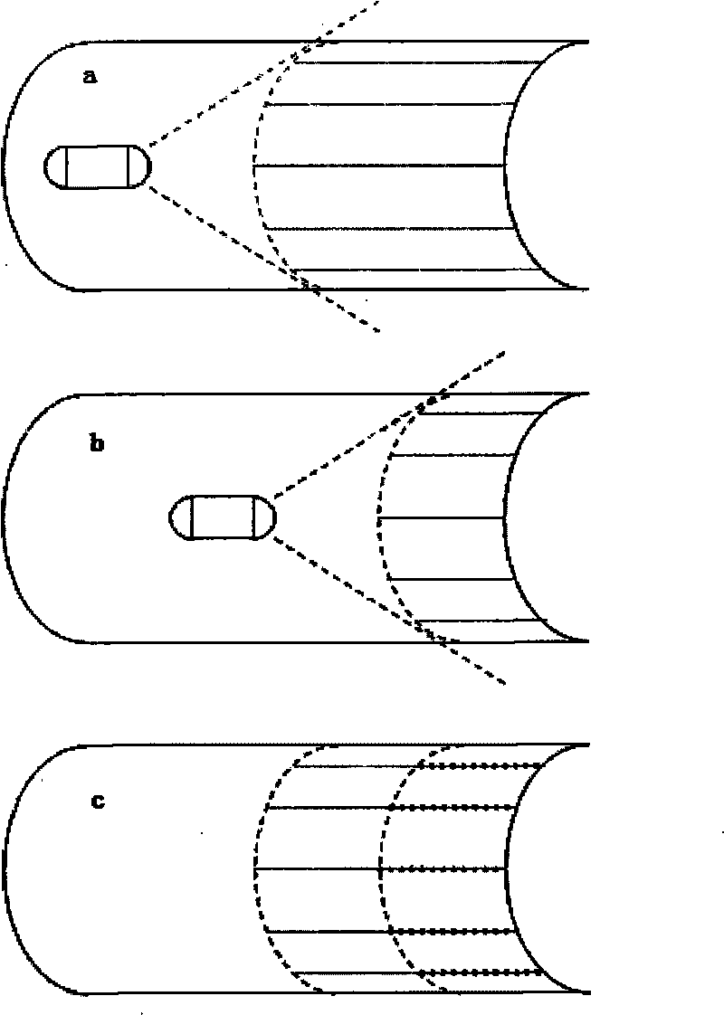
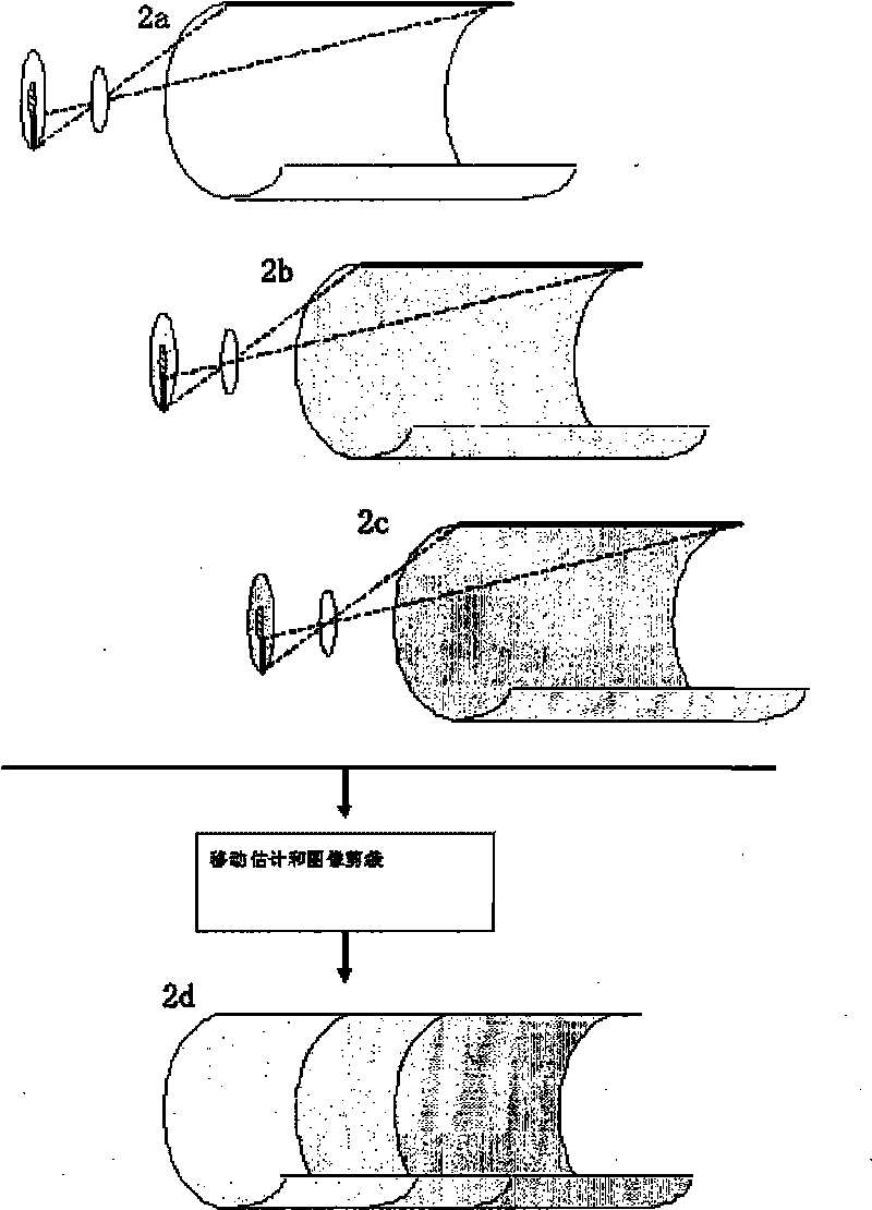
[0046] Embodiment Example of image processing based on wireless capsule endoscope camera in vivo

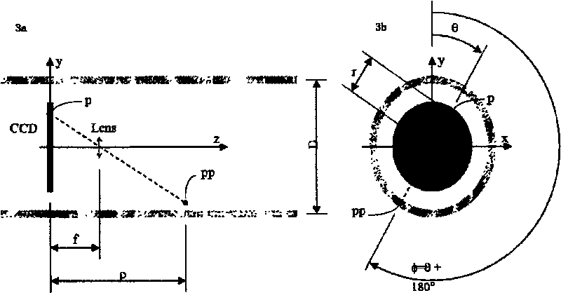
[0047] In this embodiment, the wireless capsule endoscope used is the PillCam product of Given Company; the wireless capsule endoscope includes an image sensor CCD and a wireless transmission device, and the front and rear ends of the image sensor are respectively provided with focusing lenses and batteries. Light-emitting diodes are arranged on the outside of the lens, and the light-emitting diodes, focusing lens ...


PUM


Abstract

The invention discloses a method and a system for processing images based on photographing in vivo by a wireless capsule endoscopy or a video endoscope. The method comprise the following steps of: (1) continuously capturing two-dimensional reversed images of an intestinal canal inner wall through a reversed projection of a image sensor of the wireless capsule endoscopy or the video endoscope to the intestinal wall, and sending the two-dimensional reversed images with continuous sequences by a wireless sending device of the wireless capsule endoscopy or the video endoscope to an image processing device for storage; (2) identifying a superposition part of the continuous sequences of the two-dimensional reversed images obtained from the step (1) by the image processing device; and (3) cutting and splicing the superposition part of the continuous sequences of the reversed images by the image processing device to form a three-dimensional image which presents the whole intestinal canal inner wall. The method can reduce time on studying photographed images by medical workers and greatly improve diagnosis efficiency.

Description

technical field [0001] The invention belongs to the technical field of auxiliary equipment for clinical observation and diagnosis of small intestinal mucosal diseases, and relates to a method for processing images captured by a clinical intestinal endoscope, so as to facilitate diagnosis by medical staff and reduce diagnosis time and cost. Background technique [0002] The average adult digestive tract is about 10 meters long. The upper digestive tract includes the esophagus, gastrointestinal tract and duodenum, with an average length of 1 m. Terminates in the colon and rectum, about 2 meters. Between the two is the small intestine, which is characterized by being very long (7 meters on average) and having many bends and folds. Diagnosis and treatment of digestive tract diseases generally adopts gastroscope examination, and the gastroscope in the prior art is a kind of long and flexible thin-walled flexible tube with a camera at its end; the end is swallowed into the oral ...

Claims


Application Information

Patent Timeline
IPC IPC(8): A61B5/07A61B1/00G06T15/00
Inventor 罗天明林永义
Owner XIAN JIAOTONG LIVERPOOL UNIV
Why Eureka
Try Eureka