Method for structuring ultrasound long axis image quickly with high fidelity

A high-fidelity, vascular technology, applied in the field of medical image processing, can solve the problem of insufficient precision, achieve the effect of improving quality, improving image quality, and accelerating speed

Inactive Publication Date: 2010-06-16
INST OF AUTOMATION CHINESE ACAD OF SCI
View PDF0 Cites 6 Cited by
  • Summary
  • Abstract
  • Description
  • Claims
  • Application Information

AI Technical Summary

Problems solved by technology

[0007] The purpose of the present invention is to solve the problem that the accuracy of the long-axis image construction algorithm used in the current IVUS imaging equipment or software is not high enough, for this purpose, to provide a high-fidelity real-time large data volume IVUS long-axis image construction method

Method used


Image

  • Method for structuring ultrasound long axis image quickly with high fidelity
  • Method for structuring ultrasound long axis image quickly with high fidelity
  • Method for structuring ultrasound long axis image quickly with high fidelity

Examples


Embodiment Construction

[0021] Various details involved in the technical solution of the present invention will be described in detail below in conjunction with the drawings and embodiments. It should be pointed out that the described embodiments are only intended to facilitate the understanding of the present invention, rather than limiting it in any way.

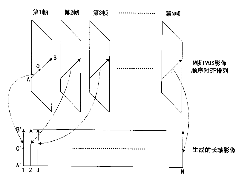
[0022] Different from the traditional method of loading three-dimensional intravascular ultrasound (IVUS) images for each frame and processing them separately, the present invention adopts a method based on three-dimensional image interpolation, so all N frames of images are regarded as a three-dimensional volume data, and the IVUS long axis Image construction can be regarded as a sampling process of three-dimensional plane to three-dimensional volume data, which requires a suitable coordinate system to ensure correct calculation.

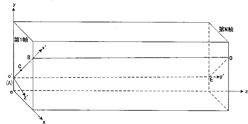
[0023] attached figure 2 A schematic diagram of the structure of the global coordinate system and the viewpoint c...


PUM


Abstract

The invention discloses a method for structuring ultrasound long axis image quickly with high fidelity. The method is implemented by using a parallel computing architecture based on a graphics processor to perform efficient management to three-dimensional IVUS data with large data volume by quadtree data structure, performing recursion traverse to the quadtree and computing the intersecting point coordinate of the plane on which the long axis image is located and the quadtree data node, decomposing the long axis image into a series of small planes, then sending to the graphics processor for mapping and interpolation computing. The invention solves the problem that the data volume is too large to be loaded display memory of the graphics processor, improves utilization efficiency of texture cache of the graphics processor, and enhances computation speed and image quality by computing the three-dimensional spline interpolation by using single instruction multidata vector dot product of the graphics processor. The invention can achieve above 30 frame per second in structuring long axis image with high fidelity on common middle / high-grade display card for large scale IVUS image data of 512*512*4000*16bit.

Description

technical field [0001] The invention relates to the technical field of medical image processing, in particular to a method for rapidly and high-fidelity constructing intravascular ultrasonic long-axis images. Background technique [0002] Worldwide, coronary heart disease (CHD for short) is currently one of the leading causes of human death, and coronary interventional surgery is the most important means of treating CHD. The recently emerging intravascular ultrasound (IVUS) imaging is considered to be the best imaging method to assist coronary interventional therapy. The acquisition of IVUS images is minimally invasive. A very small high-frequency ultrasound probe is added to the catheter, and it is placed through the patient's blood vessel into the coronary artery suspected by the doctor to have a lesion. The probe is pulled back at a uniform speed through motor control. A series of vascular endoscopic images are thus obtained. [0003] At present, doctors mainly use the ...

Claims


Application Information

Patent Timeline
Patent Type & Authority Applications(China)
IPC IPC(8): G06T1/00A61B8/12
Inventor 赵明昌何晖光
Owner INST OF AUTOMATION CHINESE ACAD OF SCI
Why Eureka
Try Eureka