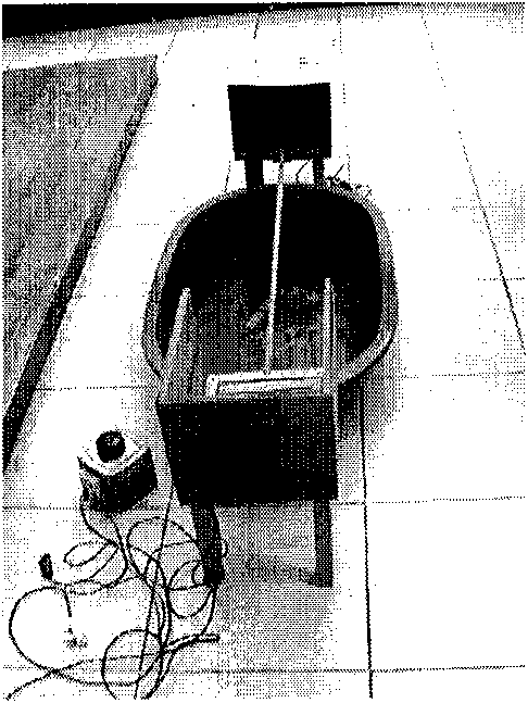
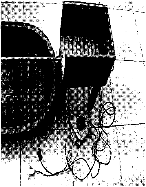
Experimental animal rat cerebellar ataxia detection model
A technique for ataxia and experimental animals, applied in diagnostic recording/measurement, medical science, sensors, etc., can solve problems such as behavioral and physiological function impairment
- Summary
- Abstract
- Description
- Claims
- Application Information
AI Technical Summary
Problems solved by technology
Method used
Image
Examples
Embodiment 1
[0017] Validity test of rat cerebellar ataxia detection model
[0018] Local anesthesia with 3% sodium pentobarbital (20mg / kg), the rat prone position, the head is fixed on the KW-II mouse brain stereotactic model. After disinfection, the occipital skin was cut along the sagittal line, the skull was exposed, and the bone surface was cleaned with hydrogen peroxide. Refer to the rat brain anatomical location map and the rat brain three-dimensional location map, take the intersection of the sagittal suture and the herringbone suture as the coordinate 0 point, 4.4mm backward, 2mm left or right side, drill a small hole with a diameter of 1mm, The 5μl microsyringe is fixed on the stereotactic model. The tip of the injection needle has a diameter of 20μ, vertically downwards 3-4mm, and a bolus speed of 0.25μl / min. Slowly inject 1μl of KA (10μg / μl) into the left and right hemispheres and vermis of the rat cerebellum After the symptoms of ataxia have stabilized (mostly 12 hours after inj...
Embodiment 2
[0024] Pathologically confirmed
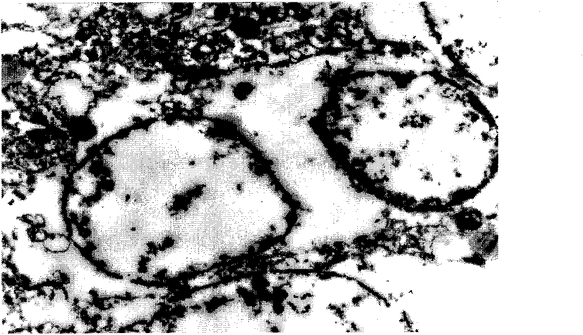
[0025] Pathology confirmed that the KA injection site was accurate. Most of the neurons at the injection site were edema, necrosis, and loss. Under light microscope, rats in the experimental group showed focal necrosis and dissolution of brain tissue, loss of a large number of neurons, brain tissue edema, granular cell nucleus condensation, nuclear fragmentation, red neuronal pyknosis, cell body shrinkage, deformation and other changes ( Picture 9 ). Under the electron microscope, the granular cells of the cerebellar fastigial nucleus in the experimental group appeared edema and large vacuoles were formed in the cytoplasm; the granular cells shrank, their size decreased, their density increased, and the cell morphology was irregular. Purkinje cell endoplasmic reticulum expands. The chromatin of some neurons is concentrated and edge-collected, showing early and mid-term apoptosis changes. The myelin sheath of the myelinated nerve fiber is sepa...
PUM
 Abstract
Description
Claims
Application Information
 - R&D
- Intellectual Property
- Life Sciences
- Materials
- Tech Scout
- Unparalleled Data Quality
- Higher Quality Content
- 60% Fewer Hallucinations
Browse by: Latest US Patents, China's latest patents, Technical Efficacy Thesaurus, Application Domain, Technology Topic, Popular Technical Reports.
© 2025 PatSnap. All rights reserved.Legal|Privacy policy|Modern Slavery Act Transparency Statement|Sitemap|About US| Contact US: help@patsnap.com


