Fluorescence microscopy method to generate multi-layer polished sections by utilizing Fresnel biprism and device

A Fresnel double prism and light sheet technology, applied in microscopes, optics, optical components, etc., can solve the problems of slow image acquisition rate, small penetration depth, uneven illumination of single-layer light microscopy technology, etc., to achieve The effect of fast image acquisition rate and large penetration depth

Inactive Publication Date: 2010-09-01
XI'AN INST OF OPTICS & FINE MECHANICS - CHINESE ACAD OF SCI
View PDF0 Cites 13 Cited by
  • Summary
  • Abstract
  • Description
  • Claims
  • Application Information

AI Technical Summary

Problems solved by technology

[0006] The present invention proposes a method and device for fluorescence microscopy using a Fresnel double prism to produce a multi-layer light sheet, which solves the problem of uneven illumination, small penetration depth in the sample, and image acquisition rate in the existing single-layer light microscopy technology. slow technical issues

Method used


Image

  • Fluorescence microscopy method to generate multi-layer polished sections by utilizing Fresnel biprism and device
  • Fluorescence microscopy method to generate multi-layer polished sections by utilizing Fresnel biprism and device
  • Fluorescence microscopy method to generate multi-layer polished sections by utilizing Fresnel biprism and device

Examples


Embodiment

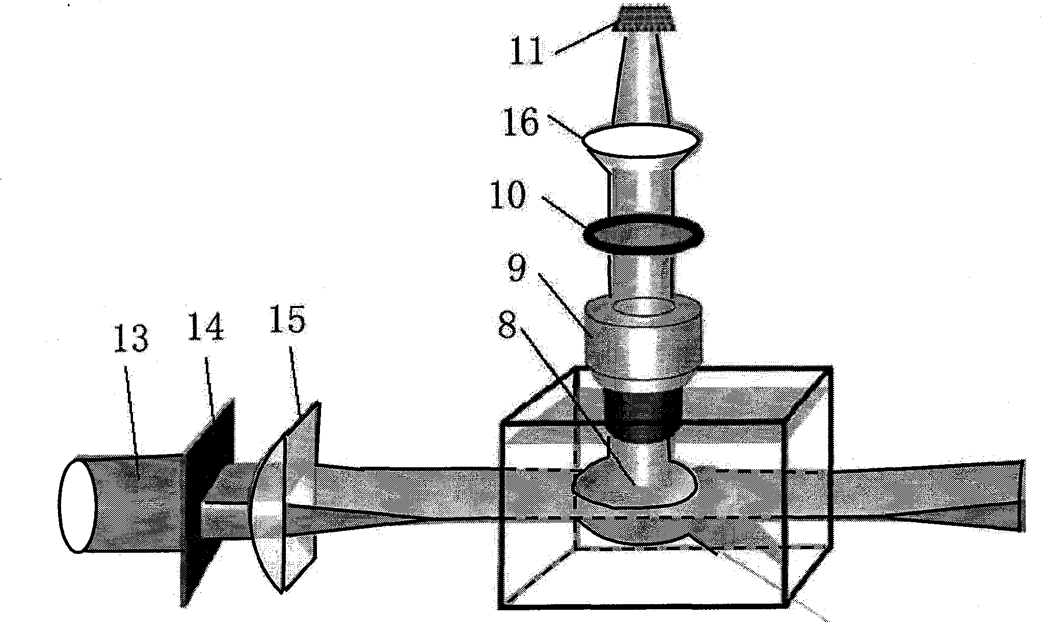
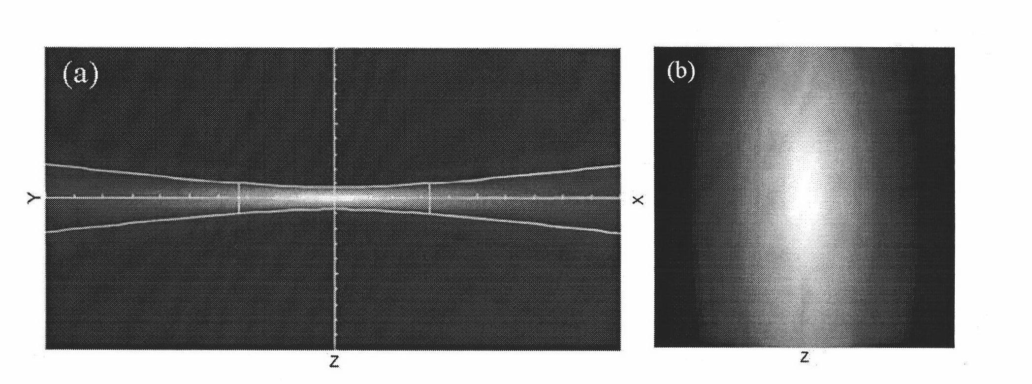
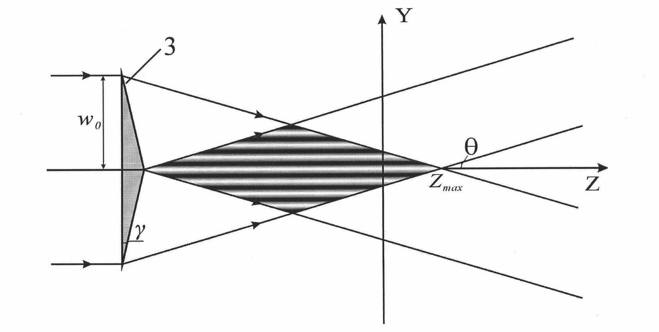
[0075] Embodiment: select the Fresnel double prism of base angle=5 ° in the experiment, material refractive index n=1.5, laser wavelength=532nm, can obtain the spacing Δ=6m of light sheet by formula (2), and the microscopic objective lens Depth of field is typically less than 6m, so there is no crosstalk between individual light sheets. The radius w of the incident laser beam 0 = 2mm, lens L 1 focal length f 1 =75mm, lens L 2 focal length f 2 =75mm, d 1 =5mm, into formula (4) can get Z max =141mm, Z min =107mm, which greatly increases the working distance of the system and facilitates the placement of components such as sample stages and microscope objectives.

[0076] A glass plate of thickness t is placed behind lens L2. The glass plate will not change the beam angle but will produce a phase difference between the two beams participating in the interference:

[0077] δ = 2 π λ ...


PUM


Abstract

The invention provides a fluorescence microscopy method to generate multi-layer polished sections by utilizing a Fresnel biprism and a device. The device comprises parallel beams, a generating system of polished sections, a sample cell and an image acquisition system. The generating system of polished sections comprises a Fresnel biprism or a system composed of a Fresnel biprism, a telescope system and a phase shifting glass sheet; the sample cell is arranged at the rear part of the Fresnel biprism or at the rear part of the phase shifting glass sheet. As the parallel beams refract after passing through the Fresnel biprism, an interference field is generated in the beam overlaying region behind the prism, thus the light field of multi-layer polished sections is obtained. The invention solves the technical problems of ununiform luminance, small penetration depth of samples and slow rate of image acquisition in the existing mono-layer microscopy technology; the obtained multi-layer polished sections have great penetration depth, can be applied to fluorescence microscopy imaging of living entity samples; and the image acquisition rate is high.

Description

technical field [0001] The invention relates to a fluorescence microscopy method and device. Background technique [0002] In life science research, in order to study the life activities of the living body at the micro level, it is required that the microscopic imaging device has high time and space resolution and three-dimensional imaging capability. In vivo optical microscopy imaging has become one of the most active fields in biomedical optics research in recent years. [0003] Fluorescence microscopy is an ideal method for in vivo imaging studies due to its ease of operation and intuitiveness. Using this imaging technique, the activities and responses of fluorescently labeled genes and cells in living animals can be observed in real time. Fluorescence microscopy mainly includes ordinary wide-field fluorescence microscopy, laser confocal fluorescence microscopy and multiphoton fluorescence microscopy. Ordinary wide-field fluorescence microscopy has the advantages of fa...

Claims


Application Information

Patent Timeline
Patent Type & Authority Applications(China)
IPC IPC(8): G02B21/16G02B21/36
Inventor 雷铭姚保利
Owner XI'AN INST OF OPTICS & FINE MECHANICS - CHINESE ACAD OF SCI
Why Eureka
Try Eureka