Rapid staining method for biopolymers

A technology of biopolymers and dyeing methods, applied in the direction of measuring devices, instruments, material analysis by electromagnetic means, etc.

Active Publication Date: 2011-01-05
NANJING GENSCRIPT BIOTECH CO LTD
View PDF6 Cites 17 Cited by
  • Summary
  • Abstract
  • Description
  • Claims
  • Application Information

AI Technical Summary

Problems solved by technology

[0009] At present, there is no report on the method of using electrophoresis technology to separ...

Method used


Image

  • Rapid staining method for biopolymers
  • Rapid staining method for biopolymers
  • Rapid staining method for biopolymers

Examples


Embodiment 1

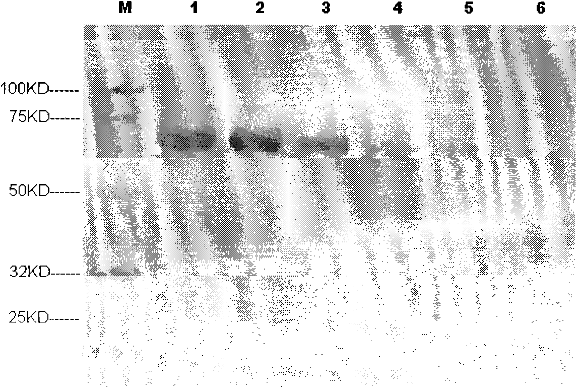
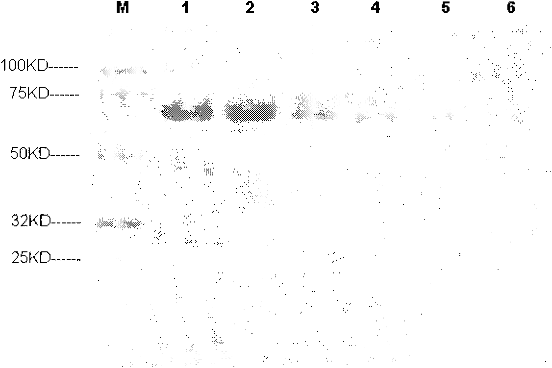
[0094] Example 1 Separation of proteins by electrophoresis and then staining.

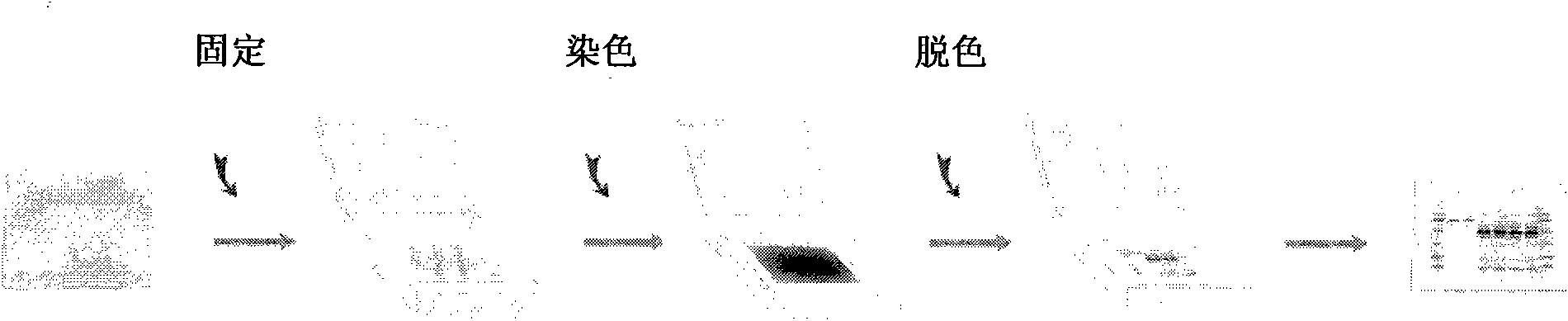
[0095] Bovine serum albumin staining. 1500ng, 750ng, 300ng, 150ng, 75ng, and 30ng of bovine serum albumin were loaded on lanes 1 to 6 of two PAGE gels (Genscript, MG008W10) respectively. After electrophoresis, one gel was used for rapid staining, specifically , after electrophoresis, take out the gel, put the gel in such as Figure 4 In the sandwich structure shown. Solution A is an aqueous solution containing 10-50% (v / v) isopropanol, 5-35% (v / v) ethanol, and 1-30mmol / L ethylenediaminetetraacetic acid, and solution B is an aqueous solution containing 0.05-0.5% ( Mass volume ratio) Coomassie brilliant blue, 20-60% (v / v) ethanol, 1-30mmol / L ethylenediaminetetraacetic acid, 2-30% (v / v) acetic acid aqueous solution, C is pH 4.0~ 10% phosphoric acid or similar buffer solution (preferably: A is an aqueous solution containing 25% isopropanol, 10% ethanol, 5mmol / L ethylenediaminetetraacetic acid, B is ...

Embodiment 2

[0099] Take DNA Marker (Genscript, 100bp ladder) respectively, and load the sample on the agarose gel with a concentration of 2%, 500ng / lane, and the nucleic acid is respectively loaded on the 1, 2, 3, 4, 5, In lanes 6 and 7, in TBE (aqueous solution containing 445 mmol / L Tris base, 445 mmol / L borate, and 10 mmol / L EDTA) buffer, constant current flow was 300 mA for 20 minutes. After electrophoresis, filter paper soaked with positively charged dye (diluted by ethidium bromide 10,000 times) was tiled on the positive plate, the agarose gel after electrophoresis was located on it, and two layers of soaked gel were tiled on top of the agarose. Wet the filter paper of TBE, add the negative electrode above to form a sandwich structure, and apply a voltage of 50V to the two electrodes for 3 minutes. After binding to DNA driven by an electric field, it is imaged under UV light. The dyeing time is 3 minutes, see the results Figure 5 .

Embodiment 3

[0100] Example 3: Combination of electrophoretic separation and staining of various biopolymers.

[0101] Take 500 μg of protein (bovine serum albumin) with a molecular weight of 67KD and 500 μg of a polypeptide with a molecular weight of 6KD, respectively, and add them to 1ml of water to prepare a mixed aqueous solution of various biopolymers. Add 1ml of solution B (containing 0.3% Coomassie brilliant blue, 46 % ethanol, 5mmol / L ethylenediaminetetraacetic acid, 8% aqueous solution of acetic acid), keep the dye in contact with the biopolymer for 1 minute, dye the biopolymer, and then take the solution and load it on one end of the cellulose acetate membrane medium (Length 10cm, immersed in pH 8.6 concentration of 0.07mol / L barbiturate buffer), medium applied voltage 200v, separation time 20 minutes, you can see that the peptide and protein can be separated and clearly colored, the result See Figure 8 .


PUM


Abstract

The invention belongs to the field of biotechnology, and discloses a rapid staining method for biopolymers. The rapid staining method comprises the steps of: combination of stain and a to-be-stained biopolymer, separation of uncombined stain and a biopolymer combined with the stain, and imaging of the biopolymer combined with the stain. The rapid staining method enables the stain to rapidly combine with the biopolymer through utilizing different mobilities of the stain and the biopolymer in a medium applied with an electric field, and can rapidly separate the uncombined stain and the biopolymer combined with the stain, shorten the whole staining time, save the cost, is simple to operate as the staining, destaining and color development can be finished in one step, and can combine the separation step and the staining step of the biopolymer together.

Description

technical field [0001] The invention belongs to the field of biotechnology, and relates to a rapid dyeing method for biopolymers with simple operation and less time consumption. Background technique [0002] The technique of using charged particles to move at different speeds in an electric field to achieve separation is called electrophoresis. Under certain conditions, the distance that charged particles move per unit time (that is, the mobility) under the action of unit electric field strength is constant. Different charged particles are electrophoresed in the same electric field due to their different charges, or the same charges but different charge-to-mass ratios. After a certain period of time, they are separated from each other due to different moving distances. The separation distance is proportional to the voltage of the applied electric field and the electrophoresis time. In 1937, the Swedish scholar A.W.K. Tiselius designed and manufactured a mobile interface el...

Claims


Application Information

Patent Timeline
IPC IPC(8): G01N1/30G01N27/447
Inventor 王珠银杨锦宇孟凡丽赖兵戴瞻章方良
Owner NANJING GENSCRIPT BIOTECH CO LTD
Why Eureka
Try Eureka